| SKU | DZ41310 |
|---|---|
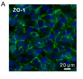
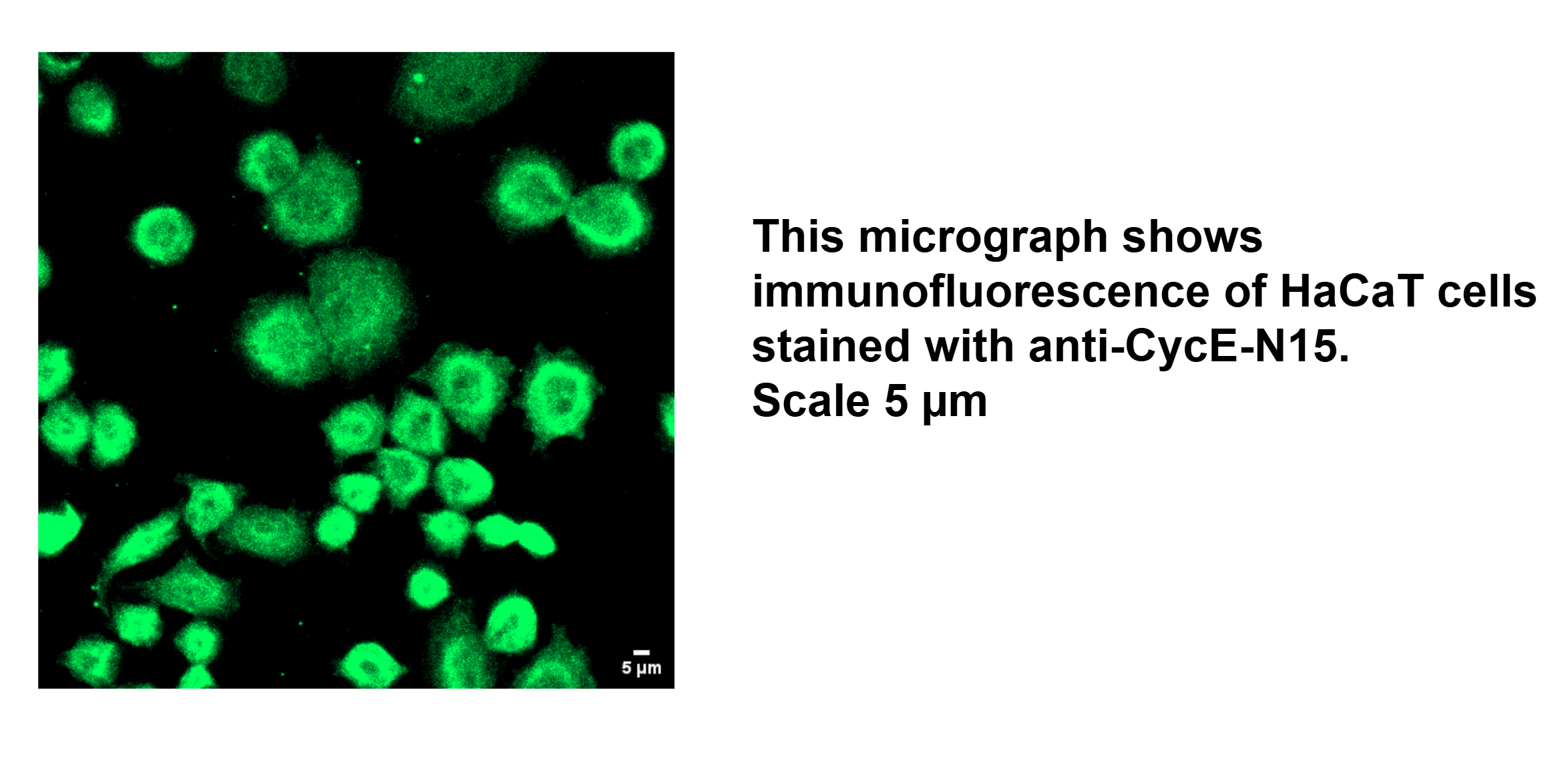
| Application | Immunofluorescence |
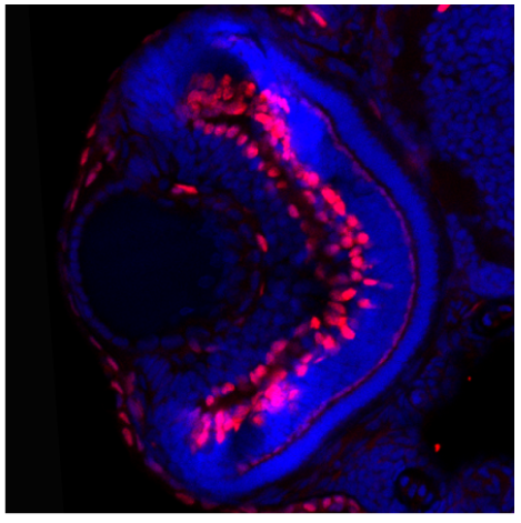
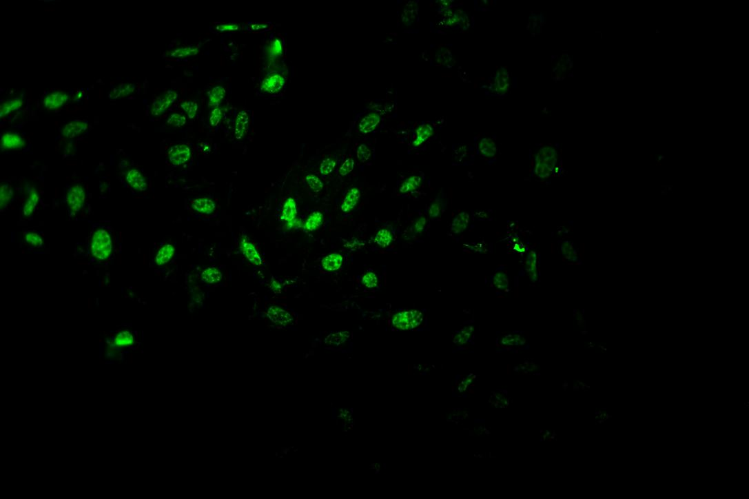
| Sample | Human HaCaT cell |
| Sample Processing Description | Cells were fixed in 4% paraformaldehyde for 15 minutes at room temperature, permeabilized with 0.1% Triton X-100 for 10 minutes and blocked in 1% BSA for 1 hour before incubation with the primary antibody. |
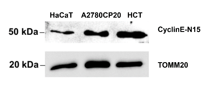
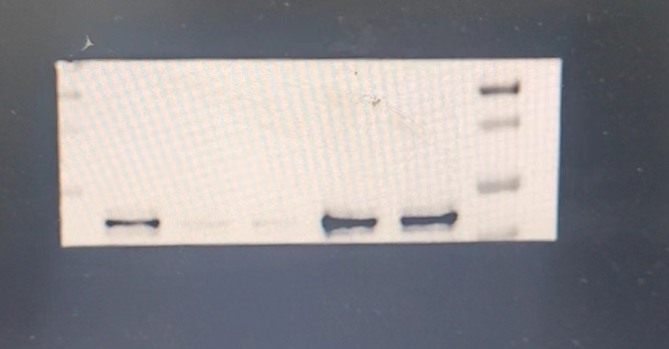
| Primary Antibody | Human CCNE1 Antibody |
| Primary Incubation | 1:100-1:200, overnight at 4 ℃ |
| Secondary Antibody | Goat anti-rabbit IgG conjugated to Alexa Fluor 488 |
| Secondary Incubation | 1:1000, 1-2 hours in room temperature |
| Other Reagents used | PBS, 0.1% Triton X-100, 1% BSA, DAPI for nuclear counterstaining |
| Detection | Fluorescence microscopy using AlexaFluor488 (excitation 488 nm, emission 519 nm) using Zeiss LSM 900 Confocal Microscope with Airyscan 2 |
| Results Summary | The CyclinE-N15 antibody (DZ41310) worked fine in IF application using human cell lines. In IF, the nuclear localization of Cyclin E matched the known expression pattern. A fluorescent secondary antibody (Alexa Fluor 488) was used for detection and gave clear signal. Overall, a reliable antibody for D15 transcript Cyclin E detection in human cell systems. |