This website uses cookies to ensure you get the best experience on our website.
- Table of Contents

7 Citations 16 Q&As


5 Citations 2 Q&As


2 Citations 10 Q&As

2 Citations 3 Q&As

2 Citations 11 Q&As

2 Citations 8 Q&As

2 Citations 10 Q&As


2 Citations 10 Q&As

2 Citations 16 Q&As
11 Citations 17 Q&As

1 Citations 3 Q&As

1 Citations 14 Q&As
1 Citations 4 Q&As


2 Citations
4 Citations
Facts about T-cell surface glycoprotein CD8 alpha chain.

Interacts concurrently with the T-cell receptor (TCR) and the MHC class I proteins presented by antigen presenting cells (APCs). Consequently, recruits the Src kinase LCK to the area of the TCR-CD3 complicated.
| Human | |
|---|---|
| Gene Name: | CD8A |
| Uniprot: | P01732 |
| Entrez: | 925 |

| Belongs to: |
|---|
| No superfamily |

53-6.7 CD8 Alpha; 53-6.7 CD8 Aplha; 53-6.7 CD8; 53-6.7 Clone; 53-6.7; CD8; CD8; CD8a molecule; CD8A; Cytotoxic T cell marker; Cytotoxic T lymphocyte marker; LEU2; p32; T cell marker
Mass (kDA):
25.729 kDA

| Human | |
|---|---|
| Location: | 2p11.2 |
| Sequence: | 2; NC_000002.12 (86784605..86808396, complement) |
CD8 on thymus-derived T-cells usually consists of a disulfide-linked alpha/CD8A and a beta/CD8B chain. Less frequently, CD8 can be expressed as a CD8A homodimer. A subset of natural killer cells, memory T-cells, intraepithelial lymphocytes, monocytes and dendritic cells expresses CD8A homo-dimers. Expressed at the cell surface of plasmacytoid dendritic cells upon herpes simplex virus-1 stimulation.
[Isoform 1]: Cell membrane; Single-pass type I membrane protein. CD8A localizes to lipid rafts only when associated with its partner CD8B.; [Isoform 2]: Secreted.


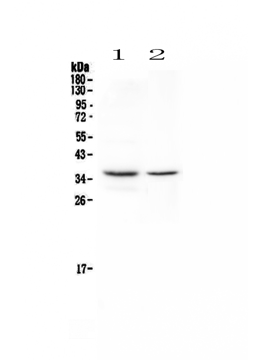
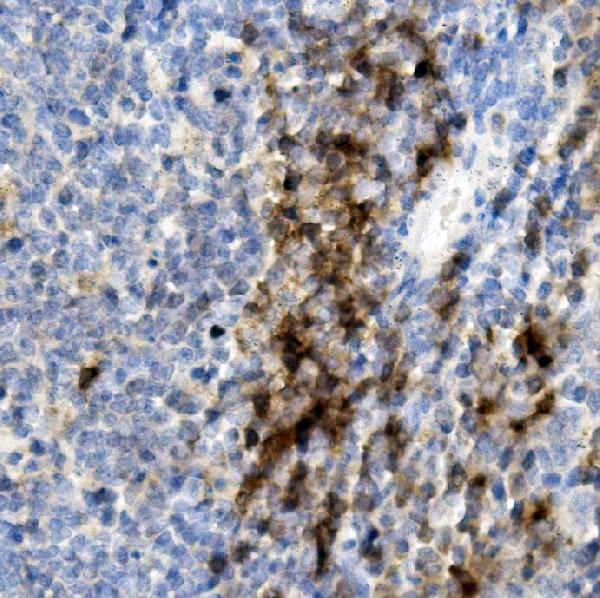
If you've ever thought about what you can do with anti-CD8A antibody You're not alone. Boster Bio offers high-affinity, primary antibodies that have been cited over the past 25 years. These antibodies are trusted by researchers because they have been validated on Western Blotting, Immunohistochemistry, and ELISA. Here's a quick review of the major features of the product.
The Boster Bio Anti-human CD8a Monoclonal antibody, CD8C, D8a by Boster Bio reacts with Human. They have been validated and flow-tested. The product can be stored at -20°C for one month or at room temperatures for up to one month. You can also freeze it and store it for up to six months. The antibodies react with human peripheral blood T cells and human Thymocytes.
Two chains comprise the molecules CD8 that are the beta and alpha chains. It is expressed on the cell surface by a subset of T cells, which are cytotoxic/suppressive. These cells are responsible for recognizing the antigens displayed by other immune cells, such as APCs and class I MHC molecules. Boster Bio Anti-human Cd8a Monoclonal Antibody recognizes this antigen in a variety of tissues. It binds to MHC class I molecules, while CD8 is expressed on the cell surface by Thymocytes and gamma delta TCR T cells, as well as dendritic cells.
The Boster Bio Anti-human CD8 C-D-Lanthan-CD8a Monoclonal Antibody is titrated and pre-tested. In the test volume of 100 uL, it stains around one million cells. Titrate the antibody in accordance with the cell number. In the test, a count of 105 to 108 cells should be used. It should be kept between 2°C and 8°C after it has been manufactured. Keep it away from long-term exposure to light and temperatures that are freezing.
The high specificity of the Boster Bio Antihuman CD8 CDLxA Monoclonal antibody is well-known, especially when administered to mice. This antibody has been proven to reduce CD8 cells in mice that have been subjected to experimental treatment according to research. The MAb's effect can be affected by the CD8 T-lymphocyte reaction. This is why it is crucial to review the citations carefully prior to purchasing this product.
Animal studies have shown that the anti-CD8 MAb caused a decrease in CD8 T-lymphocytes. It also prolongs the death of CD4 cells in acute infections. Anti-human CD8 MAb specifically reduced the production of peripheral blood CD8 T lymphocytes. The anti-CD8 MAb is safe and effective for clinical use.
A variety of studies have proven that the Boster Bio Anti-human CD8 C-DlxA Monoclonal Antibody is effective against SHIV infection. The anti-CD8 MAb significantly reduced the amount of viral load in macaques that were infected. Three macaques treated with the MAb showed p27 antigenemia, while the three control macaques didn't. The levels of PBMC-associated viral DNA in the treated macaques were also higher than those in the control group. After five weeks, the CD4 cells decreased to 88/ml blood. The macaque 565Z continued to have low levels of peripheral T cells that are CD4+.
The Boster Bio Anti-CD8 Cd88a Monoclonal Antibody is a highly specific and sensitive tool for detecting CD8A in a variety of cell types. Its specificity is demonstrated by its ability to recognize the human CD8 alpha antigen. This antibody has been flow-tested by combing it with PerCP-Cyanine5.5. It can be stored at 4°C for one year. It can be frozen in the event of need.
The CD8 molecule consists of two chains, alpha and beta. It is found on surfaces of all cell types. CD8 is expressed on the surface of a variety of cells including dendritic cells natural killer cells and cortical thymocytes. It also has important functions in recognizing foreign substances, like tumor cells. The CD8 antigen can be recognized in a variety of cell types such as thymocytes and dendritic cell as well as bone marrow cells.
Boster Bio Anti-Mouse CD8, also known as CD8a, is a monoclonal antibody. It reacts with Mouse CD8a and is PerCP-Cyanine 5.5.5 conjugated. It should be stored at a temperature of 4°C and protected from long-term light. It could contain a transporter protein. It has been tested in Immunofluorescence and ELISA.
The cell molecule of CD8a is composed of two chains: alpha and beta. It is found on T cells, which belong to the cytotoxic/suppressive cell subset. CD8 is found in 80 percent of human thymocytes. It is also found in the marrow on a variety of bone marrow cells. Boster Bio Anti-Mouse CD8 Cd8a Marker is designed to be used for research only. It is not suitable to diagnose purposes or for sale without authorization.
PMID: 3871356 by Littman D.R., et al. The isolation and sequence of the gene encoding T8: a molecule defining functional classes of T lymphocytes.
PMID: 3936473 by Parnes J.R., et al. Structure of Leu-2/T8 as deduced from the sequence of a cDNA clone.
*More publications can be found for each product on its corresponding product page