
Click image to see more details
Product Info Summary
| SKU: | MA1017 |
|---|---|
| Size: | 100 μg/vial |
| Reactive Species: | Human |
| Host: | Mouse |
| Application: | Flow Cytometry, IHC, ICC |
Customers Who Bought This Also Bought

Product info
Product Name
Anti-CD8 Cd8A Antibody (Monoclonal, CA-8)
SKU/Catalog Number
MA1017
BM0216 is an alternative SKU for this antibody, used in previous lots.
Size
100 μg/vial
Form
Lyophilized
Description
Boster Bio Anti-CD8 Cd8A Antibody (Monoclonal, CA-8) catalog # MA1017. Tested in Flow Cytometry, IHC, ICC applications. This antibody reacts with Human.
Storage & Handling
Store at -20˚C for one year from date of receipt. After reconstitution, at 4˚C for one month. It can also be aliquotted and stored frozen at -20˚C for six months. Avoid repeated freeze-thaw cycles.
Cite This Product
Anti-CD8 Cd8A Antibody (Monoclonal, CA-8) (Boster Biological Technology, Pleasanton CA, USA, Catalog # MA1017)
Host
Mouse
Contents
Mouse IgG in stabilizing components, 1.2% sodium acetate and 0.01mg NaN3.
Clonality
Monoclonal
Clone Number
CA-8
Isotype
Mouse IgG2a
Immunogen
Human thymocytes followed by peripheral blood T cells.
Cross-reactivity
No cross-reactivity with other proteins
Reactive Species
MA1017 is reactive to CD8A in Human
Calculated molecular weight
25.7 kDa
Background of CD8A
The human lymphocyte differentiation antigen CD8 is encoded by a single gene that gives rise to a 33- to 34-kDa glycoprotein expressed on the cell surface as a dimer and in higher molecular mass forms.
Antibody Validation
Boster validates all antibodies on WB, IHC, ICC, Immunofluorescence, and ELISA with known positive control and negative samples to ensure specificity and high affinity, including thorough antibody incubations.
Application & Images
Applications
MA1017 is guaranteed for Flow Cytometry, IHC, ICC Boster Guarantee
Assay Dilutions Recommendation
The recommendations below provide a starting point for assay optimization. The actual working concentration varies and should be decided by the user.
Immunohistochemistry (Frozen Section), 1μg/ml, Human
Immunocytochemistry, 0.5-1μg/ml, Human
Flow Cytometry, 1-3μg/1x106 cells, Human
Positive Control
IHC: human tonsil tissue
FCM: human PBMC cell
Validation Images & Assay Conditions
Click image to see more details
Flow Cytometry analysis of human PBMC cells using anti-CD8 antibody (MA1017).
Overlay histogram showing human PBMC cells stained with MA1017 (Blue line).The cells were blocked with 10% normal goat serum. And then incubated with mouse anti-CD8 Antibody (MA1017,1μg/1x106 cells) for 30 min at 20°C. DyLight®488 conjugated goat anti-mouse IgG (BA1126, 5-10μg/1x106 cells) was used as secondary antibody for 30 minutes at 20°C. Isotype control antibody (Green line) was mouse IgG (1μg/1x106) used under the same conditions. Unlabelled sample (Red line) was also used as a control.

Click image to see more details
IHC analysis of CD8 using anti-CD8 antibody (MA1017).
CD8 was detected in paraffin-embedded section of human tonsil tissues. Heat mediated antigen retrieval was performed in citrate buffer (pH6, epitope retrieval solution) for 20 mins. The tissue section was blocked with 10% goat serum. The tissue section was then incubated with 1μg/ml mouse anti-CD8 Antibody (MA1017) overnight at 4°C. Biotinylated goat anti-mouse IgG was used as secondary antibody and incubated for 30 minutes at 37°C. The tissue section was developed using Strepavidin-Biotin-Complex (SABC)(Catalog # SA1021) with DAB as the chromogen.

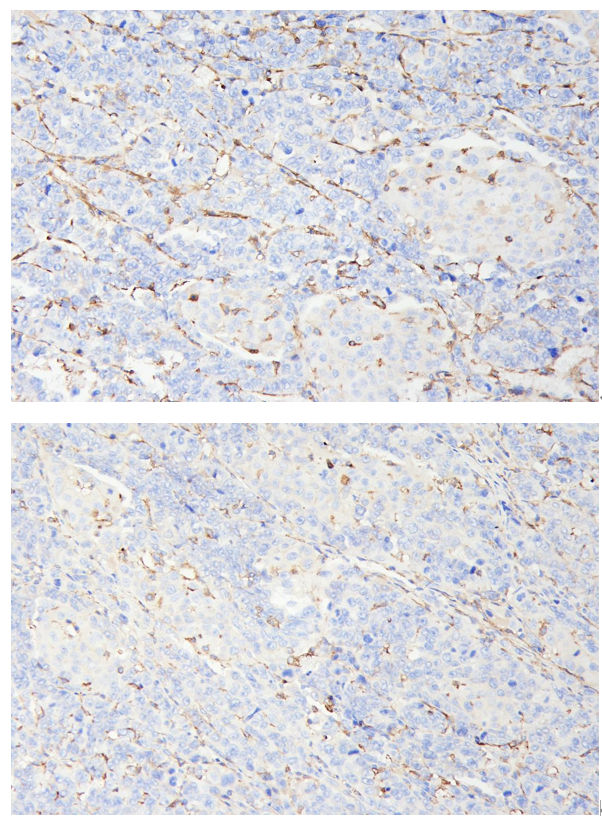
Click image to see more details
IHC analysis of CD8 using anti-CD8 antibody (MA1017).
CD8 was detected in paraffin-embedded section of HepG2 xenograft tumor in nude mouse tissues. Heat mediated antigen retrieval was performed in citrate buffer (pH6, epitope retrieval solution) for 20 mins. The tissue section was blocked with 10% goat serum. The tissue section was then incubated with 1:200 mouse anti-CD8 Antibody (MA1017) overnight at 4°C. Two step IHC detection kit was used as secondary antibody and incubated for 30 minutes at 37°C. The tissue section was developed using Strepavidin-Biotin-Complex (SABC)(Catalog # SA1021) with DAB as the chromogen.
Specific Publications For Anti-CD8 Cd8A Antibody (Monoclonal, CA-8) (MA1017)
Loading publications
Recommended Resources
Here are featured tools and databases that you might find useful.
- Boster's Pathways Library
- Protein Databases
- Bioscience Research Protocol Resources
- Data Processing & Analysis Software
- Photo Editing Software
- Scientific Literature Resources
- Research Paper Management Tools
- Molecular Biology Software
- Primer Design Tools
- Bioinformatics Tools
- Phylogenetic Tree Analysis
Customer Reviews
Have you used Anti-CD8 Cd8A Antibody (Monoclonal, CA-8)?
Share your experimental results or join a short interview to earn up to $1,000 in product credits or other rewards.
1 Reviews For Anti-CD8 Cd8A Antibody (Monoclonal, CA-8)
IHC staining with Anti-CD8a antibody (MA1017) showed strong and specific labeling of cytotoxic T cells in HepG2 xenograft tumors, with clear morphology and low background.
Excellent
| SKU | MA1017 |
|---|---|
| Application | Immunohistochemistry |
| Sample | HepG2 subcutaneous xenograft in nude mice |
| Sample Processing Description | HepG2 cells were expanded and then implanted subcutaneously into nude mice. After 2 weeks, tumors formed, which were excised, fixed in formalin for 48 hours, and processed for paraffin embedding and sectioning. |
| Other Reagents | Goat serum, DAB chromogen solution |
| Primary Antibody | CD8 Cd8A Antibody (Monoclonal, CA-8) |
| Primary Incubation | 1:200, overnight at 4 ℃ |
| Secondary Antibody | Two-step IHC detection kit |
| Secondary Incubation | 30 min in 37℃ |
| Detection | Image system: Leica DM2500 |
| Results Summary | CD8 is a marker of cytotoxic T cells; the results show clear infiltration of cytotoxic T cells in the tumor tissue, with distinct positive staining and well-preserved morphology. |
Fengtong Wang, The First Affiliated Hospital of Xinjiang Medical University
Verified customer
Submitted 2026-04-21
Customer Q&As
Have a question?
Find answers in Q&As, reviews.
Can't find your answer?
Submit your question
2 Customer Q&As for Anti-CD8 Cd8A Antibody (Monoclonal, CA-8)
Question
Has MA1017 been tested with IHC-P? What is the immunogen information?
Verified customer
Asked: 2019-07-09
Answer
The Anti-CD8 Cd8b Antibody (Monoclonal, CA-8) (MA1017) has been tested with IHC-P on human samples. It hasn't been validated with monkey samples. Please run pilot tests with monkey samples. Immunogen information: Human thymocytes followed by peripheral blood T cells.
Boster Scientific Support
Answered: 2019-07-10
Question
I would like using your anti-CD8 antibody (Monoclonal, CA-8) for adaptive immune response studies. Has this antibody been tested with western blotting on human pbmc cells? We would like to see some validation images before ordering.
T. Carter
Verified customer
Asked: 2013-10-17
Answer
I appreciate your inquiry. This MA1017 anti-CD8 antibody (Monoclonal, CA-8) is validated on human pbmc cells. It is guaranteed to work for Flow Cytometry, IHC, ICC in human. Our Boster guarantee will cover your intended experiment even if the sample type has not been be directly tested.
Boster Scientific Support
Answered: 2013-10-17
