ABI1 / Abl interactor 1 · IHC design guide

Design Immunohistochemistry for ABI1

ABI1 shows broad cytoplasmic staining in tissue IHC (HPA tissue IHC). This guide covers paraffin section controls, fixation, detection, and scoring using the catalog antibody’s IHC evidence (datasheet PB9416).

Evidence assembled Oct 2026 · For research use; verify linked source records and product datasheet before use
Immunohistochemistry protocol sheet for ABI1 (IHC for ABI1): expected localisation Cytoplasmic tissue staining (HPA tissue IHC), antibody PB9416, validated IHC image, and IHC protocol steps
Printable ABI1 IHC protocol sheet — expected localisation Cytoplasmic tissue staining (HPA tissue IHC), antibody PB9416, controls and protocol steps. Open the full ABI1 IHC guide →

ABI1 Immunohistochemistry Experimental Design Guide

Expected localisation, validated protocols, controls and antibodies — the at-a-glance facts below, then the full design guide.

Must know before staining
Expected localisation Cytoplasmic tissue staining (HPA tissue IHC)
Staining pattern Broad cytoplasmic staining; smooth muscle cells unstained (HPA tissue IHC)
Antigen retrieval EDTA pH 8.0 HIER, heat-mediated (datasheet PB9416)
Positive control ⓘ Appendix+4 more · see all
Negative control ⓘ Smooth muscle
Important caveats
Reasons your staining may differ from the expected pattern.
Fixation Keep fixation consistent across sections (standard IHC practice; not target-specific)
Caveat Staining has medium consistency with RNA data (HPA tissue IHC)
Regulation Widely expressed; highest in brain (UniProt)
Isoform / epitope 12 isoforms; epitope coverage is unspecified (UniProt)
Section 1

Recommended ABI1 IHC & IF Protocols

The catalog antibody’s paraffin-section protocol is accompanied by three published ABI1 IHC protocols from colorectal and ovarian cancer studies (PMC3393686; PMC4066275; PMC3059077).

Recommended immunohistochemistry (IHC-P) protocol parameters
SampleParaffin-embedded mouse brain tissue; fixative not specified (datasheet PB9416)
FixationImage fixative and duration unreported (datasheet PB9416); verify before use.
Sectioning4–5 µm sections on charged slides (standard)
DeparaffinisationXylene, graded ethanol series to water (standard)
Antigen retrievalHeat retrieval: EDTA pH 8.0 (datasheet PB9416); 20 min, 95–100 °C (standard)
Peroxidase block3% H2O2, 10 min, room temperature (standard)
Blocking10% goat serum (datasheet PB9416)
Primary antibodyRabbit anti-ABI1, 2-5μg/ml (datasheet PB9416)
Primary incubationOvernight at 4 °C (datasheet PB9416)
DetectionHRP-conjugated secondary, DAB chromogen (datasheet PB9416)
CounterstainHematoxylin, blue, dehydrate and mount (standard)
Expected resultABI1-positive staining in glandular cells of appendix (HPA tissue IHC: High). HPA tissue profile: Ubiquitous cytoplasmic expression. No signal in the no-primary control.
💡Decision noteStart with heat-mediated EDTA retrieval at pH 8.0 (datasheet: PB9416); these published excerpts do not specify retrieval conditions (PMC3393686; PMC4066275; PMC3059077).
Section 2

What Is the Expected ABI1 Staining Pattern?

In paraffin-section IHC, expect predominantly cytoplasmic ABI1 staining across many cell types, including glandular cells, hematopoietic cells and neurons (HPA: ubiquitous cytoplasmic expression; HPA: High in listed cells). ABI1 also localizes to the nucleus and cell projections, and has no transmembrane segment (UniProt Q8IZP0: subcellular location and topology). Treat the tissue pattern as a guide rather than a definitive specificity test: HPA rates its IHC evidence Approved, with medium consistency and pending external verification (HPA: reliability).

What am I looking at on my slide?
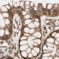
Cytoplasmic staining in appendix or colon glandular cells, with recognizable cell boundaries.This matches the reported broad cytoplasmic profile and two High-staining glandular examples (HPA: tissue IHC). Compare staining in the expected cells with surrounding tissue on the same section; intensity alone cannot establish antibody specificity (general IHC practice).
Predominantly extracellular, luminal or nuclear-only reaction product, with little cellular cytoplasm.That pattern does not match the predominant cytoplasmic IHC profile (HPA: tissue IHC). Nuclear localization is biologically possible, so nuclear signal alone is not proof of artefact (UniProt Q8IZP0: nucleus). Check morphology and controls before assigning localization (general IHC practice).
Strong staining in smooth muscle cells while nearby expected-positive cells remain unstained.HPA reports ABI1 as Not detected in smooth muscle cells and High in several listed epithelial, hematopoietic and neuronal populations (HPA: tissue IHC). Consider nonspecific antibody binding or endogenous detection activity; a single discordant section cannot identify which mechanism applies (general IHC practice).
Uniform color over tissue and empty spaces, obscuring cell outlines.A noncellular haze prevents a reliable call against HPA's cytoplasmic, cell-associated pattern (HPA: tissue IHC). Excess detection background or incomplete blocking are general possibilities; assess the no-primary control and review reagent exposure before interpreting weak cellular signal (general IHC practice).
No signal in an intact, well-preserved appendix or colon glandular compartment.Both are reported High in glandular cells, making absent staining a failed expected-positive result for this run (HPA: tissue IHC). Check the antibody's documented IHC-P conditions and the run controls before interpreting other negative tissues (general IHC practice).
💡Expected ABI1 appearanceCall a convincing positive when chromogenic staining is cell-associated and predominantly cytoplasmic in a reported High population, such as colon glandular cells; strong signal confined to smooth muscle or spread through empty spaces is suspect (HPA: tissue IHC; general IHC practice).
How each factor affects the staining
Tissue and cell selectionHPA reports High staining in appendix, breast, cervix and colon glandular cells, bone-marrow hematopoietic cells, bronchial respiratory epithelium, and selected brain cells (HPA: tissue IHC). Smooth muscle cells are reported Not detected; use these observations to choose comparison compartments, while allowing for specimen variation (HPA: tissue IHC; general IHC practice).
Compartment and topologyThe IHC profile is ubiquitous cytoplasmic expression (HPA: tissue IHC). UniProt also lists nucleus, cytoskeleton, lamellipodia, filopodia, growth cones and postsynaptic density, with no transmembrane segment (UniProt Q8IZP0). These annotations support careful review of focal cellular signal but do not require every compartment to resolve in paraffin IHC (general IHC practice).
Antibody evidenceCAB008375 has an Approved IHC assessment; the supplied record does not mark it Enhanced (HPA: antibody validation). The tissue profile has medium consistency with RNA data and awaits external verification (HPA: reliability). Interpret an unexpected pattern with tissue morphology and assay controls rather than treating approval as proof of every stained cell's identity (general IHC practice).
Isoforms and modificationsUniProt lists 12 ABI1 isoforms and multiple modified residues, including phosphorylation sites (UniProt Q8IZP0: isoforms and modified residues). The payload gives no antibody epitope or evidence that a particular isoform or modification changes this IHC signal; avoid assigning staining differences to either without separate validation.
IF/ICC Q&A: where should signal appear?HPA's ICC-IF summary reports approved plasma-membrane and cell-junction localization; HPA068407 has Approved ICC status (HPA: subcellular; HPA: antibody validation). That imaging result is application-specific and does not replace the predominantly cytoplasmic tissue-IHC expectation (HPA: tissue IHC).
Why is my staining missing, weak or wrong?
SituationLikely causeNext action
Expected-positive glandular cells show no chromogenic signal.A failed staining run is possible because these cells are reported High in appendix and colon (HPA: tissue IHC).Review the documented IHC-P antibody dilution and retrieval conditions, confirm reagent activity with a run control, then repeat if needed; no ABI1-specific retrieval setting is supplied here (general IHC practice).
Staining is weak throughout an otherwise intact section.The chosen compartment may naturally stain less strongly: HPA lists several Low populations, including glial cells in caudate and myocytes in skeletal muscle (HPA: tissue IHC).Compare with a reported High compartment in the same run before adjusting detection or antibody concentration; use the antibody's documented IHC-P conditions as the starting point (HPA: tissue IHC; general IHC practice).
Smooth muscle stains strongly.This conflicts with HPA's Not detected smooth-muscle result; nonspecific binding or endogenous detection activity is possible (HPA: tissue IHC; general IHC practice).Check the no-primary control, blocking and detection chemistry, and compare adjacent expected-positive cells; do not label the smooth-muscle signal ABI1 on intensity alone (general IHC practice).
Diffuse precipitate obscures cellular detail.Noncellular background cannot be assigned to ABI1's reported cytoplasmic distribution (HPA: tissue IHC).Inspect the no-primary control and review blocking, washes and chromogen development; repeat with conditions that preserve clear cell boundaries (general IHC practice).
Signal appears only in nuclei or along luminal surfaces.The distribution conflicts with the main tissue-IHC profile, although UniProt also lists nuclear ABI1 (HPA: tissue IHC; UniProt Q8IZP0: subcellular location).Confirm cell morphology and compare a High-staining tissue compartment with assay controls before calling artefact or a distinct localization (HPA: tissue IHC; general IHC practice).

Sample controls for ABI1 IHC & IF

🧪Run appendix first and score glandular cells, where ABI1 staining is High (HPA: High in appendix glandular cells). Run smooth muscle as the negative tissue (HPA: Not detected in smooth muscle cells); the supplied HPA rows do not identify a confirmed negative cell type within appendix, so treat unstained nonglandular areas as background references rather than validated internal negatives (HPA: appendix row).
Positive control tissue: Appendix (Glandular cells, HPA High)
Negative control tissue: Smooth muscle (HPA Not detected)
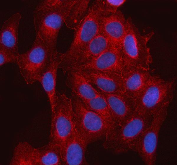
ICC-IF cell lines (HPA subcellular resource): HPA ICC-IF images show ABI1 in CACO-2, SK-MEL-30, U2OS, with annotated localisation: Plasma membrane (approved), Cell Junctions (approved) (HPA subcellular).
Technical controls: Include a no-primary, secondary-only control and a concentration-matched rabbit IgG isotype control (caption: rabbit primary antibody), plus ABI1-knockout material as a biological negative. Quench endogenous peroxidase and check inflammatory cells in appendix for misleading DAB signal (caption: HRP/DAB detection; standard IHC practice).
⚠️Feasibility: The selected PB9416 paraffin-section caption reports heat retrieval in EDTA at pH 8.0, but does not report the fixative (caption: mouse-brain tissue IHC). No target-specific fixation window or fixation effect is reported in the supplied evidence; the caption supports trying that retrieval condition, but does not establish that retrieval is required for every specimen (caption: EDTA retrieval). The supplied evidence does not establish that frozen sections or IF/ICC are easier than paraffin IHC; in appendix, check for endogenous peroxidase signal among inflammatory cells (HPA: High in appendix glandular cells; standard IHC practice).

HPA tissue IHC evidence for ABI1

Comprehensive Human Protein Atlas IHC scoring per tissue (reliability: Approved — Medium consistency between antibody staining and RNA expression data. Pending external verification.). Rows are taken directly from the HPA tissue chart — click any row's HPA link to view the source.

Positive expression · recommended positive controls

TissueCell typeLevelEvidenceSource
Appendix Glandular cells High Protein (IHC) HPA →
Bone marrow Hematopoietic cells High Protein (IHC) HPA →
Breast Glandular cells High Protein (IHC) HPA →
Bronchus Respiratory epithelial cells High Protein (IHC) HPA →
Cerebellum Cells in granular layer High Protein (IHC) HPA →

Undetected expression · recommended negative controls

TissueCell typeLevelEvidenceSource
Smooth muscle Smooth muscle cells Not detected Protein (IHC) HPA →
Section 3

Advanced ABI1 IHC Tips

Troubleshoot ABI1 staining in paraffin sections by checking retrieval, compartment, controls and scoring against the supplied tissue evidence.

What retrieval should I try first when ABI1 staining is weak?
Start with heat-mediated retrieval in EDTA at pH 8.0 for this paraffin-section assay (datasheet PB9416). The selected mouse-brain image used that retrieval before incubation with 2 μg/ml antibody overnight at 4°C, so reproduce those conditions when comparing results (datasheet PB9416). Keep section thickness, heating, cooling and detection conditions consistent across a retrieval comparison (standard IHC practice). If staining remains weak, test an alternative retrieval buffer on adjacent sections as a fallback, while checking whether tissue morphology or background deteriorates (standard IHC practice). Judge the result by cellular staining and preserved anatomy, using the reported mouse-brain staining as product-specific context (datasheet PB9416).
Could fixation explain weak or uneven ABI1 staining?
The selected paraffin-section caption does not state a fixative, so ABI1 sensitivity to fixation is unknown for this antibody (datasheet PB9416). Record the actual fixative, fixation duration and processing history for each specimen before attributing a staining difference to ABI1 biology (standard IHC practice). Compare similarly processed sections under the same EDTA pH 8.0 retrieval and antibody conditions when investigating weak signal (datasheet PB9416; standard IHC practice). Include a processed positive-control section in each run and inspect tissue preservation before adjusting detection chemistry (standard IHC practice). Neither the reported tissue distribution nor ABI1 topology establishes a target-specific fixation effect (HPA tissue IHC; UniProt Q8IZP0 topology).
Which ABI1 staining pattern should I expect in tissue sections?
Expect predominantly cytoplasmic tissue staining as the broad reference pattern, while evaluating the cell type and section anatomy (HPA tissue IHC: ubiquitous cytoplasmic expression). ABI1 is also reported in nuclei, cytoskeleton and cell projections, including lamellipodia and filopodia tips (UniProt Q8IZP0 subcellular location). Its lack of a transmembrane segment does not by itself settle how a fine tissue-section signal should appear (UniProt Q8IZP0 topology; standard IHC interpretation). High staining is reported in cerebral-cortex neuronal cells and cerebellar granular-layer cells, whereas smooth-muscle cells were not detected in the listed tissue survey (HPA tissue IHC). Check unexpected nuclear or sharply peripheral staining against morphology and controls before assigning a compartment-specific interpretation (standard IHC practice).
How could ABI1 isoforms or epitope accessibility change the result?
ABI1 has 12 listed isoforms, so an antibody may report a subset if its epitope differs among them (UniProt Q8IZP0 isoforms; standard immunostaining interpretation). The record places a coiled-coil homology region at residues 45–107 and an SH3 domain at 446–505, providing sequence regions to check against a disclosed immunogen (UniProt Q8IZP0 domains). Listed modifications include phosphorylation at residues 53, 174, 178 and 183; their effect on this antibody's staining is not established (UniProt Q8IZP0 modified residues; datasheet PB9416). Request epitope information before claiming isoform coverage or modification sensitivity for the catalog antibody (standard antibody-validation practice). Compare sections using identical retrieval and detection conditions when assessing a suspected epitope-dependent difference (standard IHC practice).
How should I adapt ABI1 assessment to multiplex immunofluorescence?
Use this as an IF-specific validation exercise: the selected antibody evidence describes chromogenic staining of a paraffin-embedded mouse-brain section (datasheet PB9416). Pair ABI1 with a separately validated marker for the expected cell population, such as a neuronal-cell marker when assessing cerebral cortex, and check each channel independently (HPA tissue IHC: high in cortical neuronal cells; standard IF practice). Select fluorophores after inspecting unstained tissue for autofluorescence and include single-stain controls to assess channel bleed-through (standard IF practice). Because ABI1 has no transmembrane segment and is reported in cytoplasm and projections, assess permeabilisation for access to the epitope's cellular compartment once that epitope is known (UniProt Q8IZP0 topology and subcellular location; standard IF practice). Keep IF fixation and permeabilisation conclusions separate from the paraffin-section caption, which does not report a fixative (datasheet PB9416).
What should I check when ABI1 DAB staining looks diffuse?
First inspect a no-primary control for detection-system staining and endogenous peroxidase, then compare it with the antibody-stained section (standard chromogenic IHC practice). The selected assay used 10% goat serum blocking, a peroxidase-conjugated secondary and DAB development, which are useful conditions to document when tracing background (datasheet PB9416). Assess whether excess primary antibody, inadequate washing or overdevelopment contributes by varying one condition across adjacent sections (standard IHC practice). Because HPA describes broad cytoplasmic ABI1 staining, diffuse color alone cannot distinguish biological signal from nonspecific background (HPA tissue IHC; standard IHC interpretation). Review tissue edges, damaged regions and no-primary staining before treating a uniform brown haze as ABI1 positive (standard IHC practice).
How should I score ABI1 staining across tissue sections? ⚠ ANSWER MARKED FOR VERIFICATION
Define the cell population and compartment before scoring, then use an H-score or percent positive cells within matched anatomical regions (standard IHC quantification practice). Report the scoring threshold, sampled area and number of evaluable cells; for counted positive cells, density per mm² can supplement the percentage (standard IHC quantification practice). Normalise cell counts to viable tissue area or the relevant cell population, and keep illumination, DAB development and image analysis settings consistent (standard IHC quantification practice). Score cytoplasmic and nuclear signal separately if both are measured, because the broad tissue reference is cytoplasmic while nuclear localisation is also reported (HPA tissue IHC; UniProt Q8IZP0 subcellular location). Interpret regional differences alongside the reported high cortical-neuronal and cerebellar granular-layer staining, without treating those categories as an assay calibration (HPA tissue IHC).
How do I distinguish genuine ABI1 staining from artefact?
A convincing result has cellular staining in preserved tissue and agrees with an independently checked compartment and cell population (standard IHC interpretation). Cytoplasmic signal fits the broad tissue survey, and cortical neuronal or cerebellar granular-layer staining has reported support (HPA tissue IHC). Isolated strong smooth-muscle staining deserves scrutiny because smooth-muscle cells were listed as not detected, though that survey is not a definitive negative control (HPA tissue IHC: smooth muscle). Check abrupt section-edge color, necrotic areas and no-primary staining before interpreting focal DAB deposits as ABI1; endogenous peroxidase can also generate color (standard chromogenic IHC practice). Treat an exclusively unfamiliar compartment cautiously, since ABI1 has reported cytoplasmic, nuclear and projection-associated locations, and the HPA tissue staining has medium consistency with RNA expression (UniProt Q8IZP0 subcellular location; HPA tissue IHC reliability).
Boster reagents

Best ABI1 / Abl interactor 1 IHC Antibodies

Anti-ABI1 antibodies have IHC images from human cancer and mouse and rat brain paraffin sections, plus IF images from human cells and cancer sections (catalog image captions).

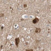
Real IHC data IHC analysis of SSH3BP1/ABI1 using anti-SSH3BP1/ABI1 antibody (PB9416). SSH3BP1/ABI1 was detected in a paraffin-embedded section of mouse brain tissue. Heat mediated antigen retrieval was performed in EDTA buffer (pH 8.0, epitope retrieval solution). The tissue section was blocked with 10% goat serum. The tissue section was then incubated with 2 μg/ml rabbit anti-SSH3BP1/ABI1 Antibody (PB9416) overnight at 4°C. Peroxidase Conjugated Goat Anti-rabbit IgG was used as secondary antibody and incubated for 30 minutes at 37°C. The tissue section was developed using HRP Conjugated Rabbit IgG Super Vision Assay Kit (Catalog # SV0002) with DAB as the chromogen.
Anti-SSH3BP1/ABI1 Antibody ®
Cat # PB9416
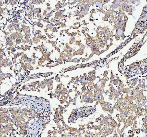
Real IHC data IHC analysis of ABI1 using anti-ABI1 antibody (PA1001). ABI1 was detected in a paraffin-embedded section of human lung cancer tissue. Heat mediated antigen retrieval was performed in EDTA buffer (pH 8.0, epitope retrieval solution). The tissue section was blocked with 10% goat serum. The tissue section was then incubated with 2 μg/ml rabbit anti-ABI1 Antibody (PA1001) overnight at 4°C. Peroxidase Conjugated Goat Anti-rabbit IgG was used as secondary antibody and incubated for 30 minutes at 37°C. The tissue section was developed using HRP Conjugated Rabbit IgG Super Vision Assay Kit (Catalog # SV0002) with DAB as the chromogen.
Anti-SSH3BP1/ABI1 Antibody ®
Cat # PA1001

PA1001 has IHC images from human lung cancer and colorectal adenocarcinoma paraffin sections and IF images from U2OS cells and human cancer sections (PA1001 image captions). PB9416 has IHC images from mouse and rat brain and human ovarian cancer paraffin sections, plus an IF image from A431 cells (PB9416 image captions).

Which to pick: For human tissue IHC, choose PA1001 for its human paraffin-section examples (PA1001 IHC captions); for mouse or rat tissue IHC, choose PB9416 for its brain-section examples (PB9416 IHC captions). For IF/ICC, PA1001 has cell and tissue IF images, while PB9416 has a cell IF image (catalog IF captions); both are rabbit antibodies listed as reactive with human, mouse and rat (catalog). Both IHC captions use EDTA retrieval at pH 8.0 and 2 μg/ml primary antibody, but neither reports the fixative (catalog IHC captions).

Each figure is that product's own IHC / IF validation image from its datasheet.

References

  1. UniProt Consortium. UniProt entry Q8IZP0 (ABI1_HUMAN, Abl interactor 1).
  2. Human Protein Atlas. ABI1 tissue IHC expression (reliability: Approved).
  3. Human Protein Atlas. ABI1 subcellular location (ICC-IF): Localized to the plasma membrane and cell junctions..
  4. Human Protein Atlas. ABI1 antibody validation summary (2 antibodies).
  5. Expression of Abelson interactor 1 (Abi1) correlates with inflammation, KRAS mutation and adenomatous change during colonic carcinogenesis. PloS one 2012 — PMC3393686.
  6. Expression and Y435-phosphorylation of Abelson interactor 1 (Abi1) promotes tumour cell adhesion, extracellular matrix degradation and invasion by colorectal carcinoma cells. Molecular cancer 2014 — PMC4066275.
  7. Integrity of SOS1/EPS8/ABI1 tri-complex determines ovarian cancer metastasis. Cancer research 2010 — PMC3059077.
  8. Fat3 acts through independent cytoskeletal effectors to coordinate asymmetric cell behaviors during polarized circuit assembly. Cell reports 2022 — PMC8865054.
  9. PubMed PMID:9010225 — UniProt-cited evidence.
  10. PubMed PMID:9593709 — UniProt-cited evidence.
  11. PubMed PMID:11418237 — UniProt-cited evidence.