Secondary Antibodies


Boster Bio Secondary Antibody


Boster offers conjugated secondary antibodies for IHC, ICC/IF, and Western Blotting. These antibodies have been referenced in over 8,000 scientific publications. Best reviews and quality guaranteed.

Browse all secondary antibodies

Browse all secondary antibodies

Product
Figures
Species
Applications
Price

1 images

Flow Cytometry
$320
Cat # BA1045-Dyl488
1 ml

1 images

ELISA, IHC, WB
$60
Cat # BA1027
0.1ml

no images

Yellowfever mosquito
$470
Cat # DZ33976
200 μl/vial

no images

Flow Cytometry, IF
$60
Cat # BA1148
100μl

no images

Flow Cytometry, IF
$60
Cat # BA1147
100μl

no images

Flow Cytometry, IF
$60
Cat # BA1145
100μl

no images

Flow Cytometry, IF
$60
Cat # BA1144
100μl

no images

Flow Cytometry, IF
$60
Cat # BA1149
100μl

no images

ELISA, IHC
$60
Cat # BA1017
100μl

Displaying Items 1-10 of Many

What is a Secondary Antibody?

Immunolabeling is critical for many areas of basic or clinical research to detect specific cell or tissue components (antigens) in a sample. This approach can be performed with direct and indirect methods by generating antigen-specific antibodies and fusing tags to antibodies.

For direct immunolabeling, the primary antibody is conjugated to a tag and directly binds to the antigen of interest. The indirect approach involves a conjugated secondary antibody that binds indirectly to the target antigen by binding to the antigen-specific primary antibody.

Secondary antibodies are generated and harvested by immunizing a host animal with an antibody from another species. For example, if the primary antibody is a rabbit polyclonal antibody, an anti-rabbit secondary antibody is raised in a different host species such as goat. The produced secondary antibody's specificity is dependent on the immunizing antibody's characteristics, such as species, subclass, fragment, etc.

Primary Antibody vs Secondary Antibody

For a successful experiment, it is important to have a good understanding of the primary and secondary antibodies. Choosing a suitable approach and/or antibodies will better serve the purpose of the experiment while saving money, time, and precious sample. Although monoclonal or polyclonal primary and secondary antibodies can be used for experiments, there are some differences between the primary and secondary antibodies that should be considered.

Primary antibodies bind directly to the antigen by recognizing a specific area/domain (epitope) of the antigen, whereas secondary antibodies bind to the primary antibody—not directly to the antigen. While primary antibodies are necessary for every immunoassay, secondary antibodies are not always required—depending on the experimental method (direct/indirect).

As previously mentioned, secondary antibodies must be made in a species different from those of the primary antibody or the specimen to minimize non-specific binding that leads to false positives and high background noise. Secondary antibodies may also require an extra purification process (pre-adsorption) with a column matrix during the immunoassay to remove non-specific antibodies and increase specificity.

Buy Primary, Get Secondary Free

You could receive a free HRP or biotin conjugated secondary antibody

Advantages of Using a Secondary Antibody

With its single labeling step, the direct method offers a shorter assay time with a simpler workflow. Since it minimizes species cross-reactivity and non-specific binding, the direct method is best used for specific targeting if multiple antibodies are raised in the same species. However, this method demands an abundant supply of expensive conjugated antibodies—with few color selections and limited range of reporter molecules available—and is much less practical than the indirect method.

Although the indirect method requires additional steps, time, and added complexity, it still offers several advantages over the direct method. More than one secondary antibody can specifically bind to different parts of the same primary antibody—increasing the versatility, antigen signal detection, and amplification. The indirect method also contributes to the detection, sorting, and purification of target antigens—providing higher degrees of specificity and sensitivity. Commercially available conjugated secondary antibodies are relatively inexpensive and available in a wider spectrum of colors compared to conjugated primary antibodies—with increased access to several different probes.

By using primary antibodies as a ‘bridge’ to bind with the target antigen, secondary antibodies reduce the possibility of the reporter molecule compromising the binding capability to the antigen epitope. If the target antigen is expressed at a low concentration, using secondary antibodies will allow for multiplexing or multi-labeling across applications (e.g. immunofluorescence, western blot) to validate target antigen detection.

Browse all secondary antibodies

How to Choose a Secondary Antibody

Selecting the right secondary antibody is essential for the successful detection of the target antigen. Based on the application, primary antibody, and experimental design, some factors should be considered when selecting a suitable secondary antibody, such as:

The host species used to generate the secondary antibody should be directed against the target species of the primary antibody to avoid cross-reactivity with endogenous immunoglobulins (Ig) in the sample material. The selected secondary antibody must be able to bind to a specific part of the targeted primary antibody.

After the host species are immunized, the antibody-containing serum is purified through a solid phase porous column with an immobilized ligand. The ligand can be either an affinity ligand that recognizes a specific antibody or the target antigen where the antibody recognizes the ligand. Following purification, pre-adsorption through a column containing immobilized antibodies from another species will remove the reactivity to a particular species, non-target Ig, or non-target Ig regions—yielding secondary antibodies with increased specificity and sensitivity.

Primary monoclonal antibodies are usually IgG isotype with a specific subclass. The chosen secondary antibody should be directed against that specific subclass (anti-IgG). For primary polyclonal antibodies with more than one subclass, the anti-IgG must recognize both the heavy and light IgG chains (anti-IgG H&L). If the class/subclass of the primary antibody is known, anti-IgG F(ab) is used.

In general, whole secondary antibodies containing both heavy (H) and light (L) chains of the Ig are more widely used. Whole secondary antibodies will give higher signal due to their stronger binding to variable regions—with sufficient regions for the attachment of enzymes and dyes. However, whole antibodies can increase cross-reactivity and lower specificity, so it may sometimes be preferable to use a fragment to eliminate non-specific binding.

There are two types of fragments, namely F(ab) and F(ab’)2. With its single binding site, F(ab) fragments (MW=~50kDa) are useful in blocking background noises caused by primary antibodies and endogenous Ig in the sample material. F(ab’)2 fragments are bigger (MW=~110 kDa) and may penetrate tissues/cells more easily. This trait—coupled with their strong divalent bonding in the variable regions—ensures the secondary does not bind to the cell surface.

Using a Secondary Antibody

For detection and visualization of the presence of the target protein, the antibodies are conjugated with probes/markers. There are different types of conjugates, depending on the application and detection technology (colorimetric, chemiluminescent, or fluorescent). Below is a description of the most commonly used secondary antibody conjugates:

Fluorophores

Fluorophores

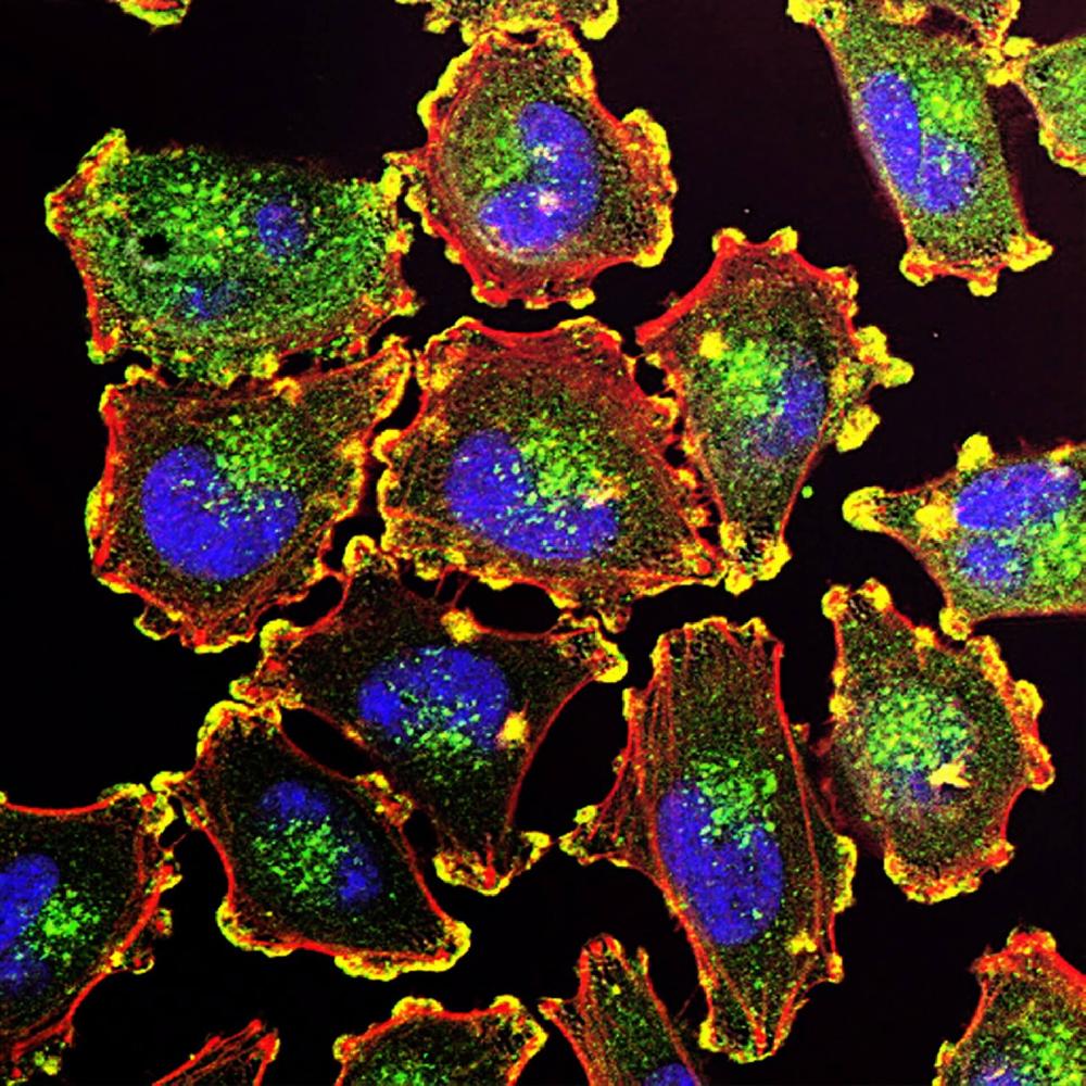
Fluorophores emit light in the visual range when excited by light at a particular wavelength—which is then detected by the fluorescent microscope. There are several types of fluorophore-conjugated secondary antibodies available (Alexa Fluor®, DyLight®, IRDye®, FITC, etc.), all with their own excitation and emission characteristics.

Enzymes

Enzymes

Enzymes such as horseradish peroxidase (HRP) and alkaline phosphatase (AP) are capable of converting soluble, colorless substrates into a water-insoluble colored precipitate, which allows visualization with colorimetric or chemiluminescent detection (western blot, immunochemistry).

Biotin

Biotin

Biotin is a small vitamin molecule that can be easily modified and/or attached to proteins, antibodies, and other biomolecular probes of interest (e.g. avidin or streptavidin protein). The signal amplification following the interaction between biotin with enzyme- or fluorochrome-labeled secondary antibodies makes it suitable for detecting proteins expressed at low levels.

Colloidal gold

Colloidal gold

Colloidal gold conjugates are primarily suitable for immunoassays using an electron microscope. However, gold conjugated secondary antibodies are also used for applications such as flow cytometry, bio-imaging, and lateral flow. The gold nanoparticles can be provided in varying sizes (e.g. 1.4nm, 5nm, 10nm, 40nm).

Secondary Antibodies

We provide several different conjugates for secondary antibodies - HRP, biotin, FITC, DyLight®, colloidal gold, etc. - as well as unconjugated secondary antibodies in our catalog

Secondary Antibodies for Different Applications

Most secondary antibodies are used in low concentrations between 1 and 10 μg/mL since excessive amount of secondary antibodies will result in high background noise from too much non-specific binding. For optimal performance with minimal background interference, a good starting concentration for a typical secondary antibody in that concentration range would be a dilution of 1:1,000. However, if the staining is extremely bright or the reading shows too much background, higher dilutions from 1:2,000 to 1:20,000 should be considered.

Incubation of the secondary antibodies for 1 hour (37°C) or 2 hours at room temperature should be sufficient. For a stronger signal, the incubation should be performed overnight with agitation at 4°C.

It should be noted that the dilution ratio and incubation time for the secondary antibodies will vary for each application. The end user will need to determine the optimal dilution ratio and incubation time for each application based on the experimental conditions, such as the antigen/antibody concentrations, pH, temperature, and buffer constituents. Optimization of the antibody dilution can be done by performing titration experiments. The table below outlines the recommended secondary antibody types for each application.

Application Secondary Antibody Types
Western Blot
  • Enzyme-conjugated (HRP or AP) secondary antibodies are commonly used for this application. They can amplify signal and more easily detect the target antigens even within a complex protein mixture. AP-conjugated secondary antibodies are mostly used at dilutions ranging from 1:5,000 to 1:50,000. The dilution range for HRP-conjugated secondary antibodies is generally between 1:2,000 to 1:20,000.
  • With advancements in fluorescent conjugate technologies, fluorescent secondary antibodies with similar sensitivity to that of chemiluminescence detection are increasingly available. They allow quantitative, sensitive, and detection of multiple proteins in the same blot simultaneously, making them a good option for quantifying relative protein abundance. Dilution range for fluorescent-conjugated secondary antibodies can be from 1:50-1:800. Optimal dilution ratios should ultimately be determined by the end user.
ELISA
  • Most commercial ELISA kits use enzyme-conjugated secondary antibodies (HRP or AP).
  • ELISA can also be performed with fluorescent-conjugated secondary antibodies. However, the detection limit is typically lower than when using an enzyme-conjugated secondary antibody.
  • For better detection, increased sensitivity, and flexibility, enzyme-conjugated secondary antibodies are often coupled with biotinylated secondary antibodies (Avidin- or Streptavidin-Biotin Complex) for ELISA to amplify the signal and achieve a better reading.
  • Monoclonal secondary antibodies have also been used, especially the subtype-specific antibodies (e.g. anti-IgG1).
  • The standard secondary antibody dilution for most ELISA kits range from 1:200 to 1:50,000.
Immunohistochemistry (IHC)
  • Affinity-purified, pre-adsorbed and fragmented F(ab’)2 secondary antibodies that can penetrate tissues more easily are recommended to reduce non-specific background.
  • Enzyme-conjugated (HRP or AP) or fluorophore-conjugated (FITC, DyLight®) secondary antibodies coupled with biotinylated secondary antibodies (Streptavidin-Biotin-Complex) detection to amplify signal reading is another common detection system that is utilized for IHC experiments.
  • Additionally, detection with light microscopy can also be done using gold-conjugated secondary antibodies with silver enhancement.
  • HRP-conjugated secondary antibodies will provide better tissue penetration and have been shown to offer high sensitivity for low-expressing proteins.
  • For IHC, the dilution factor of HRP secondary antibodies could range from 1:2,000 to 1:20,000.
Immunofluorescence (IF)
  • Immunofluorescence uses fluorophore-conjugated secondary antibodies for visualization of antigens. The fluorophore can also be combined with the gold conjugate, allowing for visualization with both the fluorescence and electron microscope. Fluorophore-gold-conjugated secondary antibodies will also provide a brighter fluorescence with a wider pH/ionic range and reduced photobleaching.
  • Popularly fluorescent used dyes for IF include FITC, TRITC, Alexa Fluor®, etc.
  • Similar to IHC, pre-adsorbed and fragmented F(ab’)2 secondary antibodies are recommended for better tissue penetration and visualization of target antigens.
  • Using biotinylated secondary antibodies will also offer good signal amplification.
  • A dilution range of 1:100 to 1:1,000 should be enough to achieve specific IF staining.

Secondary Antibodies List

Browse all the detection kits and secondary antibodies below.

Specificity Secondary Antibody
Avidin DyLight®488, HRP, TRITC, FITC, Cy3
Donkey Anti-Mouse IgG HRP, DyLight®594, DyLight®550, DyLight®488, Biotin
Donkey Anti-Mouse IgM μ Chain Biotin
Donkey Anti-Rabbit IgG HRP, DyLight®594, DyLight®550, Biotin
Donkey Anti-Rat IgG Biotin
Goat Anti Human-IgA FITC, HRP
Goat Anti-Human IgM HRP, FITC, Biotin
Goat Anti-Mouse IgG HRP, Biotin, DyLight®488, FITC, TRITC, DyLight®594, Unconjugated, FITC, Cy3
Goat Anti-Mouse IgM HRP, Biotin
Goat Anti-Rabbit IgG TRITC, Biotin, DyLight®488, FITC, HRP, DyLight®594, Unconjugated, Cy3
Mouse Anti-Human IgG FITC, Biotin
Mouse Anti-Rabbit IgG Biotin
Protein A Biotin, FITC, HRP
Protein G Biotin, FITC, Peroxidase
Rabbit Anti-Avidin HRP
Rabbit Anti-Goat IgG Biotin, FITC, HRP, TRITC, Unconjugated, Cy3
Rabbit Anti-Human IgG FITC, HRP, TRITC, Unconjugated, Biotin, Cy3
Rabbit Anti-Mouse IgG Unconjugated
Rabbit Anti-Rat IgG Biotin, FITC, HRP, Unconjugated
Streptavidin HRP

Expand your understanding of biomarker interactions with multiplex immunohistochemistry! Our advanced multiplex IHC methodology provides a comprehensive view of protein expression patterns, enabling you to uncover critical associations and potential therapeutic targets.

Buy Primary, Get Secondary Free

You could receive a free HRP or biotin conjugated secondary antibody

New Products

Popular Products

Popular Biomarkers

ALPL AQP3 BDNF BMP2
BRCA1 CCR5 CD68 COL1A1
CSF3 CTSB CTSD CXCL8
ESM1 HAVCR1 IFNG IGFBP7
IL10 IL1B IL6 LCN2

Product Categories

Primary Antibodies

Primary and Secondary Antibodies

Boster Bio offers over 16,000 antibodies that have been validated for IHC, WB, ELISA, and FC in human, mouse, and rat samples. Rabbit and mouse monoclonal antibodies as well as rabbit polyclonal antibodies are available. Buy a primary antibody and get a secondary antibody for free!

ELISA Kits

ELISA Kits

More than 1,000 ELISA kits, both singleplex and multiplex, that have been cited 6,000+ times are available at Boster. We offer our Boster-branded Picokine™ ELISA kits (sandwich ELISA) and EZ-Set™ ELISA kits (DIY antibody pairs) in addition to many multiplex ELISA kits.

Image

Recombinant Proteins

Boster provides a wide range of human, mouse, and rat recombinant proteins, which include cytokines, growth factors, chemokines, and many more. Our recombinant proteins have high stability, biological activity, and purity.

Boster Kit

Reagents

Boster offers all the reagents you need for your immunostaining and western blotting experiments. We've got everything from buffers to lysates to kits, including our popular and well-cited BCA, CCK-8, and MTT assay kits.