PCR Sample Preparation

We have a convenient Plasmid DNA Extraction and Purification MiniPrep Kit for your molecular biological experiments.

Check it out

Overview

On this page, we will cover the protocols for:

  • DNA/RNA Extraction and Purification: The genomic DNA (gDNA) and total RNA are frequently the genetic elements targeted for molecular biology experiments since these are the main sources of genetic information in an organism. Genomic DNA and total RNA extraction have a straightforward methodology, requiring only cell lysis to accomplish the task.
  • Molecular Cloning - DNA Library Preparation: Library cloning and preparation are fundamental techniques that every molecular biologist should know. See our protocols below for step-by-step instructions.

Download this comprehensive manual which discusses the basic principles and techniques of PCR and molecular biology research, including protocols and troubleshooting solutions.

Download PCR eBook

Nucleic Acids Extraction and Purification Protocols

Nucleic acid Extraction Principle

The genomic DNA (gDNA) and total RNA are frequently the genetic elements targeted for molecular biology experiments since these are the main sources of genetic information in an organism.

Genomic DNA and total RNA extraction have a straightforward methodology, requiring only cell lysis to accomplish the task. However, it is recommended to consider the type of organisms you are using in your lab research. Some organism’s cells have a cell wall structure, which confers extra protection to cells. This is the case for most yeast, plants, and bacteria species. For these organisms, it is recommended to include an extra step for cell lysis before disrupting the plasma membrane. This step is usually an enzymatic or mechanical process to disrupt the cell wall. The purification of DNA and RNA molecules is required to avoid contaminations with other intracellular components, such as proteins and metabolites. Some commonly used methods for DNA and RNA purification are precipitation with phenol-chloroform or isopropanol, or by spin columns with silica membrane.

If your research is focused on gene expression analysis, then you should target total RNA. The same procedure used to extract and purify gDNA can be used for total RNA purification, although some additional steps are required, as you will see in the following RNA extraction and purification protocol. Boster recommends the following protocols for sample preparation depending on your initial sample source.

How to separate Plasmid DNA from Genome DNA

When your target DNA is a plasmid, you should adapt your procedure to make sure the plasmid DNA (pDNA) is separated from gDNA. Alkaline lysis is the most current protocol used in the labs. The lysis buffer has in its composition sodium hydroxide and SDS, which will be responsible for denaturation of gDNA and pDNA. Subsequently, a neutralization buffer is added to renature the pDNA but not the gDNA. Because of the size of gDNA, it is much more difficult to renature again, unlike the pDNA. Then the same procedure of gDNA purification can be used for pDNA purification.
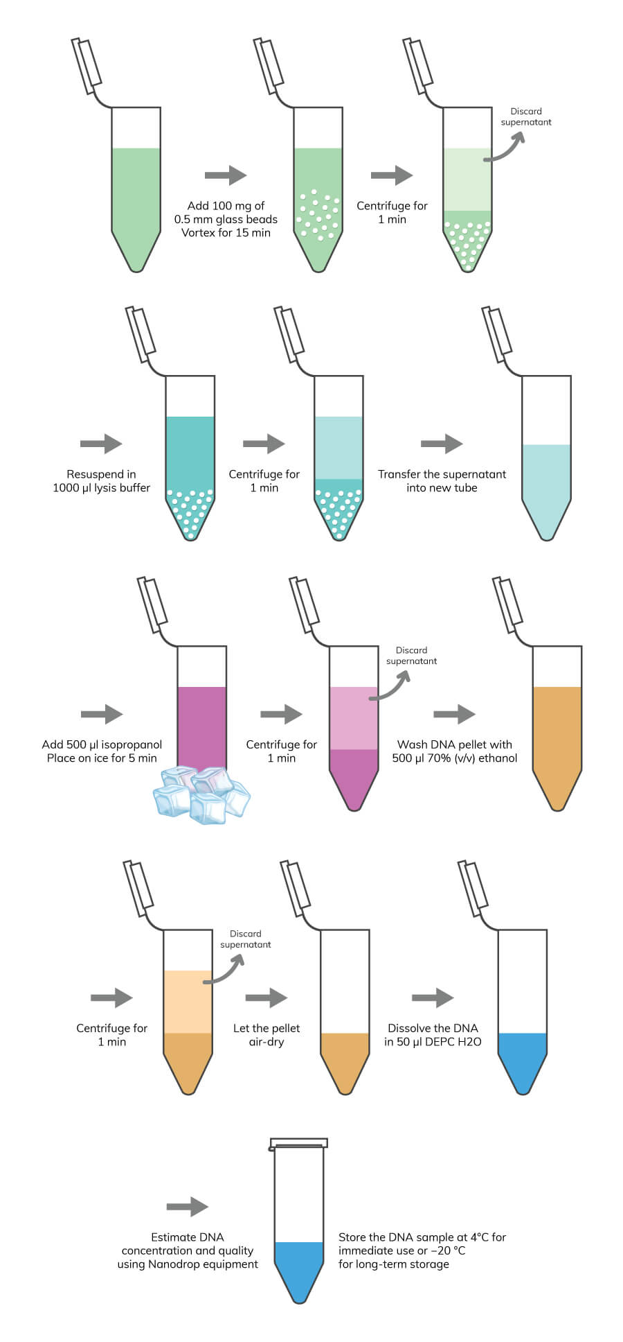
DNA Extraction and Purification Protocol

Required Reagents

  • Lysis buffer: 1% SDS, 0.5 M NaCl
  • Isopropanol
  • 70% (v/v) ethanol
  1. Collect the cells for DNA extraction into 2 ml tubes.

    Note: If you are using cell suspensions, you can move directly to the lysis procedure. If you are using samples, like cell tissues that are difficult to homogenize, you can use liquid nitrogen to create a homogeneous cell suspension. At the time you have a cell suspension, you should be careful about the membrane structure of your sample. Usually, for cells with cell walls, a pre-lysis treatment is recommended to degrade this barrier. You can use an enzymatic method or a mechanical method.

  2. Add 1ml of cell suspension into 2 ml tubes
  3. Add 100 mg of 0.5 mm glass beads and vortex for 15 min.

    Note: Alternatively, you can use 10U of lyticase (for fungi samples) or 10U of lysozyme (for gram-negative bacteria).

  4. Spin the cell suspension at top speed for 1 min using a centrifuge.
  5. Discard the supernatant and resuspend in 1000 μl of the lysis buffer.
  6. Spin at top speed for 1 min.
  7. Transfer the supernatant into a new 2ml tube.
  8. Add 500 μl of isopropanol and mix gently.
  9. Place the mixture on ice for 5 min.
  10. Spin at top speed 1min,
  11. Discard the supernatant and wash the DNA pellet with 500 μl 70% (v/v) ethanol.
  12. Spin again at top speed for 1 min.
  13. Discard the supernatant.
  14. Let the pellet air-dry, placing the 2 ml tubes upside down in a paper towel.
  15. Dissolve the DNA in 50 μl ultrapure DEPC H2O.
  16. Use the Nanodrop equipment to assess DNA concentration and quality.

    Note: Nucleic acids absorb UV light at 260 nm, whereas proteins and phenolic compounds absorb at 280 nm. Many organic compounds as well as phenol, TRIzol, and chaotropic salts have strong absorbances at around 230 nm. The A260/280 ratio is used to identify protein contamination. For RNA and DNA, A260/280 ratios should be around 2.1 and 1.8, respectively. The A260/230 ratio indicates the presence of organic contaminants, such as phenol, TRIzol, chaotropic salts and other aromatic compounds. Samples with 260/230 ratios below 1.8 have a significant amount of these contaminants.

  17. Store the DNA sample at 4°C for immediate use or −20 °C for long-term storage.

Miniprep Kit for Plasmid DNA Extraction and Purification

Fast and simple plasmid miniprep method for routine molecular biology laboratory applications.

RNA Extraction and Purification Protocol


Note:

During this protocol, always use new gloves and RNase free material.

  1. Collect the cells into 2 ml tubes.
  • Note: In the case you are using cell suspensions, you can move directly to the centrifuge step before the addition of lysis buffer. If you are using samples, like cell tissues that are difficult to homogenize, you can use liquid nitrogen to create a homogeneous cell suspension. For samples, in which cells have cell walls, add 1 ml of cell suspension into 2 ml tubes and add 100 mg of 0.5 mm glass beads and vortex for 15 minutes.
  1. Spin the cell suspension at top speed for 1 min using a centrifuge,
  2. Discard the supernatant and resuspend in 1 ml of lysis buffer prewarmed at 65ºC.
  3. Add 900 μl of acid phenol: chloroform and vortex for 10 seconds.
  4. Let the 2 ml tube on the bench for 10 min at room temperature.
  5. Spin at top speed for 10 min at 4°C.
  6. Transfer the supernatant to new 2 ml tube.
  7. Add 0.3 volume of 5 M sodium acetate and 0.7 volume of acid phenol: chloroform.
  8. Mix gently the tube and incubate on ice for 10 min.
  9. Spin for 10 min at 4°C.
  10. Transfer the supernatant to fresh 2 ml tubes.
  11. Add 0.1 volume of 3 M sodium acetate and the same volume of isopropanol.
  12. Incubate the tubes at -20 °C for 1 h.
  13. Spin at top speed for 10 min at 4°C and discard the supernatant.
  14. Wash the pellet with 500 μl ethanol (70 %).
  15. Centrifuge at 7,500 g for 5 min at 4 °C.
  16. Air-dry the pellet for 5–10 min.
  17. Dissolve in 25 μl RNase free water.
  • Note: For further use of the RNA for expression analysis, it is highly recommended to treat the RNA sample with DNase, an enzyme that digests DNA.
  1. Add into a 1ml tube 10ug of RNA.
  2. Add 5 μl of DNase buffer.
  3. Add 1 μl of DNase and complete the volume up to 50 μl with RNase free water.
  4. Incubate at 37ºC for 30 min.
  5. Add 500 μl ethanol (70 %).
  6. Centrifuge at 7,500 g for 5 min at 4°C.
  7. Air-dry the pellet for 5–10 min.
  8. Use the nanodrop equipment to access RNA concentration and quality.
  9. Store the RNA sample in 25 μl RNase free water at 4 °C for immediate use or −20°C for long-term storage.
  • Note: In order to remove DNase, which can destroy cDNA molecules in further qRT-PCR experiments, add 1ul of DNase inhibitor.

RNA Purification Kit

A simple spin column technique for preparation of high quality, high‐purity intact total RNA.

DNA Library Preparation


If the goal is to create a genomic DNA library, the first step is to extract genomic DNA (please see the protocol for DNA extraction). If the first step to generate a cDNA library relies on mRNA extraction, please see the protocol for RNA extraction. Afterwards, the mRNA is converted to cDNA through the catalytic activity of reverse transcriptase enzyme.

Once the cDNA is obtained, the use of restriction enzymes is required to create complementary ends in the vector and in the DNA fragments.

DNA Digestion

  1. In 200 μl tube, add 2 μl of cDNA or genomic DNA.
  2. Add 15 μl of DEPC-treated water.
  3. Add 2 μl of restriction enzyme buffer (10x).
  4. Add 1 μl of restriction enzyme.
  5. Incubate for 2 hours at proper temperature according to the restriction enzyme selected.
  6. Inactivate the restriction enzyme at high temperature (usually 20 min at 65ºC).

Vector Digestion

  1. In 200 μl tube, add 2 μl of vector (100 ng/μl).
  2. Add 15 μl of DEPC-treated water.
  3. Add 2 μl of restriction enzyme buffer (10x).
  4. Add 1 μl of restriction enzyme.
  5. Incubate for 2 hours at proper temperature according to the restriction enzyme selected.
  6. Inactivate the restriction enzyme at high temperature (usually 20 min at 65ºC).

Cloning

  1. In 200μl tube, add 5 μl of digested cDNA.
  2. Add 3 μl of digested vector.
  3. Add 1 μl of ligase buffer (10x).
  4. Add 1 μl of ligase enzyme.
  5. Incubate at 4ºC overnight.
  6. Use 5μl of the ligation solution to transform host competent cells.
  7. Plate the cells in media with a selection marker (e.g. ampicillin or kanamycin, depending on the selection marker of the vector used).

DNA Library Screening


To functionally characterize and identify new genes, physiological tests can be carried out to detect phenotypic differences between the host organism and the host organism carrying DNA fragments of the DNA library. Usually growth tests are the most common approach to achieve this task, either by cultivating cells in different media formulations and/or different temperatures.

One real-life example is the use of renewable carbon sources aiming to produce biofuels as a recent initiative to find alternatives to fossil fuels. Most of the organisms used in this bioprocess, like E. coli, are not able to grow in these substrates, namely xylan. But the degradation product of xylan, which is xylose, can be assimilated and metabolized by industrial microbes (and used to produce biofuels). Thus, samples of microbes growing in substrates rich in xylan, were used to extract the RNA and prepare cDNA libraries as described previously, and to be transformed in E. coli competent cells. The transformed cells are subsequently plated in minimal media with xylan as sole carbon sources. This procedure will allow positive selection of transformants. Only transformants that carry genes in the vector that code for enzymes able to degrade xylan into xylose will grow in the plate. The vector of the transformants is then extracted and sequenced. This will allow us to identify which genes are responsible to allow growth in xylan media.

FAQs

It will depend on the number of samples you intend to use. Usually for 5-10 samples, 60 minutes is enough to perform the entire protocol. Before starting, be sure that all reagents and solutions are prepared.

The lysis method that should be used will depend on the membrane and cell wall properties of the cells. If the cells do not have cell walls, you can use the lysis buffer only. If the cells have cell walls, we strongly recommend the use of enzymatic or mechanical processes to disrupt the membranes and cell walls, such as lyticase or beads respectively.

The DNA concentration will depend on the amount of DNA/RNA contained in the cell sample. Usually 1-5µg is obtained from a typical DNA/RNA extraction protocol.

Yes, it is. However, you should expect DNA concentrations to be less than 1 μg of DNA.

When you are using biological samples to perform DNA/RNA extraction, you should add a previous step in the extraction protocol. Generally, it is recommended to use liquid nitrogen to break the solid sample into small volumes in order to obtain a cell suspension after resuspension buffer is added.

When you check the DNA/RNA purity in Nanodrop, you should expect to obtain A260/280 ratios around 2.1 and 1.8 for RNA and DNA respectively. The A260/230 ratio should be close to 2 and never below 1.8.

Depending on your storage conditions, you can store at 4°C for immediate use or −20°C for 1-2 months.

One of the applications of PCR is to generate significant amounts of a specific DNA fragment to perform molecular cloning. However, it is recommended to perform a PCR cleanup before using the PCR product. PCR reagents can inhibit some enzymes used in molecular cloning.

In some situations, you do not need to perform DNA extraction, which is the case for colony PCR. Generally, colony PCR is used to check if a specific fragment is present in a sample without DNA extraction being done, typically after molecular cloning. This approach uses intact cells previously exposed to microwaves (which makes the cell membrane more permeable), and then the PCR reagents are added.

PCR Technical Resource Center

You can access more information about sample preparation, protocols, and troubleshooting in our PCR Technical Resource Center.