This website uses cookies to ensure you get the best experience on our website.
- Table of Contents
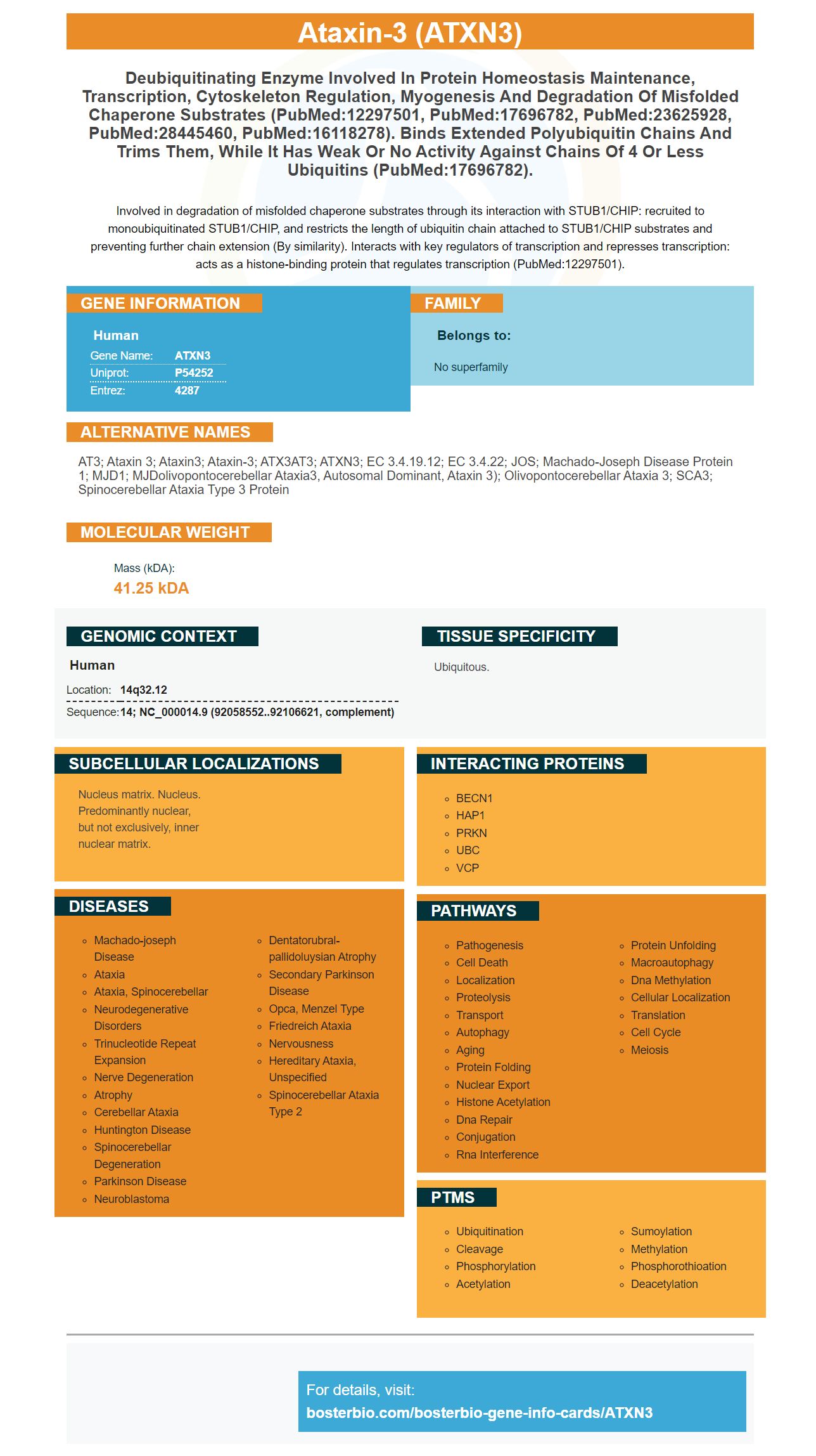
Facts about Ataxin-3.
Involved in degradation of misfolded chaperone substrates through its interaction with STUB1/CHIP: recruited to monoubiquitinated STUB1/CHIP, and restricts the length of ubiquitin chain attached to STUB1/CHIP substrates and preventing further chain extension (By similarity). Interacts with key regulators of transcription and represses transcription: acts as a histone-binding protein that regulates transcription (PubMed:12297501).
Human | |
---|---|
Gene Name: | ATXN3 |
Uniprot: | P54252 |
Entrez: | 4287 |
Belongs to: |
---|
No superfamily |
AT3; ataxin 3; Ataxin3; Ataxin-3; ATX3AT3; ATXN3; EC 3.4.19.12; EC 3.4.22; JOS; Machado-Joseph disease protein 1; MJD1; MJDolivopontocerebellar ataxia3, autosomal dominant, ataxin 3); olivopontocerebellar ataxia 3; SCA3; Spinocerebellar ataxia type 3 protein
Mass (kDA):
41.25 kDA
Human | |
---|---|
Location: | 14q32.12 |
Sequence: | 14; NC_000014.9 (92058552..92106621, complement) |
Ubiquitous.
Nucleus matrix. Nucleus. Predominantly nuclear, but not exclusively, inner nuclear matrix.
The ATXN3 marker from Boster Bio is an antigen-specific tyrosine kinase inhibitor. It can be used in research or application credits. It is used by biologists working in the biomedical and pharmaceutical fields as well as scientists. It is also used by scientists from all over the world. This Boster Bio review explains more about the ATXN3 marker.
Autophagy helps to eliminate misfolded proteins through the ATXN3 marker. It is a vital component of aggresomes which are cytoplasmic juxtanuclear structures that sequester potentially toxic proteins. ATXN3 interacts with proteins that regulate aggresome formation. Numerous studies have revealed that ATXN3 is able to facilitate autophagy.
Mutations in ATXN3 alter the endoplasmic reticulum's quality control system and alter the secretory pathway proteins. This process is known as ER-associated degradation (ERAD) and it is responsible for the degradation of proteins that are misfolded, unassembled polypeptides, as well as other undesirable substances. ATXN3 mutations inhibit ER retrotranslocation as well as ER degradation, two processes important for the development of the disease. Furthermore, the accumulation of misfolded protein could contribute to the pathogenesis SCA3.
There are many advantages to the ATXN3 marker. It can aid in early diagnosis and aid in clinical trials. It is helpful in identifying patients who might benefit from the ATXN3 mutation correction before, as the CNS changes take place before symptoms appear. The corrective intervention may not yield the full benefits because of the possibility that the mutation may have already caused irreparable changes to neurons. Doctors should focus on different treatment options that could be used to treat the neuronal dysfunctions by identifying the disease in its early stages.
A number of recent studies have shown that mutations in ATXN3 cause an increased susceptibility to toxicity by toxicants. Mutations in ATXN3 could affect the secretory pathway and the quality control mechanism for the endoplasmic-reticulum. ER-associated degradation (ERAD) is responsible for the degrading of misfolded proteins and unassembled polypeptides. This quality control system is controlled by VCP/p97, which removes proteins from the ER and transports them to proteasomes. Mutations in ATXN3 can cause retrotranslocation and degradation to ERAD substrates to be impaired, which results in the accumulation of proteins that are misfolded within the ER.
Mutant ATXN3 accumulation is associated with dysregulation of the microRNA (miRNA) machinery. These microRNAs target 3' UTR of human ATXN3. This has broad implications for cells. However, the exact mechanism that causes ATXN3 aggregation is not fully understood. In the meantime this gene expression molecule is being utilized to study different cancer-related diseases.
Mutations in ATXN3 also result in an increase in the nuclear PQC pathways' burden that results in a decrease of functional and properly folded nuclear protein levels. These malfunctioning proteins can impact the entire cellular function. Mutant ATXN3 proteins are extremely toxic. Mutant ATXN3 proteins may be artificially targeted to the nucleus. Their toxicity could increase in cells due to. These mutations could have a variety of clinical implications.
ATXN3 dysfunction can occur before symptoms become apparent. It may not be beneficial to restore ATXN3 function in the early stages of disease. It could be too late for neurons to recover from irreversible damage. The aim of this treatment is to restore the proper function of neurons through targeting the neuronal dysfunction underlying the disorder. Axonal aggregation has also been associated with neuropathy related to age. The condition is defined as metabolic disorders.
Mutants of ATXN3 were identified in the mitochondrial protein fraction. ATXN3 toxicities were previously recognized by the accumulation soluble oligomers prior to the formation of inclusion bodies. ATXN3 toxicity is more severe in certain cells than other. Patients suffering from SCA should be aware of ATXN3 toxicities.
Mutants of ATXN3 result in abnormal polyglutamine tracts. This condition affects the expression of ataxin-3. Normal people have normal levels of this protein. In patients suffering from SCA3, it is not evident if these proteins are essential for proper functioning of the brain. Application of the ATXN3 marker in patients suffering from this genetic disease is necessary to determine the role it plays in axonal development.
The use of ATXN3 as a marker during secondary antibody generation is a simple method of identifying antigens. Primary antibodies can only be produced in a limited number of host animals and are almost always IgG-class. Secondary antibodies can be manufactured easily by the manufacturers and are available for many detection systems. The kind of application and duration of the signal will determine option of the label.
Researchers have a broad selection of antibodies to choose from when they label multiple experiments with the ATXN3 marker. Secondary antibodies are usually supplied in liquid concentrate. Certain secondary antibodies might require optimization prior to being used. Product data sheets will show the correct concentrations. Liquid secondary antibodies should be stored at temperatures of 4-8 degrees Celsius to prolong their shelf lives. They can also be stored at 20degC and then thawed to make a functional solution.
Secondary antibodies can be purified using immobilized serum proteins. This method is often called cross-adsorbed (HCA). It is generally recommended for situations which require multiple primary antibodies. By preventing binding that is not specific this method is extremely specific. Boster Bio's ATXN3 marker to generate secondary antibodies is a distinct ATXN3 tag that is specifically designed for rabbit IgG light chains.
A primary antibody may be raised in a specific species, but it has to be conjugated with a secondary antibody of the same species. Secondary antibodies must recognize the primary antibody or they could be ineffective. They could also cross-react with other primary antibodies or bind to endogenous immunoglobulin in the sample. It is also important to limit the number of reactions between primary antibodies and secondary antibodies for accurate data. In addition to a primary antibody secondary antibodies must be able to recognize the target antigen to be accurate. In addition secondary antibodies must be specific to a specific fragment which allows them to be used to label multiple times.
There is a good chance that you will find a solution to your problem, whether you require it to produce secondary antibodies to one primary antibody or a number of primary antibodies. One of the best candidates for secondary antibodies is a mouse. However, if you're planning to perform multiple labeling experiments you should think about using an ATXN3-specific secondary antibody.
Affinity purification is necessary to use primary antibodies in secondary antibody generation. The primary antibody is used for the identification of a target protein. The secondary antibody is then injected into a column that is containing mouse IgG2a. This removes cross-reactive antibodies that could hinder the detection. It is possible to reduce non-specific binding by using an anti-mouse primary antibody as well as a serum from mice to produce secondary antibodies.
PMID: 7874163 by Kawaguchi Y., et al. CAG expansions in a novel gene for Machado-Joseph disease at chromosome 14q32.1.
PMID: 9274833 by Goto J., et al. Machado-Joseph disease gene products carrying different carboxyl termini.