This website uses cookies to ensure you get the best experience on our website.
- Table of Contents

1 Citations

Facts about Basal cell adhesion molecule.

.
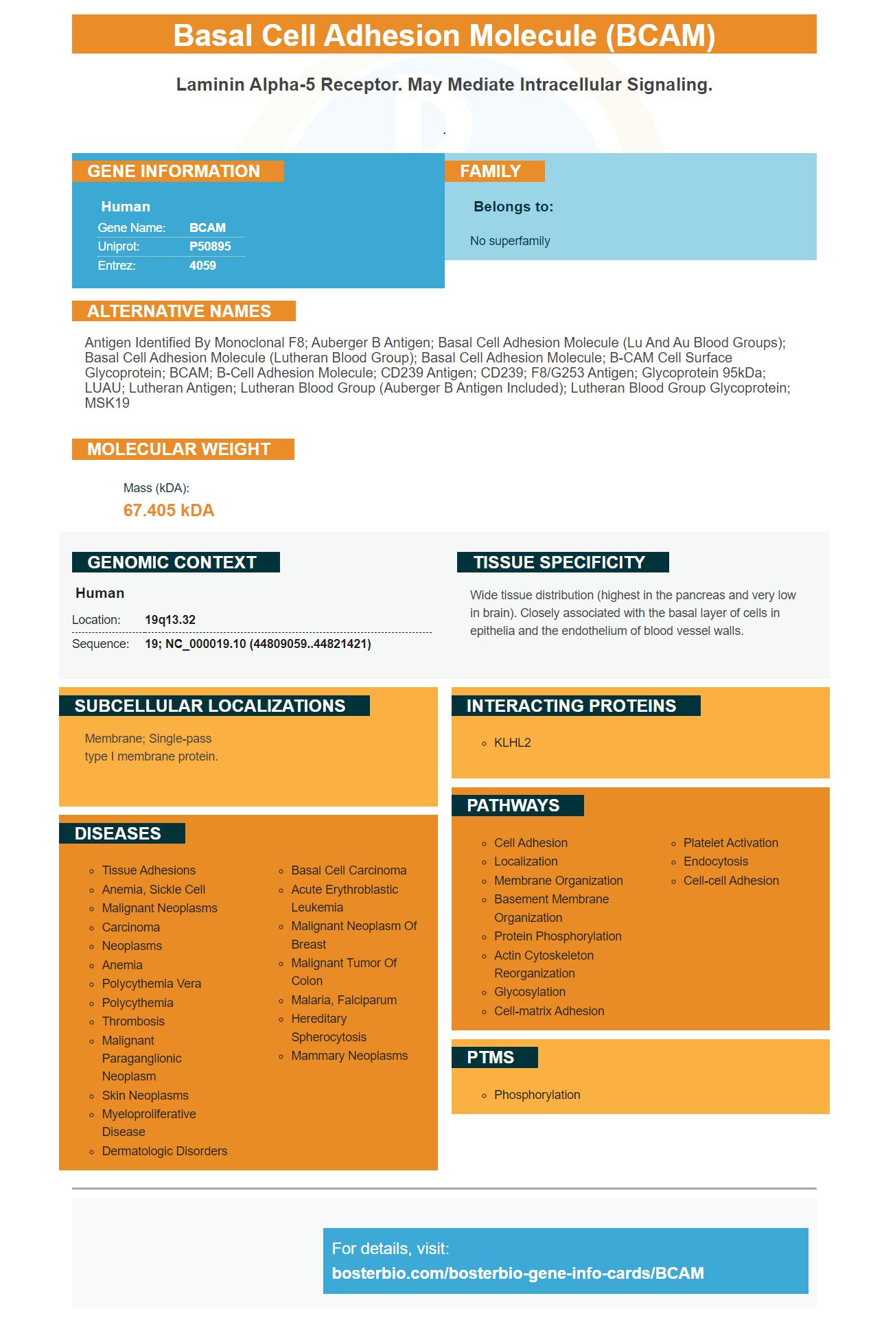
| Human | |
|---|---|
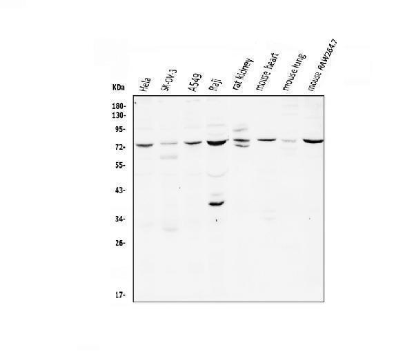
| Gene Name: | BCAM |
| Uniprot: | P50895 |
| Entrez: | 4059 |

| Belongs to: |
|---|
| No superfamily |

antigen identified by monoclonal F8; Auberger B antigen; basal cell adhesion molecule (Lu and Au blood groups); basal cell adhesion molecule (Lutheran blood group); basal cell adhesion molecule; B-CAM cell surface glycoprotein; BCAM; B-cell adhesion molecule; CD239 antigen; CD239; F8/G253 antigen; glycoprotein 95kDa; LUAU; Lutheran antigen; Lutheran blood group (Auberger b antigen included); Lutheran blood group glycoprotein; MSK19
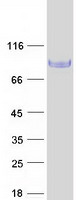
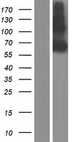
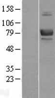
Mass (kDA):
67.405 kDA

| Human | |
|---|---|
| Location: | 19q13.32 |
| Sequence: | 19; NC_000019.10 (44809059..44821421) |
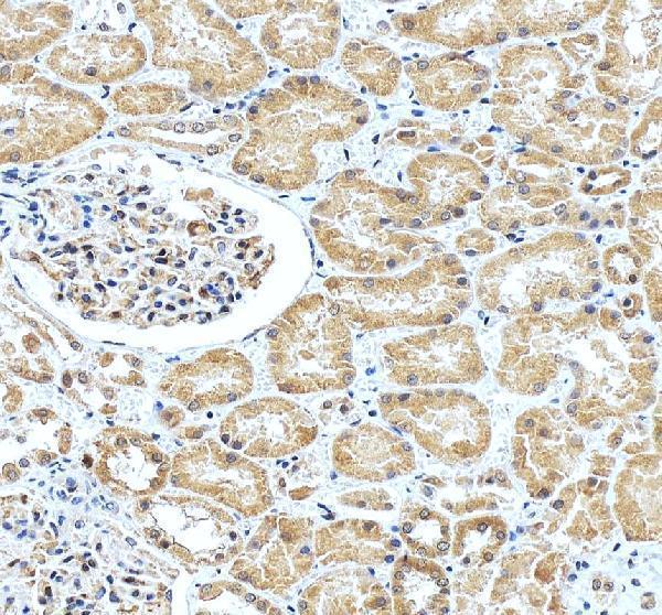
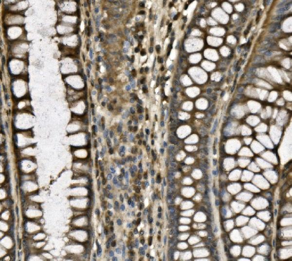
Wide tissue distribution (highest in the pancreas and very low in brain). Closely associated with the basal layer of cells in epithelia and the endothelium of blood vessel walls.
Membrane; Single-pass type I membrane protein.


PMID: 7777537 by Parsons S.F., et al. The Lutheran blood group glycoprotein, another member of the immunoglobulin superfamily, is widely expressed in human tissues and is developmentally regulated in human liver.
PMID: 7954395 by Campbell I.G., et al. Molecular cloning of the B-CAM cell surface glycoprotein of epithelial cancers: a novel member of the immunoglobulin superfamily.