This website uses cookies to ensure you get the best experience on our website.


- Table of Contents
Plan paraffin-section BCL11B IHC around nuclear staining in subsets of lymphoid, squamous epithelial and neuronal cells (HPA tissue IHC). Start within the catalog antibody’s 2–5 μg/ml IHC range (datasheet A01485-2) and score nuclear staining with appropriate controls.

Expected localisation, validated protocols, controls and antibodies — the at-a-glance facts below, then the full design guide.
| Expected localisation | Nuclear staining in subsets of tissue cells (HPA tissue IHC) | |
| Staining pattern | Nuclei in subsets of lymphoid, squamous, neuronal and testis cells (HPA tissue IHC) | |
| Antigen retrieval | EDTA pH 8.0 HIER, heat-mediated (datasheet A01485-2) | |
| Positive control | Appendix+4 more · see all | |
| Negative control | Adipose tissue+4 more · see all |
| Fixation | Matched tissue-IHC evidence does not establish the fixation claim. Validate the specimen-specific method before use. (selected-SKU IHC image A01485-2) | |
| Caveat | Subset staining; RNA concordance is medium (HPA tissue IHC) | |
| Regulation | High in brain and malignant T-cell lines (UniProt) | |
| Isoform / epitope | Two isoforms; verify epitope coverage (UniProt) |
The catalog antibody’s IHC-P protocol is accompanied by published BCL11B staining methods for HCC tissue (PMC10417273), Ewing sarcoma samples (PMC7139395), and mouse embryonic head sections (PMC3358280).
| Sample | Paraffin-embedded human bladder urothelial carcinoma with squamous differentiation tissue; fixative not specified (datasheet A01485-2) |
| Fixation | Image fixative and duration unreported (datasheet A01485-2); verify before use. |
| Sectioning | 4–5 µm sections on charged slides (standard) |
| Deparaffinisation | Xylene, graded ethanol series to water (standard) |
| Antigen retrieval | Heat retrieval: EDTA pH 8.0 (datasheet A01485-2); 20 min, 95–100 °C (standard) |
| Peroxidase block | 3% H2O2, 10 min, room temperature (standard) |
| Blocking | 10% goat serum (datasheet A01485-2) |
| Primary antibody | Rabbit anti-BCL11B, 2-5 μg/ml (datasheet A01485-2) |
| Primary incubation | Overnight at 4 °C (datasheet A01485-2) |
| Detection | HRP-conjugated secondary, DAB chromogen (datasheet A01485-2) |
| Counterstain | Hematoxylin, blue, dehydrate and mount (standard) |
| Expected result | BCL11B-positive staining in non-germinal center cells of appendix (HPA tissue IHC: High). HPA tissue profile: Nuclear expression in a subset of lymphoid cells, squamous epithelia, neurons and testis. No signal in the no-primary control. |
BCL11B is a nuclear protein with no transmembrane segment (UniProt Q9C0K0: subcellular location and topology). In paraffin-section IHC, expect staining in subsets of lymphoid cells, squamous epithelial cells and neurons (HPA: tissue IHC profile). HPA rates its tissue profile Enhanced, while reporting medium consistency with RNA data and pending external verification (HPA: tissue IHC reliability). Interpret staining by cell type and compartment together.
| Distinct nuclear staining in a subset of non-germinal center cells in appendix or lymph node (HPA: High in these cells). | This fits the strongest listed lymphoid IHC pattern. A mixture of stained and unstained cells is expected because HPA describes expression in a subset of lymphoid cells (HPA: tissue IHC profile). Assess the intended cells rather than requiring every lymphoid cell to stain. |
| Nuclear staining in neuronal cells of caudate, cerebral cortex or hippocampus, or squamous epithelial cells of cervix, esophagus or oral mucosa (HPA: Medium in each). | These are supported positive patterns at a lower reported level than the listed High lymphoid examples (HPA: tissue IHC). Compare signal within the named cell population; the HPA level does not guarantee identical chromogen intensity in every section. |
| Predominantly cytoplasmic signal, with little convincing nuclear signal (UniProt Q9C0K0: Nucleus; HPA: nuclear tissue profile). | This conflicts with the expected compartment and warrants an artefact check. Review morphology, counterstain and negative controls before scoring it as BCL11B; cytoplasmic colour alone is insufficient evidence of specific staining (general IHC practice). |
| Strong colour in a cell population HPA lists as not detected, such as adipocytes in adipose tissue (HPA: Not detected in adipocytes). | Consider cross-reactivity or endogenous detection activity, especially if the colour also appears in a no-primary control (general IHC practice). HPA reports the assessed cell population, so do not infer that every cell in that tissue must be negative. |
| Diffuse colour across cells and extracellular space, or no signal in the expected non-germinal center cells of appendix or lymph node (HPA: High in these cells). | Diffuse colour is difficult to assign to nuclei and suggests background; absent positive-control signal leaves a negative result uninterpretable (general IHC practice). Check controls and staining conditions before drawing a biological conclusion. |
| Cell distribution and level | HPA reports High staining in non-germinal center cells of appendix and lymph node, Medium in listed neurons and squamous epithelia, and a subset-based nuclear profile (HPA: tissue IHC). Choose a control by its named cell population; an adjacent cell need not match its intensity. |
| Compartment and topology | UniProt places BCL11B in the nucleus and lists no transmembrane segment (UniProt Q9C0K0: subcellular location and topology). HPA tissue IHC also reports nuclear expression (HPA: tissue IHC profile). Score nuclear signal, not membrane or cytoplasmic colour. |
| Isoforms and modifications | UniProt lists 2 isoforms and modified residues, including phosphorylation sites (UniProt Q9C0K0: isoforms and modified residues). The supplied record does not map the antibody epitope or show whether either feature changes IHC detection; do not use them to explain a weak section. |
| IHC evidence and limits | HPA lists antibody HPA049117 as IHC Enhanced; that validation category reflects reproduction by independent antibodies or orthogonal data (HPA: antibody validation). The tissue profile still has medium RNA–staining consistency and awaits external verification (HPA: tissue IHC reliability). |
| Antigen retrieval | No BCL11B-specific retrieval condition or fixation sensitivity is supplied. Select retrieval conditions using a positive tissue and appropriate controls, then record the condition used (general IHC practice). A change in signal after retrieval alone does not establish a BCL11B-specific fixation effect. |
| IF/ICC Q&A | Where should IF/ICC signal localize? Mainly to the nucleoplasm, with an additional approved nucleoli fibrillar-center location (HPA: subcellular ICC-IF). That observation supports a compartment check; this IHC section supplies no IF/ICC protocol. |
| Situation | Likely cause | Next action |
|---|---|---|
| No nuclear signal in non-germinal center cells of appendix or lymph node (HPA: High in these cells). | The run may have insufficient detection or a failed staining step; tissue identity and cell selection also need checking (general IHC practice). | Confirm the named cells on the counterstain, check the positive control and run controls, then review the recorded retrieval, antibody dilution and detection steps (general IHC practice). Do not score the sample negative until the control works. |
| Signal is mainly cytoplasmic despite preserved nuclear morphology (UniProt Q9C0K0: Nucleus). | The compartment conflicts with UniProt and HPA's nuclear tissue profile; nonspecific staining or chromogen deposition is possible (HPA: tissue IHC profile; general IHC practice). | Check a no-primary control, inspect staining at nuclear boundaries, and reassess blocking and washing (general IHC practice). Require convincing nuclear signal in an expected positive cell population before scoring. |
| Colour appears broadly in HPA-listed negative cells, such as adipocytes in adipose tissue (HPA: Not detected in adipocytes). | Cross-reactivity or endogenous detection activity is possible; the listed HPA result applies to adipocytes, not every cell in adipose tissue (HPA: tissue IHC; general IHC practice). | Compare the same cell type with no-primary and other detection controls; review endogenous-activity blocking for the chosen detection system (general IHC practice). Interpret unexpected staining cautiously if controls remain clean. |
| Diffuse background obscures nuclei in a chromogenic section. | Background can follow excessive reagent concentration, inadequate blocking or incomplete washing (general IHC practice). It cannot be assigned to BCL11B from distribution alone. | Inspect the negative controls and adjust antibody concentration, blocking or wash conditions within the validated IHC workflow (general IHC practice). Reassess whether nuclei in an expected positive population remain distinct. |
| Only some lymphoid cells stain while neighboring cells remain unstained (HPA: subset of lymphoid cells). | A subset pattern can be the expected result; HPA specifically reports High staining in non-germinal center cells of appendix and lymph node (HPA: tissue IHC). | Use morphology and the counterstain to identify the scored population, then record the fraction and intensity of nuclear-positive cells (general IHC practice). Do not treat mixed staining alone as assay failure. |
| An IF/ICC image shows nucleolar signal as well as nucleoplasmic signal (HPA: subcellular ICC-IF). | HPA approves the nucleoplasm as the main location and the nucleoli fibrillar center as an additional location (HPA: subcellular ICC-IF). | Check that nuclear structures are resolved and compare with imaging controls (general IF practice). Use the dedicated IF/ICC guide for that application; assess chromogenic paraffin-section results against the HPA tissue IHC pattern. |
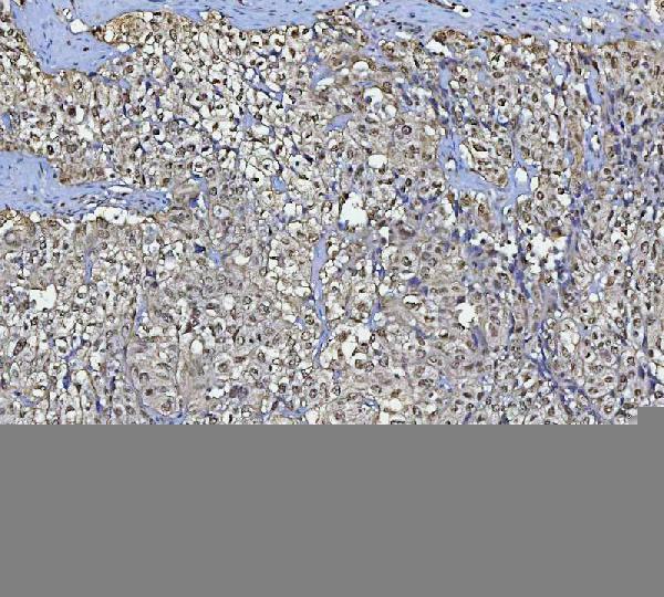
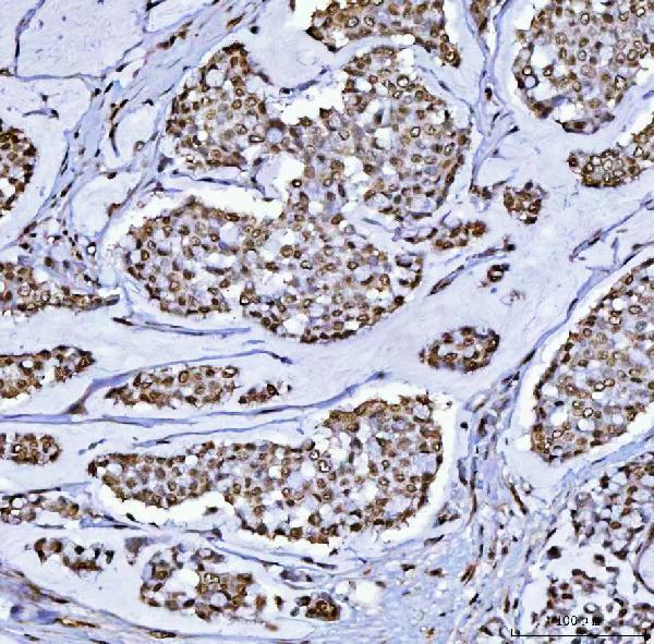
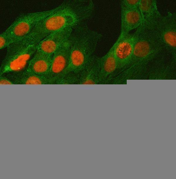
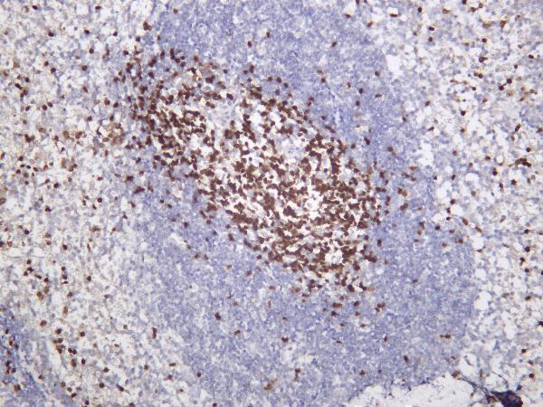
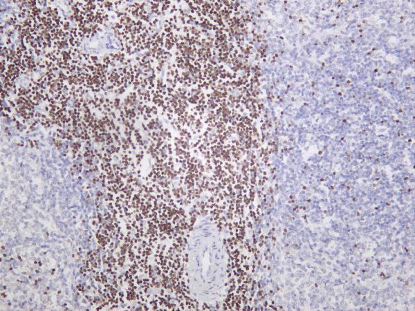


Comprehensive Human Protein Atlas IHC scoring per tissue (reliability: Enhanced — Medium consistency between antibody staining and RNA expression data. Pending external verification.). Rows are taken directly from the HPA tissue chart — click any row's HPA link to view the source.
| Tissue | Cell type | Level | Evidence | Source |
|---|---|---|---|---|
| Appendix | Non-germinal center cells | High | Protein (IHC) | HPA → |
| Lymph node | Non-germinal center cells | High | Protein (IHC) | HPA → |
| Caudate | Neuronal cells | Medium | Protein (IHC) | HPA → |
| Cerebral cortex | Neuronal cells | Medium | Protein (IHC) | HPA → |
| Cervix | Squamous epithelial cells | Medium | Protein (IHC) | HPA → |
| Tissue | Cell type | Level | Evidence | Source |
|---|---|---|---|---|
| Adipose tissue | Adipocytes | Not detected | Protein (IHC) | HPA → |
| Adrenal gland | Glandular cells | Not detected | Protein (IHC) | HPA → |
| Bone marrow | Hematopoietic cells | Not detected | Protein (IHC) | HPA → |
| Breast | Adipocytes | Not detected | Protein (IHC) | HPA → |
| Bronchus | Respiratory epithelial cells | Not detected | Protein (IHC) | HPA → |
Troubleshoot BCL11B staining in paraffin sections by checking retrieval, nuclear localisation, cell identity, and assay controls before interpreting chromogenic signal.
Both antibodies list human reactivity and have tissue IHC images (catalog reactivity; IHC captions). The images also cover mouse and rat spleen IHC (M01485-2 IHC captions) and U2OS IF (A01485-2 IF caption).
A01485-2 lists human and rat reactivity, with human paraffin-section IHC images and U2OS IF/ICC imaging (catalog reactivity and applications; A01485-2 IHC/IF captions). M01485-2 lists human, mouse and rat reactivity and IHC images of human tonsil, mouse spleen and rat spleen; IF is listed as an application (catalog reactivity and applications; M01485-2 IHC captions).
Which to pick: Choose A01485-2 for human paraffin-section IHC with a documented retrieval and detection workflow, or for IF/ICC with a documented U2OS image (A01485-2 IHC/IF captions; catalog applications). Choose M01485-2 when mouse, rat or human tissue IHC is needed: it is monoclonal and has spleen and tonsil IHC images, with an IHC starting range of 1:200–1:1000 (catalog clonality and dilution; M01485-2 IHC captions). The A01485-2 IHC captions specify paraffin sections, but neither SKU’s IHC captions report the fixative (A01485-2 and M01485-2 IHC captions).