This website uses cookies to ensure you get the best experience on our website.


- Table of Contents
Plan BRAP staining in paraffin sections with the IHC-validated antibody (datasheet A03573). This guide uses the reported cytoplasmic pattern and high staining in neurons, small-intestinal glandular cells and pachytene spermatocytes to help select tissues and interpret results (HPA tissue IHC).

Expected localisation, validated protocols, controls and antibodies — the at-a-glance facts below, then the full design guide.
| Expected localisation | Broad cytoplasmic staining (HPA tissue IHC) | |
| Staining pattern | Cytoplasmic staining across many tissue cell types (HPA tissue IHC) | |
| Antigen retrieval | Citrate pH 6.0 HIER, heat-mediated (datasheet A03573) | |
| Positive control | Cerebral cortex+4 more · see all | |
| Negative control | Adipose tissue+1 more · see all |
| Fixation | Keep paraffin-section fixation consistent across specimens. (selected-SKU IHC image A03573) | |
| Caveat | Antibody staining and RNA show low consistency (HPA tissue IHC) | |
| Regulation | Testis-enhanced RNA expression (HPA tissue RNA) | |
| Isoform / epitope | 2 isoforms; epitope coverage is unresolved (UniProt) |
Compare the catalog antibody’s IHC-P protocol (datasheet A03573) with one published BRAP tissue microarray protocol (PMC12717807).
| Sample | Paraffin-embedded human lung cancer tissue; fixative not specified (datasheet A03573) |
| Fixation | Image fixative and duration unreported (datasheet A03573); verify before use. |
| Sectioning | 4–5 µm sections on charged slides (standard) |
| Deparaffinisation | Xylene, graded ethanol series to water (standard) |
| Antigen retrieval | Heat retrieval: Citrate pH 6.0 (datasheet A03573); 20 min, 95–100 °C (standard) |
| Peroxidase block | 3% H2O2, 10 min, room temperature (standard) |
| Blocking | 10% normal serum of the secondary host, 30 min, room temperature (standard) |
| Primary antibody | Rabbit anti-BRAP, 1:50-1:100 (datasheet A03573) |
| Primary incubation | Overnight at 4 °C (standard) |
| Detection | HRP-polymer secondary, DAB chromogen 5–10 min (standard) |
| Counterstain | Hematoxylin, blue, dehydrate and mount (standard) |
| Expected result | BRAP-positive staining in neuronal cells of cerebral cortex (HPA tissue IHC: High). HPA tissue profile: Ubiquitous cytoplasmic expression. No signal in the no-primary control. |
BRAP is a cytoplasmic protein with no transmembrane segment (UniProt Q7Z569 localization and topology). In paraffin sections, expect cytoplasmic staining in neuronal cells of cerebral cortex, glandular cells of small intestine, and pachytene spermatocytes of testis (HPA: High). HPA describes the tissue pattern as ubiquitous cytoplasmic expression, but rates its IHC evidence Approved with low agreement between staining and RNA data and a possible splice or transcript discrepancy (HPA: tissue IHC reliability).
| Cytoplasmic stain is strong in the expected cells of cerebral cortex, small intestine, or testis. | This matches the reported High staining in neuronal cells, intestinal glandular cells, and pachytene spermatocytes (HPA: tissue IHC). Compare cell types within the section; HPA's Approved rating carries a low staining–RNA consistency caution (HPA: reliability). |
| Nuclear staining dominates, with little cytoplasmic signal. | Predominantly nuclear IHC conflicts with BRAP's cytoplasmic location (UniProt Q7Z569) and the HPA tissue profile. Recheck morphology and staining controls before assigning the signal to BRAP. A nuclear membrane location is reported in ICC-IF, which does not establish nuclear staining as the expected IHC pattern (HPA: subcellular ICC-IF). |
| Strong stain appears in adipocytes or ovarian stroma cells. | These cells are reported as Not detected (HPA: adipose tissue; ovary). Consider antibody cross-reactivity or endogenous detection activity, then examine a no-primary control (general IHC practice). A stain in an HPA-negative cell type alone does not identify which cause applies. |
| Color spreads across the section without clear cell boundaries. | Diffuse deposit is difficult to score as cytoplasmic BRAP even where expression is expected (UniProt Q7Z569 localization; HPA: tissue profile). Review the no-primary control, blocking, washes, and detection exposure for nonspecific background (general IHC practice). |
| No cytoplasmic signal appears in an expected positive tissue. | A blank result in HPA High cell populations warrants a technical check before a biological negative call (HPA: cerebral cortex, small intestine, testis). Verify the antibody's IHC-P validation, detection reagents, and an on-slide positive control (general IHC practice). HPA's reliability caveat also limits certainty for a single specimen (HPA: reliability). |
| Compartment and topology | BRAP is cytoplasmic and has no transmembrane segment (UniProt Q7Z569). Score cellular cytoplasm rather than treating membrane outlining or dominant nuclear staining as its expected tissue IHC pattern (HPA: tissue profile). |
| Choice of comparison cells | High HPA staining occurs in cerebral cortex neurons, small-intestine glandular cells, and testis pachytene spermatocytes; adipocytes and ovarian stroma cells are Not detected (HPA: tissue IHC). These observations support positive and negative comparison areas, subject to the HPA reliability caveat. |
| Antibody evidence | HPA040357 is Approved for IHC; HPA058820 has no IHC validation status in the supplied record (HPA: antibody validation). Use the IHC-validated antibody when comparing a new paraffin-section pattern with HPA, and do not treat Approved as Enhanced (HPA: antibody validation). |
| Isoforms and evidence limits | UniProt lists two BRAP isoforms (UniProt Q7Z569). HPA flags a possible splice or transcript discrepancy and low staining–RNA consistency (HPA: reliability); neither source identifies an isoform-specific IHC pattern. Target-specific fixation sensitivity is unreported in the supplied evidence. |
| IF/ICC: where should signal appear? | In ICC-IF, BRAP is mainly cytosolic, with an additional nuclear membrane location; the cytosol call is Supported and the nuclear membrane call Approved (HPA: subcellular ICC-IF). This localization is context for interpretation; IF/ICC methods belong to its separate guide. |
| Situation | Likely cause | Next action |
|---|---|---|
| Expected positive cells are blank. | The HPA High reference pattern was not reproduced (HPA: cerebral cortex, small intestine, testis); a failed IHC detection step is possible (general IHC practice). | Check an on-slide positive control, antibody identity and IHC-P validation, detection reagents, and the prescribed retrieval conditions if the antibody's instructions provide them (general IHC practice). Do not infer BRAP-specific retrieval sensitivity from HPA. |
| Nuclear stain is stronger than cytoplasmic stain. | That pattern conflicts with the cytoplasmic tissue expectation (UniProt Q7Z569; HPA: tissue profile). The additional nuclear membrane ICC-IF finding does not explain diffuse nuclear IHC (HPA: subcellular ICC-IF). | Inspect cell boundaries and counterstain, compare a no-primary control, and reassess antibody specificity before scoring nuclear signal as BRAP (general IHC practice). |
| Adipocytes or ovarian stroma cells stain strongly. | Both are Not detected in the HPA tissue observations (HPA: adipose tissue; ovary). Cross-reactivity or endogenous detection activity is possible (general IHC practice). | Run a no-primary control and appropriate endogenous-enzyme block for chromogenic detection; compare staining with the expected positive cell populations (general IHC practice; HPA: tissue IHC). |
| Broad haze obscures cell-level scoring. | Background can arise from nonspecific reagent binding, inadequate washing, or excessive chromogen development (general IHC practice). Haze cannot establish the cytoplasmic localization reported for BRAP (UniProt Q7Z569). | Review no-primary staining, blocking, washes, and chromogen development; adjust the general detection workflow according to the antibody and reagent instructions (general IHC practice). |
| Signal differs between HPA-positive tissues or cell populations. | Reported levels differ: High in selected neurons, glandular cells, and spermatocytes, but Medium in several other listed populations (HPA: tissue IHC). HPA also reports low staining–RNA consistency (HPA: reliability). | Score the named cell types and cytoplasmic compartment separately, document controls, and avoid equating a weaker population with technical failure solely from its tissue name (HPA: tissue IHC; general IHC practice). |
| IHC and ICC-IF images seem to disagree. | The ICC-IF summary includes an additional nuclear membrane location, whereas tissue IHC is described as ubiquitous cytoplasmic expression (HPA: subcellular ICC-IF; tissue IHC). The assays provide different localization observations. | Judge paraffin IHC against its cytoplasmic tissue pattern and IHC-validated antibody evidence (HPA: tissue IHC; antibody validation). Assess ICC-IF localization with its separate guide and controls (general IF practice). |


Comprehensive Human Protein Atlas IHC scoring per tissue (reliability: Approved — Low consistency between antibody staining and RNA expression data. Caution, Splice and/or transcript discrepancy exists. Pending external verification.). Rows are taken directly from the HPA tissue chart — click any row's HPA link to view the source.
| Tissue | Cell type | Level | Evidence | Source |
|---|---|---|---|---|
| Cerebral cortex | Neuronal cells | High | Protein (IHC) | HPA → |
| Small intestine | Glandular cells | High | Protein (IHC) | HPA → |
| Testis | Pachytene spermatocytes | High | Protein (IHC) | HPA → |
| Adrenal gland | Glandular cells | Medium | Protein (IHC) | HPA → |
| Appendix | Glandular cells | Medium | Protein (IHC) | HPA → |
BRAP staining is expected mainly in the cytoplasm; assess signal by cell type and compartment, with controls for processing and detection artefacts (UniProt Q7Z569; HPA subcellular).
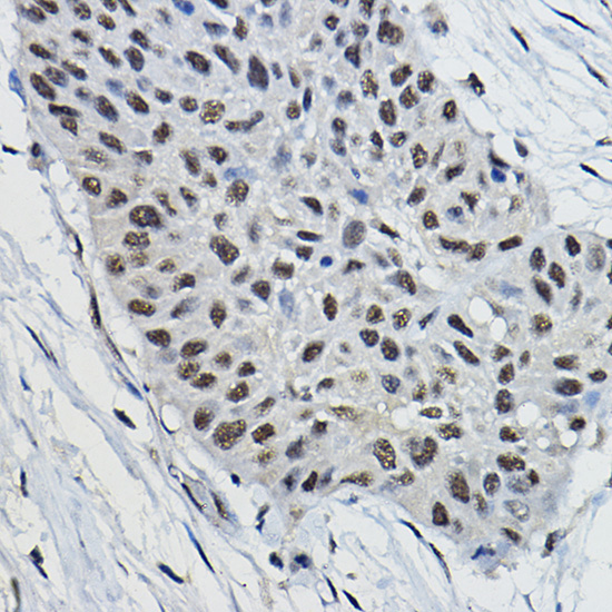
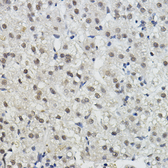
A03573 has IHC images from paraffin-embedded human lung and liver cancers (catalog IHC image captions). It is also listed for IF/ICC in human, mouse and rat (catalog applications/reactivity).
A03573 will render with its own human lung cancer IHC figure, stained at 1:100 after high-pressure retrieval in 10 mM citrate, pH 6.0 (catalog IHC image caption). Its second IHC image shows paraffin-embedded human liver cancer; IF/ICC and human, mouse and rat reactivity are listed, but no IF image is supplied (catalog IHC image captions/applications/reactivity/IF image alts).
Which to pick: Choose A03573 for tissue IHC: its own captions document paraffin-embedded human lung and liver cancer sections, with 1:100 staining after high-pressure citrate retrieval; the fixative is unreported (catalog IHC image captions). For IF/ICC, A03573 is listed at 1:50–1:100, though no IF image is supplied (catalog applications/IF dilution/IF image alts). For cross-species studies, A03573 is a rabbit polyclonal listed as reactive with human, mouse and rat; optimize staining for each specimen (catalog host/clone/reactivity; standard IHC practice).