BRI3BP / BRI3-binding protein · IHC design guide

Design Immunohistochemistry for BRI3BP

Plan BRI3BP chromogenic IHC-P around the granular cytoplasmic tissue pattern (HPA tissue IHC). The catalog antibody has a 1:100–1:300 IHC dilution range (datasheet) and a paraffin-embedded human brain example (catalog IHC caption).

Evidence assembled Oct 2026 · For research use; verify linked source records and product datasheet before use
Immunohistochemistry protocol sheet for BRI3BP (IHC for BRI3BP): expected localisation Granular cytoplasm (HPA tissue IHC), antibody A30536, validated IHC image, and IHC protocol steps
Printable BRI3BP IHC protocol sheet — expected localisation Granular cytoplasm (HPA tissue IHC), antibody A30536, controls and protocol steps. Open the full BRI3BP IHC guide →

BRI3BP Immunohistochemistry Experimental Design Guide

Expected localisation, validated protocols, controls and antibodies — the at-a-glance facts below, then the full design guide.

Must know before staining
Expected localisation Granular cytoplasm (HPA tissue IHC)
Staining pattern Granular cytoplasmic staining in most tissues (HPA tissue IHC)
Antigen retrieval Citrate pH 6.0 HIER, 95–98 °C, 20 min (rule: cytoplasmic / membrane antigen)
Positive control ⓘ Adrenal gland+4 more · see all
Negative control ⓘ Adipose tissue+4 more · see all
Important caveats
Reasons your staining may differ from the expected pattern.
Fixation Keep fixation conditions consistent across sections. (standard IHC practice; not target-specific)
Caveat Staining and RNA show medium consistency (HPA tissue IHC)
Regulation Overexpressed in various carcinomas (UniProt)
Isoform / epitope No annotated isoforms or cleavage; epitope site unspecified (UniProt)
Section 1

Recommended BRI3BP IHC & IF Protocols

The catalog antibody’s IHC-P protocol is accompanied by published BRI3BP protocols for invasive breast cancer and hepatocellular carcinoma FFPE sections (PMC11730447; PMC12578974).

Recommended immunohistochemistry (IHC-P) protocol parameters
SampleParaffin-embedded human brain tissue; fixative not specified (datasheet A30536)
FixationImage fixative and duration unreported (datasheet A30536); verify before use.
Sectioning4–5 µm sections on charged slides (standard)
DeparaffinisationXylene, graded ethanol series to water (standard)
Antigen retrievalHeat-induced epitope retrieval in citrate buffer, pH 6.0, 20 min at 95–98 °C (standard rule: cytoplasmic / membrane antigen)
Peroxidase block3% H2O2, 10 min, room temperature (standard)
Blocking10% normal serum of the secondary host, 30 min, room temperature (standard)
Primary antibodyRabbit anti-BRI3BP, 1:100 - 1:300 (datasheet A30536)
Primary incubationOvernight at 4 °C (standard)
DetectionHRP-polymer secondary, DAB chromogen 5–10 min (standard)
CounterstainHematoxylin, blue, dehydrate and mount (standard)
Expected resultBRI3BP-positive staining in glandular cells of adrenal gland (HPA tissue IHC: Medium). HPA tissue profile: Granular cytoplasmic expression in most tissues. No signal in the no-primary control.
💡Decision noteStart with citrate pH 6.0 HIER at 95–98 °C for 20 min (page antigen-retrieval rule).
Section 2

What Is the Expected BRI3BP Staining Pattern?

In paraffin-section IHC, expect granular cytoplasmic BRI3BP staining in many tissues, including glandular cells, hematopoietic cells and glial cells (HPA tissue IHC: Approved; medium consistency with RNA). This pattern is compatible with a mitochondrial outer-membrane protein containing 4 transmembrane segments (UniProt Q8WY22 topology). HPA also reports nucleoplasmic and mitochondrial localisation by ICC-IF; that result should be interpreted in its own assay context (HPA subcellular: approved).

What am I looking at on my slide?
Granular cytoplasmic staining in colon glandular cells, with a medium signal (HPA tissue IHC: colon).This matches an observed positive cell population and the predominant tissue IHC pattern (HPA tissue IHC: granular cytoplasmic expression in most tissues). Score the signal in glandular cells rather than treating every cell in the section as an expected positive (HPA tissue IHC: colon glandular cells, Medium).
Predominantly nuclear staining in a paraffin section, without a convincing granular cytoplasmic component.Investigate an IHC pattern discordant with the reported tissue pattern (HPA tissue IHC: granular cytoplasmic). Nuclear signal alone is not proof of an artefact: nucleoplasm is an approved ICC-IF location (HPA subcellular). Check controls and the antibody's IHC validation before assigning it to BRI3BP (general IHC practice).
Strong staining in adipocytes or smooth muscle cells, while expected positive cells stain (HPA tissue IHC: both Not detected).These cell types were reported as not detected by tissue IHC (HPA tissue IHC: adipocytes; smooth muscle cells). Unexpected staining warrants a check for cross-reactivity, endogenous detection activity or tissue background (general IHC practice); a single section cannot distinguish those causes.
Widespread, diffuse colour obscures the expected cellular pattern.A diffuse deposit does not establish the reported granular cytoplasmic pattern (HPA tissue IHC). Review no-primary and detection controls, blocking, washes and chromogen development to identify background from the IHC workflow (general IHC practice).
No detectable signal in colon glandular cells or bone-marrow hematopoietic cells (HPA tissue IHC: both Medium).Loss of signal in a reported positive cell population makes a negative result elsewhere difficult to interpret (HPA tissue IHC: colon; bone marrow). Check section integrity, retrieval, antibody and detection performance with a concurrently stained positive section (general IHC practice).
💡Expected BRI3BP appearanceCall a positive IHC result when glandular, hematopoietic or glial cells show a discernible granular cytoplasmic signal around the reported medium level (HPA tissue IHC); diffuse colour or staining confined to reported negative cell types needs control-based review (HPA tissue IHC; general IHC practice).
How each factor affects the staining
Compartment and assayUniProt places BRI3BP at the mitochondrial outer membrane with 4 transmembrane segments (UniProt Q8WY22 topology); HPA reports granular cytoplasm in tissue IHC and nucleoplasm plus mitochondria in ICC-IF (HPA tissue IHC; HPA subcellular). Interpret each localisation in its stated assay.
Cell population selected for scoringHPA reports Medium IHC staining in colon glandular cells, bone-marrow hematopoietic cells and cerebral-cortex glial cells, but Not detected in adipocytes and smooth muscle cells (HPA tissue IHC). Compare the named cell populations, not whole-tissue averages.
Strength of validationThe tissue IHC profile is Approved with medium consistency between antibody staining and RNA expression (HPA tissue IHC). The listed antibody, HPA014957, is IHC Approved; the supplied record does not label its IHC validation Enhanced (HPA antibodies). Treat an unexpected distribution as a finding to verify.
Expression evidence across assaysUniProt describes abundant expression in brain, liver and kidney (UniProt Q8WY22 tissue specificity), while HPA calls RNA expression tissue enhanced in intestine and reports granular protein staining in most tissues (HPA tissue IHC). RNA and protein summaries describe different measurements; use cell-level IHC observations for slide scoring.
Target-specific fixation sensitivityNo target-specific fixation effect is supplied by the UniProt or HPA records (source scope). Record retrieval and staining conditions for reproducibility (general IHC practice), without inferring that a change in fixation alters BRI3BP signal.
Why is my staining missing, weak or wrong?
SituationLikely causeNext action
Positive-control glandular cells show no stain.The control does not reproduce a reported Medium signal (HPA tissue IHC: colon glandular cells); a failed staining run is possible (general IHC practice).Repeat with a concurrently processed positive section; check retrieval, primary-antibody step and detection reagents in the IHC workflow (general IHC practice). Do not call test sections negative until the positive control works.
A reported negative cell population shows prominent colour.Adipocytes and smooth muscle cells are listed as Not detected (HPA tissue IHC); cross-reactivity or endogenous detection activity is possible (general IHC practice).Compare with a no-primary control and an appropriate detection control; reassess blocking and identify the stained cell type on the counterstained section (general IHC practice).
The slide has uniform haze or precipitate.The expected tissue pattern is granular cytoplasmic (HPA tissue IHC); diffuse deposit may arise from nonspecific staining or detection chemistry (general IHC practice).Inspect the no-primary control, wash steps and chromogen development; adjust only the implicated workflow step and compare with a positive control (general IHC practice).
Signal appears nuclear with little cytoplasmic granularity in IHC.That differs from the tissue IHC profile, although HPA reports approved nucleoplasmic localisation in ICC-IF (HPA tissue IHC; HPA subcellular).Document the assay and cell type; compare IHC controls and antibody validation before interpreting nuclear-only colour as specific (general IHC practice; HPA antibodies: HPA014957 IHC Approved).
Different tissue regions give different apparent positivity.HPA reports staining by cell population, with Medium signal in some cells and Not detected in others (HPA tissue IHC). A change in cellular composition can change the overall impression (general histology practice).Score identified cells separately and compare each population with its HPA entry; retain the counterstain so cell identity remains assessable (HPA tissue IHC; general IHC practice).
IHC and ICC-IF images appear to show different compartments.HPA describes granular cytoplasmic tissue IHC but approved nucleoplasmic and mitochondrial ICC-IF localisation (HPA tissue IHC; HPA subcellular).Report the assay used and its observed compartment. Use the tissue IHC profile to judge the paraffin-section result; consult the separate IF/ICC guide for that assay's controls and interpretation (HPA tissue IHC; HPA subcellular).

Sample controls for BRI3BP IHC & IF

🧪Run breast first and score glandular cells for BRI3BP staining (HPA: breast glandular cells, Medium). Use cervix squamous epithelium as the negative tissue (HPA: cervix squamous epithelial cells, Not detected); on the breast slide, stromal fibroblasts should provide a background reference, but verify their status rather than assuming they are negative (HPA: soft tissue fibroblasts, Not detected).
Positive control tissue: Adrenal gland (Glandular cells, HPA Medium)
Negative control tissue: Adipose tissue (HPA Not detected)
ICC-IF cell lines (HPA subcellular resource): HPA ICC-IF images show BRI3BP in RT-4, with annotated localisation: Nucleoplasm (approved), Mitochondria (approved) (HPA subcellular).
Technical controls: Include a no-primary control and a nonimmune isotype control matched to the primary antibody’s host species, immunoglobulin class, and monoclonal or polyclonal format; use a BRI3BP knockout specimen or a validated peptide block as a biological specificity control (SKU A30536 tissue-IHC caption: peptide-blocked brain section). For chromogenic IHC, block endogenous peroxidase and assess residual signal in the breast section with the no-primary control (standard IHC practice).
⚠️Feasibility: A target-specific fixation window, fixation effect, and retrieval requirement are unreported in the supplied evidence; compare retrieval conditions empirically (standard IHC practice). The selected SKU’s paraffin brain-section caption does not report a fixative (SKU A30536 tissue-IHC caption: fixative unreported), and the evidence does not establish whether frozen sections or IF are easier than paraffin IHC. If using breast tissue for IF, assess tissue autofluorescence with a no-primary control (standard IF practice); RT-4 ICC-IF images provide a localisation reference in nucleoplasm and mitochondria (HPA: subcellular localisation and RT-4 ICC-IF images).

HPA tissue IHC evidence for BRI3BP

Comprehensive Human Protein Atlas IHC scoring per tissue (reliability: Approved — Medium consistency between antibody staining and RNA expression data.). Rows are taken directly from the HPA tissue chart — click any row's HPA link to view the source.

Positive expression · recommended positive controls

TissueCell typeLevelEvidenceSource
Adrenal gland Glandular cells Medium Protein (IHC) HPA →
Appendix Glandular cells Medium Protein (IHC) HPA →
Bone marrow Hematopoietic cells Medium Protein (IHC) HPA →
Breast Glandular cells Medium Protein (IHC) HPA →
Caudate Glial cells Medium Protein (IHC) HPA →

Undetected expression · recommended negative controls

TissueCell typeLevelEvidenceSource
Adipose tissue Adipocytes Not detected Protein (IHC) HPA →
Cervix Squamous epithelial cells Not detected Protein (IHC) HPA →
Smooth muscle Smooth muscle cells Not detected Protein (IHC) HPA →
Soft tissue Fibroblasts Not detected Protein (IHC) HPA →
Vagina Squamous epithelial cells Not detected Protein (IHC) HPA →
Section 3

Advanced BRI3BP IHC Tips

Troubleshoot BRI3BP staining in paraffin sections by checking retrieval, compartment, controls and cell-specific scoring (page application: chromogenic IHC).

How should I retrieve BRI3BP in paraffin sections when staining is weak?
Start with citrate buffer at pH 6.0, heated to 95–98 °C for 20 min (page retrieval specification). Allow slides to cool consistently, then compare a known positive section across runs; variable heating can change staining intensity (standard IHC practice). The selected antibody has a paraffin-section image from human brain with peptide blocking, but its caption gives no retrieval conditions or fixative (catalog antibody A30536 caption). If staining remains weak, test an EDTA-based alkaline retrieval condition on adjacent sections while keeping antibody incubation and detection constant (standard IHC practice). Judge improvement by granular cytoplasmic signal and preserved tissue structure, since that pattern is reported across tissues (HPA: tissue IHC profile).
Could fixation explain inconsistent BRI3BP staining between paraffin blocks?
Target-specific fixation sensitivity is unknown: the selected human brain paraffin-section caption does not state a fixative or compare fixation conditions (catalog antibody A30536 caption). Record fixative, fixation duration, tissue thickness and processing history for each block before attributing intensity differences to biology (standard IHC practice). On matched tissue, compare sections cut at 4 µm and processed through the same retrieval and chromogenic detection run to reduce technical variation (standard IHC practice). Include a consistently stained reference section and a no-primary control; neither HPA staining patterns nor predicted membrane topology establishes how fixation affects this antibody (HPA: tissue IHC profile; UniProt Q8WY22 topology).
Should BRI3BP staining appear in mitochondria, cytoplasm or nuclei?
Expect predominantly granular cytoplasmic staining in tissue sections, consistent with the reported IHC profile (HPA: granular cytoplasmic expression in most tissues). BRI3BP is annotated at the mitochondrial outer membrane, while approved subcellular imaging also reports mitochondria and nucleoplasm (UniProt Q8WY22 localisation; HPA: subcellular localisation). Assess nuclear signal separately from cytoplasmic granules instead of combining both into a single intensity score; the two compartments have different supporting evidence (standard IHC scoring practice; HPA: subcellular localisation). If staining is diffuse across every compartment, repeat the no-primary and peptide-blocked comparisons before assigning localisation (standard IHC practice; catalog antibody A30536 caption). Preserve morphology with a counterstain so granules can be assigned to cells (standard IHC practice).
How can I assess whether the antibody detects the intended BRI3BP epitope?
The record lists one chain of 251 amino acids, no annotated isoforms and four transmembrane segments, but does not identify this antibody’s epitope (UniProt Q8WY22 record). Accordingly, do not infer whether retrieval exposes an external or cytosolic sequence from topology alone (UniProt Q8WY22 topology; standard antibody-validation practice). The selected brain image includes a synthesized-peptide blocking comparison, which supports epitope-dependent staining in that image but cannot establish specificity in every tissue (catalog antibody A30536 caption; standard IHC interpretation). Compare staining with and without blocking peptide under identical retrieval and detection conditions, and record any residual cell-specific signal (standard IHC practice). Acetyllysine 229 and phosphoserine 248 are annotated modifications; their effects on antibody binding are unknown (UniProt Q8WY22 modified residues).
How should I adapt BRI3BP localisation checks to multiplex immunofluorescence?
For a brain-cell multiplex comparison, pair BRI3BP with a glial-cell marker because glial staining is reported in caudate and cerebral cortex (HPA: tissue IHC). Choose spectrally separated fluorophores, favouring a far-red channel for weaker signal, and image an unstained section to measure tissue autofluorescence (standard IF practice). Permeabilise according to the epitope’s membrane-facing side; because the catalog epitope is unspecified, compare a gentle condition with 0.1% Triton X-100 for 5 min while checking mitochondrial morphology (UniProt Q8WY22 topology; standard IF practice). Score mitochondrial and nuclear channels separately, as both localisations are reported in subcellular imaging (HPA: approved mitochondria and nucleoplasm). The selected paraffin IHC caption does not establish IF performance for this antibody (catalog antibody A30536 caption).
What controls distinguish BRI3BP signal from chromogenic background?
Use a no-primary section to reveal secondary-reagent or detection background, and apply a peroxidase block before HRP-based chromogen development (standard chromogenic IHC practice). For an initial comparison, 3% hydrogen peroxide for 10 min is a general workflow condition that should be checked against tissue preservation (standard IHC practice). Titrate the catalog antibody and a protein blocking step together on adjacent sections, keeping retrieval and development time identical (standard IHC practice). The selected human brain image includes synthesized-peptide blocking, so compare the blocked section with its unblocked counterpart when investigating diffuse signal (catalog antibody A30536 caption). Accept cell-associated granules more readily than uniform haze, consistent with the reported tissue pattern (HPA: granular cytoplasmic expression).
How should I quantify BRI3BP staining across cell types and sections? ⚠ ANSWER MARKED FOR VERIFICATION
Define the tissue region and cell type before scoring; HPA reports medium staining in cerebral-cortex glial cells and no detected staining in adipocytes (HPA: tissue IHC). For cell-based scoring, record the percentage of positive cells and an H-score from 0–300, calculated as the sum of each intensity grade multiplied by its cell percentage (standard IHC scoring practice). Alternatively, report positive-cell density per mm² when cell counts vary across regions (standard image-analysis practice). Normalise chromogen measurements to eligible cell number or analysed tissue area, and exclude folds, edges and necrotic regions (standard IHC quantification practice). Analyse nuclear and granular cytoplasmic staining separately because both subcellular locations have evidence (HPA: subcellular localisation; HPA: tissue IHC profile).
When is apparent BRI3BP positivity more likely to be an artefact?
Interpret cell-associated granular cytoplasmic signal as plausible, especially in reported positive populations such as brain glial cells or colon glandular cells (HPA: tissue IHC). Mitochondrial and nucleoplasmic localisation are both reported, so nuclear staining alone requires compartment-specific validation instead of automatic rejection (HPA: approved subcellular localisation). Suspect an artefact when colour concentrates at section edges, folds or necrosis, or appears equally in the no-primary control after chromogenic detection (standard IHC practice). Check for residual endogenous peroxidase signal with the detection control and repeat the peroxidase block if needed (standard chromogenic IHC practice). A matching peptide-blocked comparison strengthens interpretation, although the published comparison is limited to the selected human brain image (catalog antibody A30536 caption).
Boster reagents

Best BRI3BP / BRI3-binding protein IHC Antibodies

A30536 has a paraffin-section human brain IHC figure and listed IF/ICC applications; its catalog lists human and mouse reactivity (A30536 image caption; applications; reactivity).

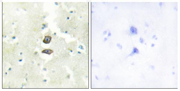
Real IHC data Immunohistochemistry analysis of paraffin-embedded human brain tissue, using BRI3B Antibody. The picture on the right is blocked with the synthesized peptide.
Anti-BRI3B BRI3BP Antibody
Cat # A30536

A30536 will render with an IHC image of paraffin-embedded human brain tissue and a peptide-blocked companion image (A30536 IHC image caption). IF and ICC are listed applications, but no IF image is supplied; human and mouse are listed as reactive species (catalog applications; IF image alts; reactivity).

Which to pick: For tissue IHC, choose A30536: its own figure shows paraffin-embedded human brain tissue, though the fixative is unreported (A30536 IHC image caption). For IF/ICC, A30536 is the listed rabbit polyclonal option with a 1:200–1:1000 catalog dilution; no IF figure is supplied (catalog host; applications; dilution; IF image alts). For cross-species work, A30536 lists human and mouse reactivity, while its depicted tissue IHC is human only (catalog reactivity; A30536 IHC image caption).

Each figure is that product's own IHC / IF validation image from its datasheet.

References

  1. UniProt Consortium. UniProt entry Q8WY22 (BRI3B_HUMAN, BRI3-binding protein).
  2. Human Protein Atlas. BRI3BP tissue IHC expression (reliability: Approved).
  3. Human Protein Atlas. BRI3BP subcellular location (ICC-IF): Localized to the nucleoplasm and mitochondria..
  4. Human Protein Atlas. BRI3BP antibody validation summary (1 antibodies).
  5. The clinicopathological significance of BRI3BP in women with invasive breast cancer. Translational cancer research 2024 — PMC11730447.
  6. Multi-omic analysis reveals elevated BRI3BP expression associated with hepatocellular carcinoma progression and poor prognosis. Scientific reports 2025 — PMC12578974.
  7. PubMed PMID:11860200 — UniProt-cited evidence.
  8. PubMed PMID:16541075 — UniProt-cited evidence.
  9. PubMed PMID:15489334 — UniProt-cited evidence.