This website uses cookies to ensure you get the best experience on our website.


- Table of Contents
Plan chromogenic BTD IHC on paraffin sections using catalog antibody A03020 at 1:100–1:200 (datasheet: 1:100–1:200). Assess endothelial membranous staining alongside high staining in breast myoepithelial cells and kidney glomerular cells, while accounting for uncertain agreement between staining and RNA expression (HPA tissue IHC).

Expected localisation, validated protocols, controls and antibodies — the at-a-glance facts below, then the full design guide.
| Expected localisation | Endothelial membranes (HPA tissue IHC); extracellular space (UniProt) | |
| Staining pattern | Membranous staining in endothelial cells (HPA tissue IHC) | |
| Antigen retrieval | Citrate pH 6.0 HIER, 95–98 °C, 20 min (rule: cytoplasmic / membrane antigen) | |
| Positive control | Breast+4 more · see all | |
| Negative control | Adipose tissue+4 more · see all |
| Fixation | Keep fixation consistent across paraffin sections. (standard IHC practice; not target-specific) | |
| Caveat | Secreted BTD may separate RNA and staining sites (HPA tissue IHC) | |
| Regulation | Liver-enhanced RNA expression (HPA tissue RNA) | |
| Isoform / epitope | 4 isoforms; mature secreted chain 22–523; epitope unknown (UniProt) |
The catalog antibody’s IHC-P protocol is accompanied by one published BTD protocol using breast cancer tissue sections (PMC12183389).
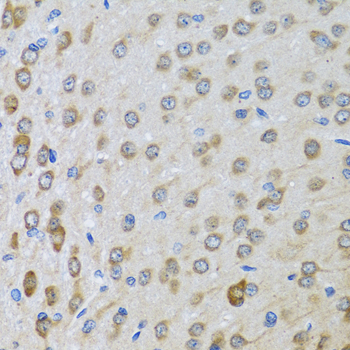
| Sample | Paraffin-embedded rat brain tissue; fixative not specified (datasheet A03020) |
| Fixation | Image fixative and duration unreported (datasheet A03020); verify before use. |
| Sectioning | 4–5 µm sections on charged slides (standard) |
| Deparaffinisation | Xylene, graded ethanol series to water (standard) |
| Antigen retrieval | Heat-induced epitope retrieval in citrate buffer, pH 6.0, 20 min at 95–98 °C (standard rule: cytoplasmic / membrane antigen) |
| Peroxidase block | 3% H2O2, 10 min, room temperature (standard) |
| Blocking | 10% normal serum of the secondary host, 30 min, room temperature (standard) |
| Primary antibody | Rabbit anti-BTD, 1:100-1:200 (datasheet A03020) |
| Primary incubation | Overnight at 4 °C (standard) |
| Detection | HRP-polymer secondary, DAB chromogen 5–10 min (standard) |
| Counterstain | Hematoxylin, blue, dehydrate and mount (standard) |
| Expected result | BTD-positive staining in myoepithelial cells of breast (HPA tissue IHC: High). HPA tissue profile: Membranous expression in endothelial cells. No signal in the no-primary control. |
BTD is a secreted protein with no transmembrane segment (UniProt P43251). HPA describes membranous staining in endothelial cells, with high staining in breast myoepithelial cells and cells in kidney glomeruli, and medium staining in hepatocytes (HPA tissue IHC). Interpret that distribution cautiously: HPA rates the tissue staining evidence Uncertain because antibody staining and RNA expression have low consistency, and secreted protein may appear away from its producing cells (HPA tissue IHC).
| Membranous endothelial staining, with stronger signal in the reported breast or kidney cell populations. | This is compatible with the observed HPA profile: membranous endothelial expression, high staining in breast myoepithelial cells and high staining in glomerular cells (HPA tissue IHC). Check cell identity and local tissue architecture before scoring; HPA calls the overall tissue evidence Uncertain (HPA tissue IHC). |
| Predominantly nuclear staining in otherwise intact cells. | A nuclear pattern does not match BTD's secreted annotation or HPA's reported membranous endothelial profile (UniProt P43251; HPA tissue IHC). Treat it as suspect and compare with a control omitting the primary antibody and with the reported positive tissues (general IHC practice; HPA tissue IHC). |
| Strong staining in an unexpected cell population, especially one HPA reports as not detected. | Consider antibody cross-reactivity or endogenous chromogenic detection activity before assigning BTD expression (general IHC practice). HPA reports no detection in adipocytes, bone marrow hematopoietic cells and bronchial respiratory epithelial cells, among other listed populations; its overall rating remains Uncertain (HPA tissue IHC). |
| Diffuse colour across tissue and spaces, with little distinction between structures. | A secreted protein could contribute extracellular signal, so diffuse staining alone does not establish an artefact (UniProt P43251). Broad colour that also appears in a control omitting the primary antibody supports background from the detection workflow; reassess blocking, washing and chromogen development (general IHC practice). |
| No visible signal in breast myoepithelial cells or kidney glomerular cells. | These are high-staining populations in HPA's tissue record, so an absent signal prompts a check of the IHC run, section quality and scoring location (HPA tissue IHC; general IHC practice). Absence does not by itself overturn the HPA pattern or prove a failed assay, because that pattern has Uncertain reliability (HPA tissue IHC). |
| Secretion and topology (UniProt P43251) | BTD has a signal peptide at residues 1–21, a mature chain at 22–523 and no transmembrane segment; do not interpret an apparent membrane outline as proof that BTD is membrane anchored (UniProt P43251; HPA tissue IHC). |
| Location of production versus detection (UniProt P43251; HPA tissue IHC) | BTD is secreted, and HPA warns that tissue RNA and protein locations may differ; use the observed staining and cell morphology to interpret a section, rather than treating liver-enhanced RNA as a promise of strongest liver IHC (UniProt P43251; HPA tissue IHC). |
| Reported cell-level contrast (HPA tissue IHC) | HPA reports high staining in breast myoepithelial and kidney glomerular cells, medium staining in hepatocytes, and no detection in several listed cell populations. These are useful comparison points, subject to HPA's Uncertain reliability rating (HPA tissue IHC). |
| Antibody evidence (HPA antibody and tissue IHC records) | The listed antibody HPA052275 has an Uncertain IHC assessment; HPA also rates the tissue pattern Uncertain. A visually convincing slide therefore needs appropriate run controls and cautious interpretation, rather than an assumption of independently verified specificity (HPA antibody and tissue IHC records; general IHC practice). |
| Processing and variants (UniProt P43251) | UniProt lists a cleaved signal peptide, six glycosylation sites and four isoforms. The supplied sources do not locate the antibody epitope or show how these features affect IHC staining; avoid assigning a compartment or intensity change to any one of them (UniProt P43251; HPA antibody record). |
| Situation | Likely cause | Next action |
|---|---|---|
| The HPA-reported high-staining cells are blank. | The comparison population may have been misidentified, or the IHC run may have lost detectable signal (HPA tissue IHC; general IHC practice). | Locate breast myoepithelial cells or kidney glomerular cells by morphology; review section integrity, retrieval, antibody incubation and detection controls as general IHC checks. Do not infer a BTD-specific retrieval requirement from HPA (HPA tissue IHC; general IHC practice). |
| Nuclei dominate the stain. | That pattern conflicts with the secreted annotation and reported membranous endothelial profile; nonspecific staining is a possibility (UniProt P43251; HPA tissue IHC; general IHC practice). | Compare a primary-omission control and a reported positive tissue, then score only anatomically credible signal. HPA's Uncertain rating limits how firmly any single pattern can be declared specific (general IHC practice; HPA tissue IHC). |
| Colour appears broadly, including where no cell pattern is discernible. | Extracellular BTD is possible, while diffuse detection background is also possible; appearance alone cannot distinguish them (UniProt P43251; general IHC practice). | Check the primary-omission control and inspect whether colour tracks extracellular structures or persists throughout the section. Review blocking, washing and chromogen development as general IHC steps (general IHC practice). |
| An HPA-listed negative population stains strongly. | Cross-reactivity or endogenous detection activity could produce colour, and the HPA negative call is itself within an Uncertain tissue profile (general IHC practice; HPA tissue IHC). | Confirm the cell type, compare the primary-omission control, and check the same run's reported positive population. Avoid declaring a new BTD-positive cell type from a single discordant slide (general IHC practice; HPA tissue IHC). |
| Liver stains less strongly than breast or kidney. | That order is compatible with HPA's medium hepatocyte staining versus high staining in breast myoepithelial and glomerular cells; liver-enhanced RNA does not settle protein location (HPA tissue IHC). | Score each named cell population separately and record intensity with the HPA reliability caveat. Do not use liver RNA enrichment alone to call the chromogenic run a failure (HPA tissue IHC). |
| Can IF/ICC establish BTD's subcellular pattern? | HPA labels BTD secreted but provides no main ICC-IF location or cell-line ICC-IF images; the listed antibody has no ICC validation entry (HPA subcellular and antibody records). | Treat IF/ICC localization as unresolved from these sources and use its separate guide for assay design. Keep this page's pattern call tied to the reported tissue IHC observations (HPA subcellular, antibody and tissue IHC records). |
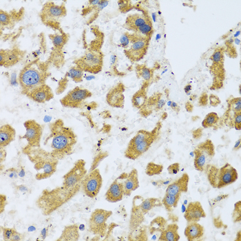


Comprehensive Human Protein Atlas IHC scoring per tissue (reliability: Uncertain — Low consistency between antibody staining and RNA expression data. Pending external verification. At least one protein variant secreted, tissue location of RNA and protein might differ and correlation is complex. Antibody staining in cells/structures not annotated, view images.). Rows are taken directly from the HPA tissue chart — click any row's HPA link to view the source.
| Tissue | Cell type | Level | Evidence | Source |
|---|---|---|---|---|
| Breast | Myoepithelial cells | High | Protein (IHC) | HPA → |
| Kidney | Cells in glomeruli | High | Protein (IHC) | HPA → |
| Liver | Hepatocytes | Medium | Protein (IHC) | HPA → |
| Parathyroid gland | Glandular cells | Medium | Protein (IHC) | HPA → |
| Stomach | Glandular cells | Medium | Protein (IHC) | HPA → |
| Tissue | Cell type | Level | Evidence | Source |
|---|---|---|---|---|
| Adipose tissue | Adipocytes | Not detected | Protein (IHC) | HPA → |
| Appendix | Glandular cells | Not detected | Protein (IHC) | HPA → |
| Bone marrow | Hematopoietic cells | Not detected | Protein (IHC) | HPA → |
| Bronchus | Respiratory epithelial cells | Not detected | Protein (IHC) | HPA → |
| Caudate | Glial cells | Not detected | Protein (IHC) | HPA → |
Troubleshoot BTD staining in paraffin sections by checking retrieval, signal location, controls and scoring before interpreting chromogenic IHC (UniProt P43251; HPA tissue IHC).
Anti-BTD antibody A03020 has IHC images from paraffin sections of rat brain, human liver cancer and human prostate (catalog: A03020 image captions); its listed reactivity includes Human, Mouse and Rat (catalog: A03020 reactivity).
A03020 will render with its rat brain IHC figure from a paraffin section (catalog: A03020 figure caption). Its other IHC images show paraffin sections of human liver cancer and human prostate (catalog: A03020 image captions).
Which to pick: Choose A03020 for tissue IHC: it is a rabbit polyclonal antibody listed for IHC, with a 1:100–1:200 IHC dilution; its paraffin-section captions specify microwave antigen retrieval in 10 mM PBS at pH 7.2, but do not report the fixative (catalog: A03020 applications, dilution and image captions). For cross-species work, A03020 lists Human, Mouse and Rat reactivity, while its IHC images document human and rat samples only (catalog: A03020 reactivity and image captions). No IF/ICC choice is supported because A03020 has no listed IF application, dilution or image (catalog: A03020 applications, IF dilution and IF images).