This website uses cookies to ensure you get the best experience on our website.


- Table of Contents
Plan BTRC chromogenic IHC in paraffin sections around the general cytoplasmic tissue pattern (HPA tissue IHC), while recognising that nuclear localisation is also annotated at the molecular level (UniProt). The IHC-validated antibody has a stated dilution range of 1:50–1:200 (datasheet: A01747).

Expected localisation, validated protocols, controls and antibodies — the at-a-glance facts below, then the full design guide.
| Expected localisation | Predominantly cytoplasmic in tissue (HPA tissue IHC) | |
| Staining pattern | Cytoplasmic staining across multiple cell types (HPA tissue IHC) | |
| Antigen retrieval | Citrate pH 6.0 HIER, heat-mediated (datasheet A01747) | |
| Positive control | Appendix+4 more · see all | |
| Negative control | Spleen |
| Fixation | Keep formalin fixation consistent across paraffin sections. (standard IHC practice; not target-specific) | |
| Caveat | Staining has medium consistency with RNA data (HPA tissue IHC) | |
| Regulation | Low tissue specificity; no defined inducer (HPA tissue IHC) | |
| Isoform / epitope | 2 isoforms; antibody epitope coverage is unspecified (UniProt) |
Start with the catalog antibody’s citrate pH 6.0 retrieval protocol (datasheet A01747). The published option below describes staining of FFPE sections (PMC13280727).
| Sample | Paraffin-embedded mouse spleen tissue; fixative not specified (datasheet A01747) |
| Fixation | Image fixative and duration unreported (datasheet A01747); verify before use. |
| Sectioning | 4–5 µm sections on charged slides (standard) |
| Deparaffinisation | Xylene, graded ethanol series to water (standard) |
| Antigen retrieval | Heat retrieval: Citrate pH 6.0 (datasheet A01747); 20 min, 95–100 °C (standard) |
| Peroxidase block | 3% H2O2, 10 min, room temperature (standard) |
| Blocking | 10% normal serum of the secondary host, 30 min, room temperature (standard) |
| Primary antibody | Rabbit anti-BTRC, 1:50-1:200 (datasheet A01747) |
| Primary incubation | Overnight at 4 °C (standard) |
| Detection | HRP-polymer secondary, DAB chromogen 5–10 min (standard) |
| Counterstain | Hematoxylin, blue, dehydrate and mount (standard) |
| Expected result | BTRC-positive staining in glandular cells of appendix (HPA tissue IHC: Medium). HPA tissue profile: General cytoplasmic expression. No signal in the no-primary control. |
BTRC should appear chiefly as cytoplasmic staining across several cell types in tissue IHC, including glandular, respiratory epithelial, glial, and endothelial cells (HPA tissue IHC: general cytoplasmic expression; medium staining in listed cells). Nuclear staining is biologically plausible (UniProt Q9Y297: cytoplasm and nucleus), but should be assessed against the tissue pattern. BTRC has no transmembrane segment (UniProt Q9Y297: topology). HPA rates the tissue IHC evidence Approved, with medium consistency between staining and RNA data (HPA tissue IHC: reliability).
| Moderate cytoplasmic signal in appendix or breast glandular cells, or bronchial respiratory epithelial cells. | This matches the reported tissue IHC pattern (HPA tissue IHC: Medium in these cells; general cytoplasmic expression). Judge signal in the named cells and compartment; staining elsewhere on the section does not by itself establish a BTRC-positive result. |
| Dominant nuclear-only or sharply membranous staining in a tissue section. | Check for an assay-dependent pattern or artefact before calling it positive: tissue IHC is described as generally cytoplasmic (HPA tissue IHC: profile). Nuclear localization remains possible (UniProt Q9Y297: nucleus); plasma-membrane localization was observed in ICC-IF, a different assay (HPA subcellular: additional location). |
| Strong signal in an unexpected cell population, especially when the expected cells are unstained. | Consider antibody cross-reactivity or endogenous chromogenic detection activity (standard IHC practice). HPA reports low tissue specificity and several stained cell types, so an unlisted population is a reason to verify specificity, not proof of a false positive (HPA tissue IHC: RNA specificity and positive-cell list). |
| Diffuse color across cells, stroma, or the whole section without a clear cellular pattern. | Treat this as background until controls resolve it (standard IHC practice). Excess detection signal, inadequate blocking, or insufficient washing can obscure compartment boundaries (standard IHC practice). A diffuse field cannot be matched confidently to HPA's general cytoplasmic profile (HPA tissue IHC: profile). |
| No signal in appendix glandular cells or bronchial respiratory epithelial cells. | First check section quality, retrieval, antibody incubation, and detection controls (standard IHC practice). These are useful reference cells because HPA reports Medium staining, although its Approved rating has only medium consistency with RNA data (HPA tissue IHC: positive cells and reliability). A failed reference section cannot establish that the test tissue lacks BTRC. |
| Compartment and assay | Tissue IHC is generally cytoplasmic (HPA tissue IHC: profile); ICC-IF is mainly nucleoplasmic, with cytosol and plasma membrane also reported (HPA subcellular: approved locations). Interpret each assay against its own evidence. |
| Reference-cell selection | Appendix and breast glandular cells, bronchial epithelium, and selected neural or endothelial cells show Medium staining (HPA tissue IHC: positive cells). Spleen red-pulp cells are reported Not detected; that finding does not classify the entire spleen (HPA tissue IHC: negative cell). |
| Antibody validation | CAB032986 is Approved for IHC; HPA031156 is Approved for ICC, with no IHC status listed for it (HPA antibodies: assay-specific status). An Approved label is not an Enhanced validation claim (HPA antibodies: validation status). |
| Protein features | BTRC has two isoforms, no listed signal peptide or propeptide, and no transmembrane segment (UniProt Q9Y297: isoforms, processing, topology). The supplied record gives no antibody epitope, so isoform coverage cannot be inferred. |
| Evidence limit for paraffin sections | No target-specific fixation or retrieval effect is provided (supplied UniProt and HPA records). Retrieval may be optimized as general IHC practice, but these sources cannot predict which condition improves BTRC staining. |
| Situation | Likely cause | Next action |
|---|---|---|
| Reference cells are blank and the test section is blank. | A failed staining run or unsuitable assay conditions are possible (standard IHC practice); HPA reports Medium staining in several reference cells (HPA tissue IHC: positive cells). | Confirm the IHC antibody identity and assay validation, then check retrieval, incubation, detection reagents, and the positive control (standard IHC practice; HPA antibodies: CAB032986 IHC Approved). |
| Color is widespread, with little contrast between cells and surrounding tissue. | Nonspecific background or endogenous detection activity may overwhelm cellular signal (standard IHC practice). | Review the no-primary control; optimize blocking, washes, antibody concentration, and detection exposure as needed (standard IHC practice). Reassess whether identifiable cells show the HPA cytoplasmic pattern (HPA tissue IHC: profile). |
| Only nuclei stain in tissue IHC. | BTRC can occur in the nucleus (UniProt Q9Y297: subcellular location), but a nuclear-only result differs from HPA's general tissue IHC profile (HPA tissue IHC: profile). | Compare a reference tissue and appropriate controls before scoring; record the nuclear pattern separately from the expected cytoplasmic tissue IHC signal (standard IHC practice; HPA tissue IHC: profile). |
| Spleen red-pulp cells stain strongly. | This conflicts with the reported Not detected result for those cells (HPA tissue IHC: spleen red pulp); cross-reactivity or background is possible (standard IHC practice). | Verify cell identity and inspect no-primary and positive-reference controls (standard IHC practice). Do not extend the red-pulp finding to every spleen cell (HPA tissue IHC: negative cell). |
| Epididymis glandular cells stain weakly despite the UniProt tissue note. | UniProt reports protein expression in epididymis, while HPA lists Low glandular staining there (UniProt Q9Y297: tissue specificity; HPA tissue IHC: low group). | Use an HPA Medium-staining cell population as the run reference, and interpret weak epididymal staining within its reported range (HPA tissue IHC: positive and low groups). |
| ICC-IF shows nucleoplasmic signal while paraffin IHC looks cytoplasmic. | The reported profiles differ by assay (HPA subcellular: mainly nucleoplasmic ICC-IF; HPA tissue IHC: general cytoplasmic expression). The listed Approved ICC and IHC antibodies also differ (HPA antibodies: assay-specific status). | Evaluate ICC-IF against its separate guide and assay controls (standard ICC-IF practice); retain the tissue IHC profile when scoring paraffin sections (HPA tissue IHC: profile). |
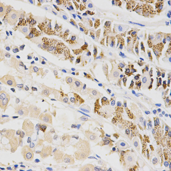
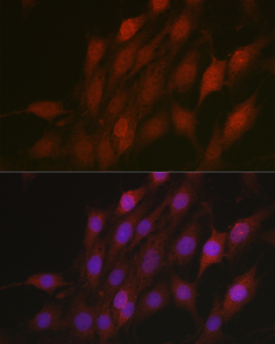

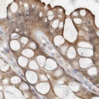


Comprehensive Human Protein Atlas IHC scoring per tissue (reliability: Approved — Medium consistency between antibody staining and RNA expression data.). Rows are taken directly from the HPA tissue chart — click any row's HPA link to view the source.
| Tissue | Cell type | Level | Evidence | Source |
|---|---|---|---|---|
| Appendix | Glandular cells | Medium | Protein (IHC) | HPA → |
| Breast | Glandular cells | Medium | Protein (IHC) | HPA → |
| Bronchus | Respiratory epithelial cells | Medium | Protein (IHC) | HPA → |
| Caudate | Glial cells | Medium | Protein (IHC) | HPA → |
| Cerebellum | Cells in granular layer | Medium | Protein (IHC) | HPA → |
| Tissue | Cell type | Level | Evidence | Source |
|---|---|---|---|---|
| Spleen | Cells in red pulp | Not detected | Protein (IHC) | HPA → |
Use compartment, cell type, and matched controls to interpret BTRC staining in paraffin sections; the selected tissue image reports retrieval conditions but not fixation.
A01747 has IHC data from paraffin-embedded mouse spleen and IF data from C6 cells (catalog image captions); listed reactivity includes human, mouse, and rat (catalog reactivity).
A01747 will render with an IHC image of paraffin-embedded mouse spleen stained at 1:100 after citrate retrieval (A01747 IHC image caption). A01747 also has an IF image of C6 cells stained at 1:100 with DAPI nuclear staining (A01747 IF image caption).
Which to pick: Choose A01747 for paraffin-section IHC; its own mouse spleen image documents that preparation, while the fixative is unreported (A01747 IHC image caption). Choose A01747 for IF/ICC because both applications are listed and its IF image shows C6 cells (catalog applications; A01747 IF image caption). For work across species, A01747 is listed as reactive with human, mouse, and rat and is described as a rabbit polyclonal antibody in its IHC caption (catalog reactivity; A01747 IHC image caption).