CADM2 / Cell adhesion molecule 2 · IHC design guide

Design Immunohistochemistry for CADM2

Plan CADM2 paraffin IHC using cerebellar synaptic glomeruli and cortical neuropil as positive staining references (HPA tissue IHC). The guide covers fixation consistency, epitope choice across 5 isoforms (UniProt), and interpretation of neural staining.

Evidence assembled Oct 2026 · For research use; verify linked source records and product datasheet before use
Immunohistochemistry protocol sheet for CADM2 (IHC for CADM2): expected localisation Synaptic glomeruli and cortical neuropil (HPA tissue IHC), antibody A06688-1, validated IHC image, and IHC protocol steps
Printable CADM2 IHC protocol sheet — expected localisation Synaptic glomeruli and cortical neuropil (HPA tissue IHC), antibody A06688-1, controls and protocol steps. Open the full CADM2 IHC guide →

CADM2 Immunohistochemistry Experimental Design Guide

Expected localisation, validated protocols, controls and antibodies — the at-a-glance facts below, then the full design guide.

Must know before staining
Expected localisation Synaptic glomeruli and cortical neuropil (HPA tissue IHC)
Staining pattern Strong staining in cerebellar glomeruli and cortical neuropil (HPA tissue IHC)
Antigen retrieval Tris-EDTA pH 9.0 HIER, heat-mediated (datasheet A06688-1)
Positive control ⓘ Cerebellum+1 more · see all
Negative control ⓘ Adipose tissue+4 more · see all
Important caveats
Reasons your staining may differ from the expected pattern.
Fixation Keep fixation consistent across paraffin specimens (selected-SKU IHC image A06688-1)
Caveat Neuropil staining can complicate cell-level scoring (HPA tissue IHC)
Regulation Expression regulation is not annotated (UniProt)
Isoform / epitope 5 isoforms; check extracellular vs cytoplasmic epitope (UniProt)
Section 1

Recommended CADM2 IHC & IF Protocols

The catalog antibody’s IHC-P protocol (datasheet A06688-1) is accompanied by two published CADM2 paraffin-section IHC methods (PMC8081990; PMC11239024).

Recommended immunohistochemistry (IHC-P) protocol parameters
SampleParaffin-embedded human tonsil tissue; fixative not specified (datasheet A06688-1)
FixationImage fixative and duration unreported (datasheet A06688-1); verify before use.
Sectioning4–5 µm sections on charged slides (standard)
DeparaffinisationXylene, graded ethanol series to water (standard)
Antigen retrievalHeat retrieval: Tris-EDTA pH 9.0 (datasheet A06688-1); 20 min, 95–100 °C (standard)
Peroxidase block3% H2O2, 10 min, room temperature (standard)
Blocking10% normal serum of the secondary host, 30 min, room temperature (standard)
Primary antibodyRabbit anti-CADM2, 1:100-1:300 (datasheet A06688-1)
Primary incubationOvernight at 4 °C (standard)
DetectionHRP-polymer secondary, DAB chromogen 5–10 min (standard)
CounterstainHematoxylin, blue, dehydrate and mount (standard)
Expected resultCADM2-positive staining in synaptic glomeruli - core of cerebellum (HPA tissue IHC: High). HPA tissue profile: Distinct expression in CNS and peripheral nerve fibers. No signal in the no-primary control.
💡Decision noteStart with heat-mediated Tris-EDTA pH 9.0 retrieval (datasheet A06688-1); the published citrate method offers a comparison at 95°C for 10 minutes (PMC11239024).
Section 2

What Is the Expected CADM2 Staining Pattern?

CADM2 is a membrane protein also annotated at synapses and in axons, including the axoplasm of myelinated axons (UniProt Q8N3J6 localization and topology). In paraffin section IHC, expect strong staining in cerebellar synaptic glomerular cores and cerebral cortical neuropil, with a broader pattern in CNS and peripheral nerve fibers (HPA: High in both listed structures; HPA: tissue profile). HPA rates its tissue staining reliability Enhanced (HPA: tissue IHC).

What am I looking at on my slide?
Strong staining in cerebellar synaptic glomerular cores or cortical neuropil.This matches the reported positive structures (HPA: High in both). Assess the named structures rather than requiring uniformly stained neuronal cell bodies; the tissue observations specify glomerular cores and neuropil, while UniProt places CADM2 at synapses, membranes and axons (HPA: tissue IHC; UniProt Q8N3J6 localization).
Predominantly nuclear staining in an otherwise positive section.Nuclear localization is unsupported by the supplied protein record, which places CADM2 at the membrane, synapse and axon (UniProt Q8N3J6 localization). Treat a nuclear dominant result as suspect; compare it with the expected tissue structures and a no primary control before assigning specificity (HPA: tissue IHC; standard IHC practice).
Strong staining of adrenal glandular cells or adipocytes.Those cells are listed as Not detected in the sampled HPA tissues (HPA: adrenal gland and adipose tissue IHC). The result warrants a specificity check; cross reactivity or endogenous detection activity are possibilities, but this observation alone cannot establish which caused it (standard IHC practice).
Diffuse color across tissue and empty spaces, obscuring local structures.That distribution cannot be scored as the discrete glomerular core or neuropil pattern reported for CADM2 (HPA: cerebellum and cerebral cortex IHC). Examine a no primary control for detection background, and review washing, blocking and chromogen development as general IHC checks (standard IHC practice).
No staining in an adequately preserved cerebellum or cerebral cortex section.An absent signal conflicts with the supplied High staining observations for synaptic glomerular cores and neuropil (HPA: tissue IHC). First verify that the relevant structures are present, then check the staining run and antibody conditions; one negative section does not establish biological absence (standard IHC practice).
💡Expected CADM2 appearanceA convincing positive shows strong signal in cerebellar synaptic glomerular cores or cortical neuropil, consistent with CADM2's membrane and synaptic localization; dominant nuclear color or strong staining in listed Not detected cells is suspect (HPA: tissue IHC; UniProt Q8N3J6 localization).
How each factor affects the staining
Protein orientation and epitope positionCADM2 has an extracellular region at residues 25–367, one transmembrane segment at 368–388, and a cytoplasmic region at 389–435 (UniProt Q8N3J6 topology). The supplied evidence does not map the antibody epitope, so it cannot predict which side its staining reports.
Maturation and glycosylationThe annotated signal peptide is residues 1–24; the mature chain begins at residue 25, with glycosylation sites at 31, 51 and 291 (UniProt Q8N3J6 processing and glycosylation). No supplied evidence links these features to IHC retrieval or staining intensity.
Isoform coverageUniProt lists 5 CADM2 isoforms: 1, 2, 3, 5 and 6 (UniProt Q8N3J6 isoforms). Because neither the antibody epitope nor isoform specific tissue staining is supplied, an IHC pattern cannot be assigned to a particular isoform from this record.
Tissue and antibody evidenceHPA reports High staining in two named neural structures and rates tissue staining reliability Enhanced, citing consistency between staining and RNA expression (HPA: tissue IHC). Antibody HPA010024 is listed as IHC Enhanced (HPA: antibodies); that designation does not establish a retrieval condition or universal tissue positivity.
Antigen retrievalNo target specific retrieval condition or CADM2 fixation sensitivity is supplied (HPA: tissue IHC; UniProt Q8N3J6). If signal is weak, assess retrieval alongside a known positive tissue using ordinary IHC controls; interpret any change as an assay observation, not a documented CADM2 specific fixation effect (standard IHC practice).
IF/ICC expectation?HPA summarizes the subcellular pattern as Membrane, but supplies no ICC/IF images or main location, and lists no ICC validation for HPA010024 (HPA: subcellular; HPA: antibodies). UniProt also annotates synapses and axons (UniProt Q8N3J6 localization); these records do not establish an IF/ICC protocol or a validated cell line.
Why is my staining missing, weak or wrong?
SituationLikely causeNext action
Positive neural tissue is blank.The sampled section may lack the named structure, or the staining run may have failed; the supplied record identifies High signal specifically in glomerular cores and neuropil (HPA: tissue IHC).Confirm those structures on the section and review the run's positive control, antibody application and detection steps (standard IHC practice). Repeat before calling the sample CADM2 negative.
Only nuclei stain strongly.A nuclear dominant pattern conflicts with CADM2's annotated membrane, synapse and axon locations (UniProt Q8N3J6 localization); nonspecific staining or detection background is possible (standard IHC practice).Compare the no primary control and the positive tissue's glomerular core or neuropil pattern; score the nuclear signal cautiously until its source is resolved (HPA: tissue IHC; standard IHC practice).
Adipocytes or adrenal glandular cells stain strongly.HPA lists those sampled cells as Not detected (HPA: adipose tissue and adrenal gland IHC). Cross reactivity and endogenous detection activity remain possible explanations (standard IHC practice).Run a no primary control and compare staining with a listed High neural tissue in the same run; investigate detection background before interpreting the unexpected cells as CADM2 positive (HPA: tissue IHC; standard IHC practice).
Color is diffuse across the section.Background from incomplete washing, blocking or detection control may obscure the localized neural pattern (standard IHC practice; HPA: cerebellum and cerebral cortex IHC).Inspect the no primary control and review washing, blocking and chromogen development; then reassess whether glomerular cores or neuropil remain distinguishable (standard IHC practice; HPA: tissue IHC).
Signal appears in the no primary control.The primary antibody cannot account for color produced without it; endogenous activity or detection reagent background should be considered (standard IHC practice).Check the detection system and its control steps; for a peroxidase based chromogenic assay, assess endogenous peroxidase blocking before attributing the color to CADM2 (standard IHC practice).
The positive tissue stains weakly after a workflow change.The record gives no CADM2 specific fixation sensitivity or retrieval requirement, so it does not identify the changed step as the cause (HPA: tissue IHC; UniProt Q8N3J6).Compare matched positive tissue sections across the changed and prior conditions, with the same detection controls; document the observed difference as run specific evidence (standard IHC practice).

Sample controls for CADM2 IHC & IF

🧪Run cerebellum first and expect staining in synaptic glomeruli cores (HPA: High in synaptic glomeruli cores); run adipose tissue as the negative and expect adipocytes to lack detectable staining (HPA: Not detected in adipocytes). On the cerebellum slide, use areas outside the stained cores to assess background, without assuming every other cell is CADM2-negative (HPA: High in synaptic glomeruli cores).
Positive control tissue: Cerebellum (Synaptic glomeruli - core, HPA High)
Negative control tissue: Adipose tissue (HPA Not detected)
ICC-IF cell lines (HPA subcellular resource): HPA carries no ICC-IF cell line for CADM2; derive a cell-line control from the positive tissue's cell type (Synaptic glomeruli - core) and confirm it by RNA or western blot first.
Technical controls: Include no-primary (secondary-only) and control antibody slides, using a host- and isotype-matched control for a monoclonal primary or host-matched nonimmune IgG for a polyclonal primary; confirm specificity with CADM2 knockout tissue or peptide blocking if the immunizing peptide is available (standard IHC practice). For chromogenic IHC, quench endogenous peroxidase; for IF, assess cerebellar autofluorescence, including lipofuscin (standard IHC/IF practice).
⚠️Feasibility: No target-specific fixation window or fixation effect is reported in the supplied evidence, and the fixative in the selected paraffin-section caption is unreported (selected-SKU tissue-IHC caption: fixative not stated). That caption uses Tris-EDTA retrieval at pH 9.0 and a 1:200 primary dilution overnight at 4°C; a requirement for that retrieval condition is unreported (selected-SKU tissue-IHC caption). Whether frozen sections or IF are easier is unestablished, and dense synaptic-core staining can complicate cell-level scoring in cerebellum (HPA: High in synaptic glomeruli cores).

HPA tissue IHC evidence for CADM2

Comprehensive Human Protein Atlas IHC scoring per tissue (reliability: Enhanced — High consistency between antibody staining and RNA expression data.). Rows are taken directly from the HPA tissue chart — click any row's HPA link to view the source.

Positive expression · recommended positive controls

TissueCell typeLevelEvidenceSource
Cerebellum Synaptic glomeruli - core High Protein (IHC) HPA →
Cerebral cortex Neuropil High Protein (IHC) HPA →

Undetected expression · recommended negative controls

TissueCell typeLevelEvidenceSource
Adipose tissue Adipocytes Not detected Protein (IHC) HPA →
Adrenal gland Glandular cells Not detected Protein (IHC) HPA →
Appendix Glandular cells Not detected Protein (IHC) HPA →
Bone marrow Hematopoietic cells Not detected Protein (IHC) HPA →
Breast Adipocytes Not detected Protein (IHC) HPA →
Section 3

Advanced CADM2 IHC Tips

Use the catalog antibody’s paraffin section image as a processing reference, then judge CADM2 staining against its documented neural distribution and membrane topology.

Which retrieval condition should I try first for CADM2 in paraffin sections?
Start with heat mediated retrieval in Tris-EDTA at pH 9.0 (datasheet A06688-1). The catalog image used paraffin embedded human tonsil, a 1:200 primary dilution, and incubation at 4°C overnight; its fixative and retrieval heating time were not reported (datasheet A06688-1). Keep heating and cooling conditions consistent while assessing whether staining is localized and tissue architecture remains intact (standard IHC practice). If staining is weak, adjust retrieval duration cautiously before testing another buffer, and compare each change against a concurrently processed section (standard IHC practice). Include a neural reference section because stronger staining is documented in cerebral cortex neuropil and cerebellar synaptic glomeruli (HPA tissue IHC).
Could fixation explain weak CADM2 staining in my paraffin sections?
Target specific fixation sensitivity is unknown: the catalog paraffin section caption does not identify its fixative (datasheet A06688-1). Record fixation type and duration for each specimen, then compare similarly processed sections before attributing a staining difference to CADM2 abundance (standard IHC practice). In the reported catalog workflow, Tris-EDTA at pH 9.0 preceded a 1:200 primary incubation at 4°C overnight (datasheet A06688-1). If signal remains weak, check section integrity and retrieval consistency on a reference section processed alongside the sample (standard IHC practice). HPA tissue staining patterns establish neither fixation sensitivity nor a preferred fixative for this antibody (HPA tissue IHC).
Where should convincing CADM2 staining appear in neural tissue?
Look for staining associated with neural processes and synaptic regions: CADM2 is annotated at the cell membrane, synapse, and axon, including axoplasm of myelinated axons (UniProt Q8N3J6 localisation). HPA reports high staining in cerebral cortex neuropil and cerebellar synaptic glomerulus cores, with an overall profile in CNS and peripheral nerve fibers (HPA tissue IHC). Interpret fine process staining in its anatomical context rather than requiring every positive structure to have a clearly outlined cell membrane (UniProt Q8N3J6 localisation; HPA tissue IHC). Compare the same compartment across matched sections and keep counterstain intensity consistent (standard IHC practice). Predominantly nuclear staining warrants scrutiny because the documented locations are membrane, synapse, and axon (UniProt Q8N3J6 localisation).
How could an unknown epitope affect CADM2 staining across samples?
CADM2 has 5 listed isoforms, so determine whether the antibody’s documented immunogen is shared before interpreting differences as changes in total CADM2 (UniProt Q8N3J6 isoforms). Its extracellular segment spans residues 25–367, its transmembrane segment 368–388, and its cytoplasmic segment 389–435 (UniProt Q8N3J6 topology). The extracellular region includes three annotated glycosylation sites at residues 31, 51, and 291, which make epitope location relevant to assay interpretation (UniProt Q8N3J6 glycosylation and topology). Without an epitope map, do not assign staining to a particular isoform or membrane face (standard IHC interpretation). Compare staining after the same retrieval and detection conditions, and document the antibody identifier with every image set (standard IHC practice).
How should I check CADM2 localisation in a multiplex fluorescence experiment?
Treat fluorescence as a separate assay to optimize; the supplied catalog image documents paraffin section chromogenic IHC, not IF performance (datasheet A06688-1). Pair CADM2 with a validated neuronal or axonal marker when examining the neural processes and synaptic regions where CADM2 is documented (UniProt Q8N3J6 localisation; HPA tissue IHC). Choose well separated fluorophores after inspecting unstained tissue for autofluorescence, and include single color controls to assess channel bleed through (standard IF practice). If the antibody recognizes the extracellular region at residues 25–367, assess access without permeabilisation; if it recognizes the cytoplasmic region at 389–435, optimize gentle permeabilisation (UniProt Q8N3J6 topology; standard IF practice). The epitope is unspecified in the supplied evidence, so test access empirically (standard IF practice).
How can I reduce diffuse or misleading CADM2 chromogenic background?
First compare the suspect section with a no primary control and a concurrently processed reference section to separate detection background from candidate CADM2 signal (standard IHC practice). Apply the peroxidase block required by the chromogenic detection workflow, then check blocking, washes, and DAB development time if staining spreads across unrelated structures (standard IHC practice). The catalog caption reports a 1:200 primary incubation at 4°C overnight and a 1:200 secondary incubation for 45 minutes at room temperature (datasheet A06688-1). Use those values as the documented starting point for that image, while checking whether shorter development improves contrast in your sections (datasheet A06688-1; standard IHC practice). Background in glandular cells from an HPA negative reference should prompt scrutiny of detection conditions (HPA tissue IHC).
What should I measure when CADM2 stains neuropil or nerve fibers? ⚠ ANSWER MARKED FOR VERIFICATION
Define the anatomical region and analysis threshold before scoring, and acquire all chromogenic images with consistent illumination and exposure settings (standard IHC practice). For process rich regions, report CADM2 positive area fraction or positive fiber density per mm², normalized to the sampled region or available neural process area (standard IHC practice). In a section with individually interpretable cells, report percent positive cells or an H-score, but specify the cellular compartment used (standard IHC practice). Keep cerebral cortex neuropil and cerebellar synaptic glomerulus cores as separate regions because HPA identifies both as high staining sites (HPA tissue IHC). Compare matched regions and processing batches, and record the threshold and exclusion rules so measurements can be reproduced (standard IHC practice).
What distinguishes true CADM2 signal from a staining artefact?
Give greater weight to reproducible staining in neural processes and synaptic regions, consistent with CADM2 localisation and the HPA tissue profile (UniProt Q8N3J6 localisation; HPA tissue IHC). High signal in cerebral cortex neuropil or cerebellar synaptic glomerulus cores has supporting tissue evidence, whereas HPA reports no detection in adipocytes or several glandular cell populations (HPA tissue IHC). Scrutinize predominantly nuclear or widespread glandular staining because it conflicts with the documented membrane, synaptic, and axonal pattern (UniProt Q8N3J6 localisation; HPA tissue IHC). Exclude torn edges and necrotic areas, and compare a no primary control to identify edge deposits or endogenous enzyme signal (standard IHC practice). Confirm the pattern across intact, similarly processed sections before assigning biological meaning (standard IHC practice).
Boster reagents

Best CADM2 / Cell adhesion molecule 2 IHC Antibodies

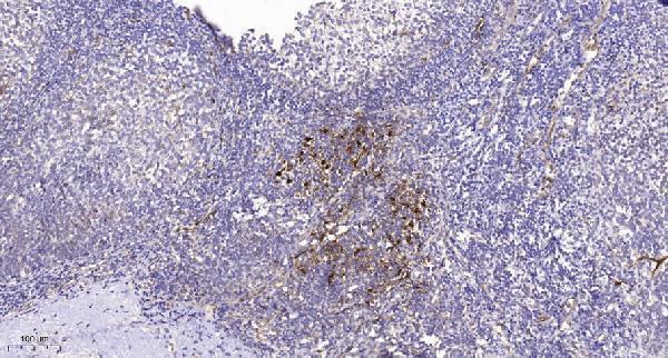
A06688-1 has a human tonsil paraffin-section IHC image (A06688-1 image caption) and is listed for IHC and IF in human, mouse and rat (catalog: applications/reactivity).

Real IHC data Immunohistochemical analysis of paraffin-embedded human tonsil. 1, Tris-EDTA, pH9.0 was used for antigen retrieval. 2 Antibody was diluted at 1:200 (4° overnight.3, Secondary antibody was diluted at 1:200 (room temperature, 45min).
Anti-IGSF4D CADM2 Antibody
Cat # A06688-1

A06688-1 is listed for IHC and IF with human, mouse and rat reactivity (catalog: applications/reactivity). Its IHC figure shows paraffin-embedded human tonsil with Tris-EDTA pH 9 retrieval and 1:200 primary dilution (A06688-1 image caption); no IF figure is supplied (catalog: IF image alts).

Which to pick: For tissue IHC, choose A06688-1 for paraffin sections: its image documents human tonsil at 1:200, with the fixative unreported (A06688-1 image caption). For IF, A06688-1 is listed at 1:200–1:1000, but ICC validation and an IF figure are unreported (catalog: applications/IF dilution/IF image alts). For mouse or rat samples, A06688-1 is the listed reactive choice, though its pictured IHC sample is human tonsil (catalog: reactivity; A06688-1 image caption).

Each figure is that product's own IHC / IF validation image from its datasheet.

References

  1. UniProt Consortium. UniProt entry Q8N3J6 (CADM2_HUMAN, Cell adhesion molecule 2).
  2. Human Protein Atlas. CADM2 tissue IHC expression (reliability: Enhanced).
  3. Human Protein Atlas. CADM2 subcellular location (ICC-IF): Membrane.
  4. Human Protein Atlas. CADM2 antibody validation summary (1 antibodies).
  5. FOXO3a inhibits the EMT and metastasis of breast cancer by regulating TWIST-1 mediated miR-10b/CADM2 axis. Translational oncology 2021 — PMC8081990.
  6. CircDDX17 inhibits invasive progression of pituitary adenomas by sponging miR-1279 and regulating CADM2 expression. Frontiers in oncology 2023 — PMC10646483.
  7. Regulation of body weight and energy homeostasis by neuronal cell adhesion molecule 1. Nature neuroscience 2017 — PMC5533218.
  8. Quercetin inhibits the epithelial-mesenchymal transition and reverses CDK4/6 inhibitor resistance in breast cancer by regulating circHIAT1/miR-19a-3p/CADM2 axis. PloS one 2024 — PMC11239024.
  9. PubMed PMID:21864505 — UniProt-cited evidence.
  10. PubMed PMID:17974005 — UniProt-cited evidence.
  11. PubMed PMID:15489334 — UniProt-cited evidence.