This website uses cookies to ensure you get the best experience on our website.


- Table of Contents
Plan CERS6 IHC on paraffin sections using the reported nuclear tissue pattern (HPA tissue IHC) and the ER membrane annotation (UniProt). The guide helps select tissue controls and interpret staining in light of low antibody–RNA concordance (HPA tissue IHC).

Expected localisation, validated protocols, controls and antibodies — the at-a-glance facts below, then the full design guide.
| Expected localisation | Nuclear in tissues (HPA tissue IHC); ER membrane (UniProt) | |
| Staining pattern | Nuclear staining in several tissues (HPA tissue IHC) | |
| Antigen retrieval | Citrate pH 6.0 HIER, 95–98 °C, 20 min (rule: cytoplasmic / membrane antigen) | |
| Positive control | Cerebellum+4 more · see all | |
| Negative control | Adipose tissue+4 more · see all |
| Fixation | Keep fixation consistent across sections. (standard IHC practice; not target-specific) | |
| Caveat | Antibody staining has low concordance with RNA (HPA tissue IHC) | |
| Regulation | Staining induction unreported (UniProt) | |
| Isoform / epitope | 2 isoforms; epitope coverage needs checking (UniProt) |
The catalog antibody’s IHC-P protocol is accompanied by published CERS6 staining methods for ESCC tissue arrays (PMC12594893) and mouse leukemia xenografts (PMC4695011).
| Sample | Tissue sections; selected-image fixative not specified (standard IHC workflow) |
| Fixation | Image fixative and duration unreported (datasheet A07033); verify before use. |
| Sectioning | 4–5 µm sections on charged slides (standard) |
| Deparaffinisation | Xylene, graded ethanol series to water (standard) |
| Antigen retrieval | Heat-induced epitope retrieval in citrate buffer, pH 6.0, 20 min at 95–98 °C (standard rule: cytoplasmic / membrane antigen) |
| Peroxidase block | 3% H2O2, 10 min, room temperature (standard) |
| Blocking | 10% normal serum of the secondary host, 30 min, room temperature (standard) |
| Primary antibody | Rabbit anti-CERS6, 2.5 μg/mL (datasheet A07033) |
| Primary incubation | Overnight at 4 °C (standard) |
| Detection | HRP-polymer secondary, DAB chromogen 5–10 min (standard) |
| Counterstain | Hematoxylin, blue, dehydrate and mount (standard) |
| Expected result | CERS6-positive staining in cells in granular layer of cerebellum (HPA tissue IHC: High). HPA tissue profile: Nuclear expression in several tissues. No signal in the no-primary control. |
CERS6 is a seven-pass endoplasmic reticulum (ER) membrane protein, so target-consistent staining would be intracellular and ER-associated (UniProt Q6ZMG9 topology). For tissue selection, HPA reports high staining in cerebellar granular-layer cells and medium staining in several other cell types (HPA tissue IHC). HPA also describes nuclear expression; its IHC reliability is Approved, with low consistency between antibody staining and RNA expression (HPA tissue IHC).
| Granular-layer cells stain strongly; signal appears cytoplasmic or perinuclear (HPA: High in cerebellar granular-layer cells; UniProt Q6ZMG9: ER membrane). | The cell type and ER-associated distribution fit the strongest supplied tissue observation and the annotated location. Use the cerebellar section to judge whether staining is cellular and patterned, while retaining the HPA reliability caveat (HPA tissue IHC: Approved; low staining–RNA consistency). |
| Staining is exclusively nuclear, including in granular-layer cells (HPA tissue IHC: nuclear expression in several tissues). | This matches HPA’s reported IHC profile but conflicts with the UniProt ER-membrane annotation (HPA tissue IHC; UniProt Q6ZMG9). Treat nuclear-only staining as unresolved; examine cellular distribution and controls before assigning it to CERS6. Nuclear location alone cannot establish specificity. |
| Strong staining appears in adipocytes or cardiomyocytes (HPA tissue IHC: Not detected in these cells). | This differs from the supplied cell-specific IHC observations and raises concern for cross-reactivity or endogenous chromogenic activity (HPA tissue IHC; general IHC practice). Check cell identity, compare the no-primary control, and interpret only staining attributable to the primary antibody. |
| Color spreads across stroma or tissue without clear cellular boundaries (general IHC practice). | A diffuse deposit is difficult to assign to the ER or to HPA-reported positive cells (UniProt Q6ZMG9; HPA tissue IHC). Assess the no-primary control and review blocking, washes, detection conditions, and counterstain as general chromogenic IHC checks. |
| Granular-layer cells show no detectable signal (HPA: High in cerebellar granular-layer cells). | The expected positive tissue has failed to reproduce the supplied HPA observation; this does not by itself prove CERS6 is absent (HPA tissue IHC; general IHC practice). Review the antibody’s validated IHC-P conditions, detection reagents, and tissue integrity before interpreting a negative result. |
| Membrane topology (UniProt Q6ZMG9 topology) | CERS6 has 7 transmembrane segments with lumenal and cytoplasmic regions (UniProt Q6ZMG9). Epitope location could affect antibody access, but the supplied record does not identify the catalog antibody’s epitope or establish a CERS6-specific antigen-retrieval requirement. |
| IHC validation (HPA antibodies; HPA tissue IHC) | HPA044683 is listed as IHC Approved; the tissue summary reports low consistency between staining and RNA expression (HPA antibodies; HPA tissue IHC). Approved status supports use of the reported pattern for comparison but does not resolve its nuclear-versus-ER discrepancy. |
| Cell-specific tissue contrast (HPA tissue IHC) | Cerebellar granular-layer cells are High; bone-marrow hematopoietic cells, cortical neurons, hippocampal glia, fallopian-tube glandular cells, and smooth-muscle cells are Medium (HPA tissue IHC). Adipocytes and cardiomyocytes are Not detected, providing cell-specific comparisons within the supplied observations. |
| Isoforms and epitope coverage (UniProt Q6ZMG9) | UniProt lists 2 isoforms (UniProt Q6ZMG9). The payload gives no antibody epitope or isoform coverage, so differing staining cannot be assigned to one isoform. The listed glycosylation site at residue 18 likewise does not establish an effect on IHC staining. |
| IF evidence boundary (HPA subcellular; HPA antibodies) | HPA supplies no CERS6 ICC-IF image, main subcellular location, or ICC validation entry for HPA044683 (HPA subcellular; HPA antibodies). Its ICC-IF record therefore cannot independently confirm the compartment seen in paraffin-section IHC. |
| Situation | Likely cause | Next action |
|---|---|---|
| Nuclear-only signal dominates the IHC section (HPA tissue IHC: nuclear expression). | HPA’s reported nuclear pattern conflicts with the ER-membrane annotation; the supplied evidence does not identify the reason (HPA tissue IHC; UniProt Q6ZMG9). | Record nuclear and cytoplasmic staining separately, inspect the no-primary control, and avoid scoring nuclear signal as confirmed ER-localized CERS6 (general IHC practice; UniProt Q6ZMG9). |
| Cerebellar granular-layer cells are blank (HPA: High in these cells). | A failed stain or detection step is possible; absence of CERS6 cannot be inferred from one blank section (HPA tissue IHC; general IHC practice). | Confirm tissue integrity and primary-antibody application, then check the catalog antibody’s IHC-P instructions and the chromogenic detection run (general IHC practice). |
| Diffuse color obscures cell boundaries (general IHC practice). | Background from detection reagents, incomplete blocking, or insufficient washing is possible in chromogenic IHC (general IHC practice). | Compare a no-primary control; review blocking, washes, and detection conditions before judging cellular staining (general IHC practice). |
| HPA-negative cells stain strongly (HPA: adipocytes and cardiomyocytes Not detected). | Cross-reactivity or endogenous detection activity is possible, especially if staining persists without primary antibody (HPA tissue IHC; general IHC practice). | Inspect the no-primary control and compare the same cell type across sections; report the discrepancy rather than treating it as established CERS6 expression (general IHC practice; HPA tissue IHC). |
| Medium-level tissues vary across sections (HPA: Medium in several listed cell types). | Section quality or staining-run variation may affect the comparison; HPA also reports low staining–RNA consistency (general IHC practice; HPA tissue IHC). | Compare equivalent cell types and a cerebellar positive section within the run; score cell type, compartment, and intensity separately (HPA tissue IHC; general IHC practice). |
| Can IF/ICC settle the nuclear-versus-ER question? (HPA tissue IHC; UniProt Q6ZMG9). | The supplied HPA ICC-IF record has no images or assigned main location, and HPA044683 has no ICC validation entry (HPA subcellular; HPA antibodies). | Treat IF/ICC as a separate validation question; this IHC-P evidence provides no supported IF/ICC protocol or confirmed IF localization (HPA subcellular; HPA antibodies). |
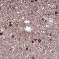


Comprehensive Human Protein Atlas IHC scoring per tissue (reliability: Approved — Low consistency between antibody staining and RNA expression data.). Rows are taken directly from the HPA tissue chart — click any row's HPA link to view the source.
| Tissue | Cell type | Level | Evidence | Source |
|---|---|---|---|---|
| Cerebellum | Cells in granular layer | High | Protein (IHC) | HPA → |
| Bone marrow | Hematopoietic cells | Medium | Protein (IHC) | HPA → |
| Cerebral cortex | Neuronal cells | Medium | Protein (IHC) | HPA → |
| Fallopian tube | Glandular cells | Medium | Protein (IHC) | HPA → |
| Hippocampus | Glial cells | Medium | Protein (IHC) | HPA → |
| Tissue | Cell type | Level | Evidence | Source |
|---|---|---|---|---|
| Adipose tissue | Adipocytes | Not detected | Protein (IHC) | HPA → |
| Adrenal gland | Glandular cells | Not detected | Protein (IHC) | HPA → |
| Breast | Adipocytes | Not detected | Protein (IHC) | HPA → |
| Epididymis | Glandular cells | Not detected | Protein (IHC) | HPA → |
| Heart muscle | Cardiomyocytes | Not detected | Protein (IHC) | HPA → |
Troubleshoot CERS6 staining in paraffin section IHC using the page retrieval method, the catalog antibody’s tissue image, and compartment aware controls.
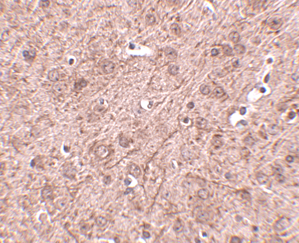
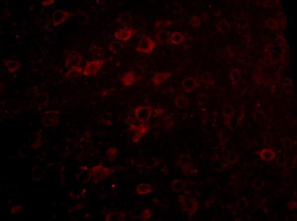
A07033 has IHC-P data from mouse brain tissue and IF data from mouse brain cells (catalog image captions). Its listed reactivity covers human, mouse and rat (catalog: reactivity).
A07033 lists IHC-P and IF applications, with an IHC image of mouse brain tissue at 2.5 μg/mL (catalog: applications; IHC image caption). Its IF image shows mouse brain cells at 5 μg/mL; the catalog lists human, mouse and rat reactivity (catalog: IF image caption; reactivity).
Which to pick: Choose A07033 for paraffin-section IHC because IHC-P is listed and its own IHC image shows staining in mouse brain tissue; the image caption does not report the fixative (catalog: applications; IHC image caption). For IF, A07033 has a mouse brain-cell image, but ICC validation is unreported (catalog: IF image caption; applications). For work across species, A07033 lists human, mouse and rat reactivity; the supplied IHC and IF images show mouse samples only, and clonality is unreported (catalog: reactivity; image captions; clone field).