This website uses cookies to ensure you get the best experience on our website.


- Table of Contents
Plan CKMT2 paraffin-section IHC around cytoplasmic staining in heart and skeletal muscle (HPA tissue IHC). This guide covers fixation consistency, antibody dilution, controls and interpretation of possible off-target staining (HPA tissue IHC; datasheet: 1:50–1:200).

Expected localisation, validated protocols, controls and antibodies — the at-a-glance facts below, then the full design guide.
| Expected localisation | Cytoplasmic staining (HPA tissue IHC); inner mitochondrial membrane target (UniProt) | |
| Staining pattern | High cardiomyocyte and medium skeletal myocyte cytoplasmic staining (HPA tissue IHC) | |
| Antigen retrieval | Citrate pH 6.0 HIER, 95–98 °C, 20 min (rule: cytoplasmic / membrane antigen) | |
| Positive control | Heart muscle+2 more · see all | |
| Negative control | Adipose tissue+4 more · see all |
| Fixation | Keep fixation consistent across sections (standard IHC practice; not target-specific) | |
| Caveat | Presumed off-target binding complicates interpretation (HPA tissue IHC) | |
| Regulation | Sarcomere-specific expression (UniProt) | |
| Isoform / epitope | No listed isoforms; mature chain spans aa 40–419 (UniProt) |
The catalog antibody’s IHC-P protocol is accompanied by two published CKMT2 staining methods for paraffin sections (PMC10764887; PMC13006575).
| Sample | Paraffin-embedded human tonsil cancer tissue; fixative not specified (datasheet A11457-1) |
| Fixation | Image fixative and duration unreported (datasheet A11457-1); verify before use. |
| Sectioning | 4–5 µm sections on charged slides (standard) |
| Deparaffinisation | Xylene, graded ethanol series to water (standard) |
| Antigen retrieval | Heat-induced epitope retrieval in citrate buffer, pH 6.0, 20 min at 95–98 °C (standard rule: cytoplasmic / membrane antigen) |
| Peroxidase block | 3% H2O2, 10 min, room temperature (standard) |
| Blocking | 10% normal serum of the secondary host, 30 min, room temperature (standard) |
| Primary antibody | Rabbit anti-CKMT2, 1:50-1:200 (datasheet A11457-1) |
| Primary incubation | Overnight at 4 °C (standard) |
| Detection | HRP-polymer secondary, DAB chromogen 5–10 min (standard) |
| Counterstain | Hematoxylin, blue, dehydrate and mount (standard) |
| Expected result | CKMT2-positive staining in cardiomyocytes of heart muscle (HPA tissue IHC: High). HPA tissue profile: Cytoplasmic expression mainly in heart and skeletal muscle. No signal in the no-primary control. |
CKMT2 localises to the mitochondrial inner membrane without a transmembrane segment (UniProt P17540 topology). In paraffin section IHC, expect cytoplasmic staining in cardiomyocytes and skeletal myocytes (HPA tissue IHC). HPA reports high staining in cardiomyocytes and medium staining in skeletal myocytes, with Enhanced IHC reliability; its summary also notes presumed off target binding (HPA tissue IHC; HPA antibodies).
| Cardiomyocytes show strong cytoplasmic staining; skeletal myocytes show moderate cytoplasmic staining. | This matches the reported cell types, compartments and relative levels (HPA tissue IHC). Chromogenic IHC shows a cytoplasmic pattern; it does not resolve the mitochondrial inner membrane identified by UniProt (UniProt P17540 topology; general IHC practice). |
| Nuclei stain prominently while the expected cytoplasmic pattern is absent. | Nuclear localisation conflicts with the mitochondrial inner membrane assignment (UniProt P17540 topology). Review the counterstain, detection controls and antibody specificity before scoring nuclear colour as CKMT2 (general IHC practice). |
| Unexpected cell types stain, including distal tubules in kidney. | HPA reports high distal tubular staining, although UniProt describes CKMT2 as found only in heart and skeletal muscle (HPA tissue IHC; UniProt P17540 tissue specificity). Given HPA's off target caveat, treat this as unresolved staining, not confirmed CKMT2 expression (HPA tissue IHC). |
| Colour spreads across tissue structures or appears in negative controls. | A poorly confined signal cannot establish the reported cytoplasmic pattern (HPA tissue IHC; general IHC practice). Background may reflect nonspecific binding or endogenous detection activity; compare controls and examine where the colour is deposited (general IHC practice). |
| Cardiomyocytes show no signal in an otherwise interpretable section. | This differs from HPA's high cardiomyocyte staining (HPA tissue IHC). Check section quality, antibody and detection controls, and the laboratory's retrieval conditions before interpreting the result as absent CKMT2 (general IHC practice). |
| Compartment and resolution | CKMT2 is assigned to the mitochondrial inner membrane and has no transmembrane segment (UniProt P17540 topology). Interpret chromogenic staining at the cytoplasmic level; this method does not by itself verify inner membrane localisation (general IHC practice). |
| Tissue and antibody evidence | HPA reports Enhanced IHC for HPA051880 and medium consistency between staining and RNA data, while noting presumed off target binding (HPA antibodies; HPA tissue IHC). These qualifications matter when judging unexpected staining. |
| Processing and isoforms | UniProt lists a mature chain spanning residues 40–419 and no isoforms (UniProt P17540 processing; UniProt P17540 isoforms). The supplied record gives no antibody epitope, so it cannot establish whether processing changes antibody recognition. |
| Retrieval and detection | Antigen retrieval and checks for endogenous chromogenic activity are general paraffin IHC considerations (general IHC practice). The supplied HPA and UniProt records give no CKMT2 specific retrieval condition or fixation sensitivity. |
| IF/ICC Q&A: can its localisation be scored here? | HPA supplies no main ICC/IF location or cell line images for CKMT2 (HPA subcellular ICC-IF). UniProt supports a mitochondrial expectation, but this record provides no observed ICC/IF pattern to score against (UniProt P17540 topology; HPA subcellular ICC-IF). |
| Situation | Likely cause | Next action |
|---|---|---|
| Heart muscle is negative. | The result conflicts with high cardiomyocyte staining in HPA (HPA tissue IHC); the failed step is undetermined. | Confirm tissue identity and section integrity, then review retrieval, antibody application, detection and a working positive control (general IHC practice). |
| Skeletal muscle is weaker than heart muscle. | HPA grades skeletal myocytes medium and cardiomyocytes high (HPA tissue IHC). | Judge each tissue against its reported level and cytoplasmic distribution before calling skeletal muscle negative (HPA tissue IHC; general IHC practice). |
| Kidney distal tubules stain strongly. | HPA reports this staining despite UniProt's heart and skeletal muscle tissue specificity; HPA also notes presumed off target binding (HPA tissue IHC; UniProt P17540 tissue specificity). | Record the discrepancy, review controls and seek independent specificity evidence before attributing tubular colour to CKMT2 (general IHC practice). |
| Nuclear staining dominates. | This conflicts with the mitochondrial inner membrane assignment (UniProt P17540 topology); the source of the nuclear colour is undetermined. | Inspect a negative detection control, counterstain and antibody specificity, then score only interpretable compartmental staining (general IHC practice). |
| Diffuse staining obscures cell boundaries. | Background or endogenous detection activity can make cytoplasmic staining hard to distinguish (general IHC practice). | Compare negative controls and review blocking, antibody concentration, washing and detection conditions (general IHC practice). |
| A presumed negative tissue stains. | HPA lists adipocytes in adipose tissue as not detected, but its tissue summary notes presumed off target binding (HPA tissue IHC). A single unexpected positive does not identify its cause. | Check the matching cell type, negative controls and signal distribution; seek independent specificity evidence before revising the expected pattern (HPA tissue IHC; general IHC practice). |


Comprehensive Human Protein Atlas IHC scoring per tissue (reliability: Enhanced — Medium consistency between antibody staining and RNA expression data. Presumed off target binding observed and disregarded.). Rows are taken directly from the HPA tissue chart — click any row's HPA link to view the source.
| Tissue | Cell type | Level | Evidence | Source |
|---|---|---|---|---|
| Heart muscle | Cardiomyocytes | High | Protein (IHC) | HPA → |
| Kidney | Distal tubules | High | Protein (IHC) | HPA → |
| Skeletal muscle | Myocytes | Medium | Protein (IHC) | HPA → |
| Tissue | Cell type | Level | Evidence | Source |
|---|---|---|---|---|
| Adipose tissue | Adipocytes | Not detected | Protein (IHC) | HPA → |
| Appendix | Glandular cells | Not detected | Protein (IHC) | HPA → |
| Bone marrow | Hematopoietic cells | Not detected | Protein (IHC) | HPA → |
| Breast | Adipocytes | Not detected | Protein (IHC) | HPA → |
| Bronchus | Respiratory epithelial cells | Not detected | Protein (IHC) | HPA → |
Troubleshoot CKMT2 staining in paraffin sections by checking retrieval, compartment, tissue context and controls before assigning a chromogenic signal to the target.
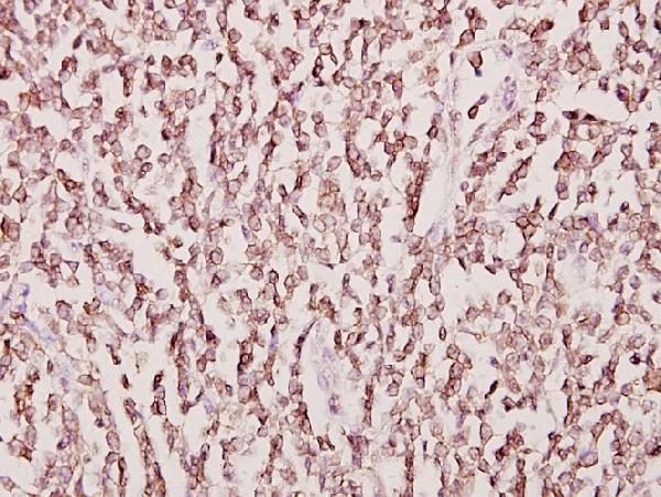
Anti-CKMT2 A11457-1 has a paraffin-section IHC image from human tonsil cancer tissue; IF is listed without an image (catalog image caption; catalog applications). Human, mouse and rat reactivity is listed (catalog reactivity).
A11457-1 has an IHC image of paraffin-embedded human tonsil cancer tissue at 1:50 (catalog image caption). A11457-1 lists IHC and IF applications and human, mouse and rat reactivity; no IF image is supplied (catalog applications; catalog reactivity; catalog IF images).
Which to pick: Choose A11457-1 for paraffin-section tissue IHC because its own image shows that preparation at 1:50; the fixative is unreported (catalog image caption). For IF/ICC, A11457-1 lists IF at 1:50–1:200, but supplies no IF image or ICC validation (catalog applications; catalog IF dilution; catalog IF images). It is the listed cross-species option for human, mouse and rat and is polyclonal (catalog reactivity; catalog dilution record).