CLDN18 / Claudin-18 · IHC design guide

Design Immunohistochemistry for CLDN18

Plan CLDN18 paraffin IHC around membranous staining in gastric epithelium and alveolar cells (HPA tissue IHC). The catalog antibody has an IHC dilution range of 2–5 μg/ml (datasheet A09129-1).

Evidence assembled Oct 2026 · For research use; verify linked source records and product datasheet before use
Immunohistochemistry protocol sheet for CLDN18 (IHC for CLDN18): expected localisation Epithelial membrane staining (HPA tissue IHC), antibody A09129-1, validated IHC image, and IHC protocol steps
Printable CLDN18 IHC protocol sheet — expected localisation Epithelial membrane staining (HPA tissue IHC), antibody A09129-1, controls and protocol steps. Open the full CLDN18 IHC guide →

CLDN18 Immunohistochemistry Experimental Design Guide

Expected localisation, validated protocols, controls and antibodies — the at-a-glance facts below, then the full design guide.

Must know before staining
Expected localisation Epithelial membrane staining (HPA tissue IHC)
Staining pattern Membranous in gastric glands, gallbladder and alveolar cells (HPA tissue IHC)
Antigen retrieval EDTA pH 8.0 HIER, heat-mediated (datasheet A09129-1)
Positive control ⓘ Stomach+1 more · see all
Negative control ⓘ Adipose tissue+4 more · see all
Important caveats
Reasons your staining may differ from the expected pattern.
Fixation Keep fixation consistent across sections (standard IHC practice; not target-specific)
Caveat Stomach staining differs from the lung-only annotation (HPA tissue IHC; UniProt)
Regulation Lung and stomach RNA group-enriched (HPA RNA specificity)
Isoform / epitope 2 isoforms (A1/A2); antibody epitope and isoform coverage unspecified (UniProt)
Section 1

Recommended CLDN18 IHC & IF Protocols

The catalog antibody uses EDTA pH 8.0 heat retrieval (datasheet A09129-1). Published IHC protocols below cover tubo-ovarian carcinoma and cervical adenocarcinoma (PMC11271439; PMC12975664).

Recommended immunohistochemistry (IHC-P) protocol parameters
SampleParaffin-embedded human gastric cancer tissue; fixative not specified (datasheet A09129-1)
FixationImage fixative and duration unreported (datasheet A09129-1); verify before use.
Sectioning4–5 µm sections on charged slides (standard)
DeparaffinisationXylene, graded ethanol series to water (standard)
Antigen retrievalHeat retrieval: EDTA pH 8.0 (datasheet A09129-1); 20 min, 95–100 °C (standard)
Peroxidase block3% H2O2, 10 min, room temperature (standard)
Blocking10% goat serum (datasheet A09129-1)
Primary antibodyRabbit anti-CLDN18, 2-5 μg/ml (datasheet A09129-1)
Primary incubationOvernight at 4 °C (datasheet A09129-1)
DetectionHRP-conjugated secondary, DAB chromogen (datasheet A09129-1)
CounterstainHematoxylin, blue, dehydrate and mount (standard)
Expected resultCLDN18-positive staining in glandular cells of stomach (HPA tissue IHC: High). HPA tissue profile: Distinct membranous expression in stomach, gallbladder epithelium and alveolar cells. No signal in the no-primary control.
💡Decision noteStart with EDTA pH 8.0 heat retrieval for the catalog antibody (datasheet A09129-1). The tubo-ovarian protocol specifies CC1 retrieval at 100 °C for 64 min (PMC11271439).
Section 2

What Is the Expected CLDN18 Staining Pattern?

CLDN18 is a four-pass membrane protein that localizes to epithelial tight junctions and lateral membranes (UniProt P56856 topology and subcellular location). On paraffin sections, expect membranous staining in stomach glandular cells and lung alveolar type II cells, with weaker staining in gallbladder glandular cells (HPA tissue IHC). HPA rates its tissue pattern Enhanced for consistency between antibody staining and RNA expression (HPA tissue IHC).

What am I looking at on my slide?
Crisp staining outlines stomach glandular cells or lung alveolar type II cells.This fits the expected membranous pattern (HPA tissue IHC) and junctional localization (UniProt P56856). HPA reports High staining in stomach glandular cells and Medium staining in alveolar type II cells (HPA tissue IHC).
Signal is mainly nuclear or diffusely cytoplasmic, without a discernible membrane outline.That distribution conflicts with the reported membrane and tight-junction locations (UniProt P56856; HPA subcellular). Treat it as suspect and compare it with a known-positive section and a detection control before assigning CLDN18 positivity (general IHC practice).
Strong staining appears in an unexpected cell population while expected epithelial membranes are unstained.Check for cross-reactivity or endogenous detection activity (general IHC practice). HPA reports CLDN18 staining in specific epithelial populations, rather than every cell within a positive organ (HPA tissue IHC); tissue identity alone does not establish a positive result.
Color spreads across the section, obscuring cell borders and tissue structure.This is uninterpretable background rather than a clear junctional pattern (UniProt P56856; general IHC practice). Review the detection control, blocking and wash steps, and antibody concentration using the selected antibody's instructions (general IHC practice).
The known-positive stomach control has no interpretable glandular-cell membrane signal.HPA reports High stomach glandular-cell staining (HPA tissue IHC). A negative control section cannot validate a negative study specimen; first check section quality, antibody suitability and detection performance, then assess the specimen (general IHC practice).
💡Expected CLDN18 appearanceCall a result positive when glandular-cell or alveolar type II-cell borders show distinct membranous staining at the tissue-appropriate level (HPA tissue IHC); isolated nuclear color or widespread color without membrane definition is suspect (UniProt P56856; general IHC practice).
How each factor affects the staining
Membrane topologyCLDN18 has 4 transmembrane segments, extracellular loops and cytoplasmic regions (UniProt P56856 topology). Membrane-associated staining is expected, but the supplied record does not identify the epitope of a chosen antibody or establish which retrieval condition exposes it.
Tissue evidenceHPA observes High stomach glandular-cell, Medium alveolar type II-cell and Low gallbladder glandular-cell staining (HPA tissue IHC). UniProt describes expression as restricted to lung (UniProt P56856 tissue specificity); use the stated HPA IHC observations when assessing these sections.
IsoformsUniProt lists 2 isoforms, A1 and A2 (UniProt P56856). The supplied evidence does not show which isoform an IHC antibody recognizes, so a positive section cannot be assigned to either isoform from staining pattern alone.
Antibody validationHPA lists Enhanced IHC status for HPA018446 and CAB013243, and Supported status for CAB013010 (HPA antibodies). These are antibody-specific validation summaries; select and document the antibody actually used before applying their status to an experiment.
Antigen retrievalRetrieval conditions can be optimized with positive and negative controls in paraffin-section IHC (general IHC practice). Neither the HPA tissue pattern nor UniProt topology establishes CLDN18-specific retrieval sensitivity or a preferred condition; report the condition actually tested.
Why is my staining missing, weak or wrong?
SituationLikely causeNext action
No membrane staining appears in the stomach positive control.The control failed to show the High glandular-cell signal reported by HPA (HPA tissue IHC); the cause remains undetermined.Check antibody identity and IHC suitability, then review retrieval, primary-antibody application and detection with a fresh positive control (general IHC practice). Do not interpret study sections until the control works.
Only nuclear or broadly cytoplasmic staining appears.This distribution disagrees with CLDN18 membrane and junction localization (UniProt P56856; HPA subcellular); nonspecific staining is possible (general IHC practice).Compare a known-positive section and a detection control, then reassess antibody concentration and washes (general IHC practice). Require a discernible epithelial membrane pattern before calling CLDN18 positive (HPA tissue IHC).
Unexpected cells stain strongly in an otherwise negative tissue.HPA reports no detection in several listed cell populations, including adipocytes in adipose tissue and respiratory epithelial cells in bronchus (HPA tissue IHC). Cross-reactivity or endogenous detection activity should be investigated (general IHC practice).Confirm the cell type and compare a no-primary detection control with the stained section; use an independently validated antibody if the unexpected pattern persists (general IHC practice).
Background color obscures the epithelial borders.The section cannot be scored for the distinct membranous pattern described by HPA (HPA tissue IHC). Excess detection signal or inadequate blocking or washing are general IHC possibilities (general IHC practice).Inspect the detection control, blocking and wash steps; adjust antibody concentration according to the selected antibody's instructions and repeat alongside a positive section (general IHC practice).
Stomach signal is stronger than lung signal.That ordering matches HPA's High stomach glandular-cell and Medium alveolar type II-cell observations (HPA tissue IHC). UniProt's lung-restricted tissue statement differs from the HPA IHC observations (UniProt P56856 tissue specificity; HPA tissue IHC).Score the expected cell populations and membrane pattern in each tissue separately (HPA tissue IHC); do not require equal intensity across the two controls.
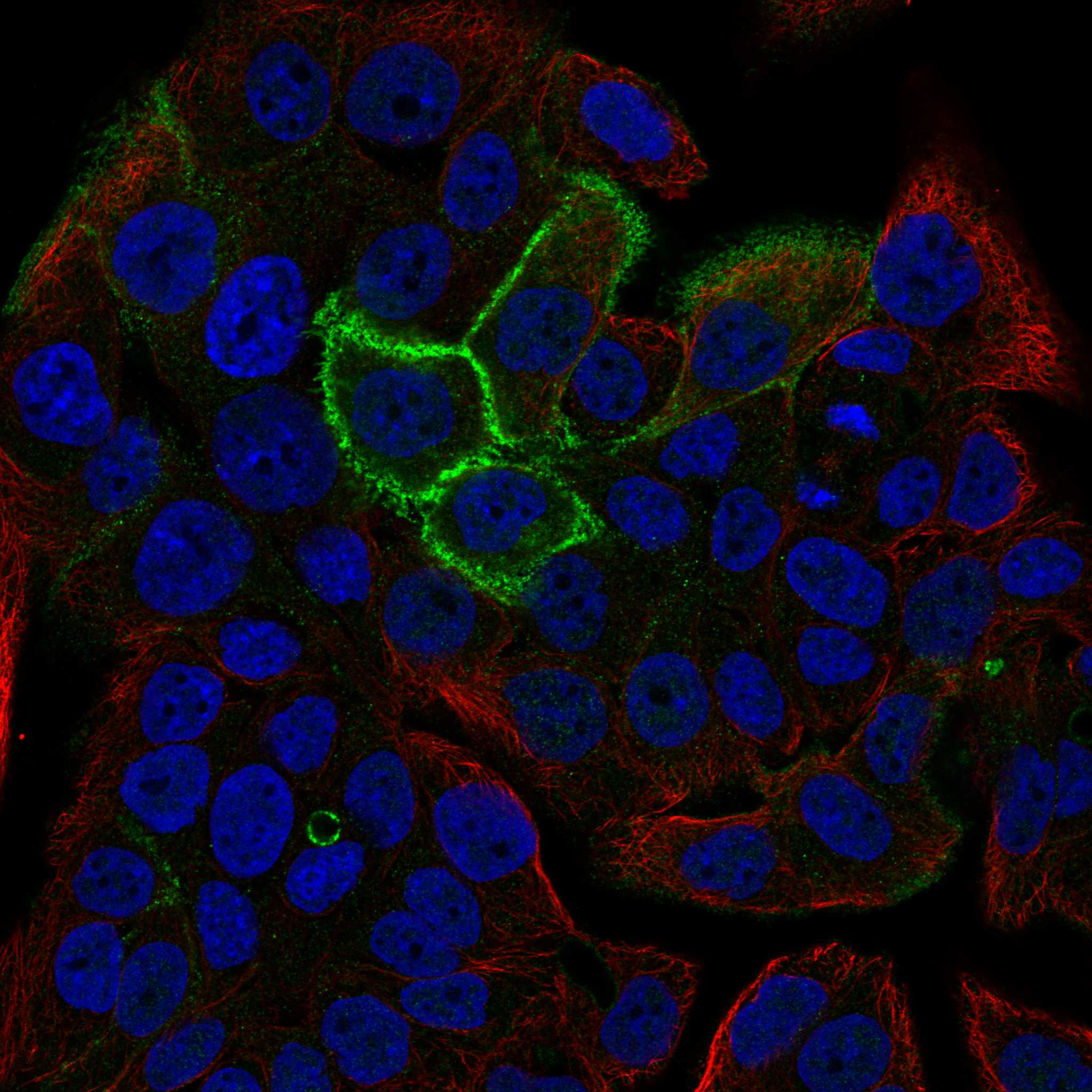
IF/ICC Q: Where should CLDN18 signal appear?HPA reports an approved cell-junction localization in ICC-IF (HPA subcellular). That localization agrees with epithelial tight-junction placement (UniProt P56856).A: Assess whether signal follows cell junctions (HPA subcellular). Consult the separate IF/ICC guide for experimental conditions; the HPA localization record supplies no IF/ICC protocol.

Sample controls for CLDN18 IHC & IF

🧪Run stomach first: glandular cells should stain (HPA: Stomach glandular cells, High), with junctional localization expected (UniProt P56856: tight junction localization). Use adipose tissue as a negative, with adipocytes lacking specific staining (HPA: Adipose tissue adipocytes, Not detected); surrounding nonglandular cells on the stomach slide should lack specific junctional signal (standard IHC control interpretation).
Positive control tissue: Stomach (Glandular cells, HPA High)
Negative control tissue: Adipose tissue (HPA Not detected)
ICC-IF cell lines (HPA subcellular resource): HPA ICC-IF images show CLDN18 in OE19, U-251MG, U2OS, with annotated localisation: Cell Junctions (approved) (HPA subcellular).
Technical controls: Include no-primary (secondary-only) and rabbit IgG isotype controls matched to the primary antibody’s clonality if known (product caption: rabbit primary; standard IHC practice). Use CLDN18 knockout tissue or a peptide-block control if a suitable immunogen is available, and block endogenous peroxidase before HRP/DAB detection in stomach sections (standard IHC practice; product caption: HRP/DAB detection).
⚠️Feasibility: Paraffin-section IHC is documented with EDTA retrieval at pH 8.0, but whether retrieval is required has not been established (product caption). The selected A09129-1 caption does not report a fixative, and a target-specific fixation window or fixation effect is unreported (product caption). The supplied evidence does not establish that frozen sections or IF are easier; assess gastric background from endogenous peroxidase during chromogenic detection (standard IHC practice).

HPA tissue IHC evidence for CLDN18

Comprehensive Human Protein Atlas IHC scoring per tissue (reliability: Enhanced — High consistency between antibody staining and RNA expression data.). Rows are taken directly from the HPA tissue chart — click any row's HPA link to view the source.

Positive expression · recommended positive controls

TissueCell typeLevelEvidenceSource
Stomach Glandular cells High Protein (IHC) HPA →
Lung Alveolar cells type II Medium Protein (IHC) HPA →

Undetected expression · recommended negative controls

TissueCell typeLevelEvidenceSource
Adipose tissue Adipocytes Not detected Protein (IHC) HPA →
Adrenal gland Glandular cells Not detected Protein (IHC) HPA →
Appendix Glandular cells Not detected Protein (IHC) HPA →
Bone marrow Hematopoietic cells Not detected Protein (IHC) HPA →
Breast Adipocytes Not detected Protein (IHC) HPA →
Section 3

Advanced CLDN18 IHC Tips

Use membrane pattern, tissue controls, and the catalog antibody’s paraffin-section conditions to troubleshoot CLDN18 staining (UniProt P56856; datasheet A09129-1).

What retrieval conditions should I try when CLDN18 staining is weak?
Start with heat-mediated antigen retrieval in EDTA at pH 8.0 for paraffin sections (datasheet A09129-1). Compare the test section with a processed stomach control, where glandular cells show high CLDN18 staining, and check for a distinct membrane pattern (HPA: stomach glandular cells, High; HPA: distinct membranous expression). Keep section thickness, heating, cooling, antibody concentration, and DAB development consistent while assessing retrieval, so their variation does not obscure the comparison (standard IHC practice). If staining remains weak, test another retrieval condition on serial sections as an empirical fallback and assess both signal and tissue preservation (standard IHC practice).
Could fixation explain inconsistent CLDN18 staining between paraffin blocks?
Target-specific fixation sensitivity is unknown: the selected paraffin-section caption does not state a fixative, and the supplied evidence gives no CLDN18-specific fixation comparison (caption A09129-1). Record each block’s fixative and processing history before comparing staining, because those conditions can affect antigen accessibility in IHC generally (standard IHC practice). Process a known positive stomach control alongside the samples and use the same retrieval and detection conditions across blocks (HPA: stomach glandular cells, High; datasheet A09129-1; standard IHC practice). If blocks differ, interpret a weaker membrane signal cautiously and assess tissue preservation before attributing the difference to CLDN18 abundance (UniProt P56856 localization; standard IHC practice).
Which staining pattern should count as CLDN18 localisation in chromogenic IHC?
Prioritize a crisp epithelial membrane pattern, especially at cell contacts, because CLDN18 localizes to tight junctions and lateral cell membranes (UniProt P56856 localization). The protein has 4 transmembrane segments, so diffuse nuclear DAB is inconsistent with its annotated topology (UniProt P56856 topology; standard IHC interpretation). Use stomach glandular cells or lung alveolar type II cells as tissue-context checks, while allowing for their different reported staining levels (HPA: stomach glandular cells, High; lung alveolar type II cells, Medium). Examine counterstained morphology at high magnification and score membrane staining separately from diffuse cytoplasmic pigment or staining over damaged tissue (standard IHC practice).
How can epitope position change interpretation of CLDN18 IHC?
Check the antibody’s stated immunogen or epitope before claiming isoform specificity: CLDN18 has 2 annotated isoforms, A1 and A2, but the supplied caption gives no epitope mapping (UniProt P56856 isoforms; caption A09129-1). Its extracellular loops span residues 28–80 and 144–174, whereas residues 196–261 form a cytoplasmic tail (UniProt P56856 topology). Accessibility and the meaning of membrane staining depend on which region the antibody recognizes and how processing exposes it (standard IHC practice). Until isoform-selective recognition is established, report the signal as CLDN18 immunoreactivity rather than assigning it to A1 or A2 (UniProt P56856 isoforms; standard IHC interpretation).
How should I plan a CLDN18 IF follow-up to the IHC result?
Treat IF as a separately optimized application, since the supplied paraffin-section caption documents chromogenic IHC conditions (caption A09129-1). Multiplex CLDN18 with a validated marker of the expected cell type, such as a type II alveolar-cell marker in lung, and inspect cell-border overlap rather than pooled field intensity (HPA: lung alveolar type II cells, Medium; UniProt P56856 localization). Choose a fluorophore in a channel with low measured tissue autofluorescence and include single-color controls for bleed-through (standard IF practice). If the epitope is extracellular, compare staining without permeabilisation; if cytoplasmic, optimize permeabilisation while preserving junctions, since the antibody epitope is unspecified (UniProt P56856 topology; caption A09129-1; standard IF practice).
How do I reduce diffuse DAB background without losing junctional signal?
Compare background with a no-primary control, then inspect whether DAB follows epithelial borders or spreads across tissue and stroma (UniProt P56856 localization; standard IHC practice). The selected image used 10% goat serum blocking, 2 μg/ml primary antibody overnight at 4°C, and a peroxidase-linked detection system (caption A09129-1). Use a peroxidase block before HRP detection and control DAB development time as general chromogenic IHC measures (standard IHC practice). If diffuse signal persists, titrate primary antibody on serial sections while retaining a positive stomach control and compare membrane contrast at matched development times (HPA: stomach glandular cells, High; standard IHC practice).
What is a defensible way to score CLDN18 across IHC samples? ⚠ ANSWER MARKED FOR VERIFICATION
Define the target compartment as evaluable epithelial cells with membrane staining, consistent with CLDN18 junctional localization (UniProt P56856 localization; HPA: distinct membranous expression). Record the percentage of positive cells and membrane intensity by category, then calculate an H-score if that scoring system is prespecified (standard IHC practice). Normalize counts to evaluable epithelial cells, or report positive membrane area per mm² of viable epithelial tissue when area is the study endpoint (standard IHC practice). Exclude necrosis and folds, keep retrieval and DAB development comparable, and document whether glandular or alveolar cells were assessed because their reported levels differ (standard IHC practice; HPA: stomach High, lung Medium).
When is a CLDN18-positive IHC result likely to be artefactual?
A convincing result shows membrane staining in the relevant epithelial cells, consistent with CLDN18 localization and the HPA tissue pattern (UniProt P56856 localization; HPA: distinct membranous expression). Question isolated nuclear signal, diffuse staining in the wrong cell population, or pigment concentrated at section edges or necrotic areas (UniProt P56856 topology; standard IHC interpretation). Compare no-primary and peroxidase-block controls when brown precipitate might reflect endogenous enzyme activity rather than antibody-dependent DAB deposition (standard IHC practice). Interpret tissue identity with care: HPA reports high staining in stomach glandular cells and medium staining in lung alveolar type II cells, while the supplied UniProt tissue-specificity statement says expression is restricted to lung (HPA: tissue IHC; UniProt P56856 tissue specificity).
Boster reagents

Best CLDN18 / Claudin-18 IHC Antibodies

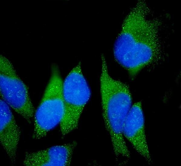
A09129-1 has real IHC images from paraffin-embedded human gastric cancer and rat stomach, and IF images from those tissues and HeLa cells (catalog: image captions).

Real IHC data IHC analysis of Claudin18/CLDN18 using anti-Claudin18/CLDN18 antibody (A09129-1). Claudin18/CLDN18 was detected in a paraffin-embedded section of human gastric cancer tissue. Heat mediated antigen retrieval was performed in EDTA buffer (pH 8.0, epitope retrieval solution). The tissue section was blocked with 10% goat serum. The tissue section was then incubated with 2 μg/ml rabbit anti-Claudin18/CLDN18 Antibody (A09129-1) overnight at 4°C. Peroxidase Conjugated Goat Anti-rabbit IgG was used as secondary antibody and incubated for 30 minutes at 37°C. The tissue section was developed using HRP Conjugated Rabbit IgG Super Vision Assay Kit (Catalog # SV0002) with DAB as the chromogen.
Anti-Claudin18/CLDN18 Antibody ®
Cat # A09129-1

A09129-1 is listed for IHC, IF and ICC in human and rat samples (catalog: applications/reactivity). Its IHC images show paraffin-embedded human gastric cancer and rat stomach; its IF images show those tissues and HeLa cells (catalog: image captions).

Which to pick: For tissue IHC, choose A09129-1: its own IHC captions document paraffin sections, EDTA pH 8.0 retrieval and 2 μg/ml primary antibody (A09129-1: IHC image captions). For IF/ICC and human or rat work, A09129-1 is also the listed choice; it is a rabbit antibody, and clonality is unreported (catalog: applications, reactivity, host and clone). The IHC captions do not report the fixative (A09129-1: IHC image captions).

Each figure is that product's own IHC / IF validation image from its datasheet.

References

  1. UniProt Consortium. UniProt entry P56856 (CLD18_HUMAN, Claudin-18).
  2. Human Protein Atlas. CLDN18 tissue IHC expression (reliability: Enhanced).
  3. Human Protein Atlas. CLDN18 subcellular location (ICC-IF): Localized to the cell junctions..
  4. Human Protein Atlas. CLDN18 antibody validation summary (3 antibodies).
  5. Claudin 18.2 Expression in 1404 Digestive Tract Adenocarcinomas Including 1175 Colorectal Carcinomas: Distinct Colorectal Carcinoma Subtypes Are Claudin 18.2 Positive. Modern pathology : an official journal of the United States and Canadian Academy of Pathology, Inc 2025 — PMC12103281.
  6. Claudin-18.2 immunohistochemical evaluation in pancreatic cancer specimens: review and recommendations for routine testing and scoring. Virchows Archiv : an international journal of pathology 2025 — PMC12488767.
  7. Spatial expression of claudin 18.2 in matched primaries and metastases of tubo-ovarian carcinoma of all subtypes. Virchows Archiv : an international journal of pathology 2024 — PMC11271439.
  8. Claudin-18 expression in gastric type adenocarcinoma and HPV-associated adenocarcinoma of the uterine cervix. Histopathology 2026 — PMC12975664.
  9. PubMed PMID:11585919 — UniProt-cited evidence.
  10. PubMed PMID:12975309 — UniProt-cited evidence.
  11. PubMed PMID:15489334 — UniProt-cited evidence.