This website uses cookies to ensure you get the best experience on our website.
- Table of Contents
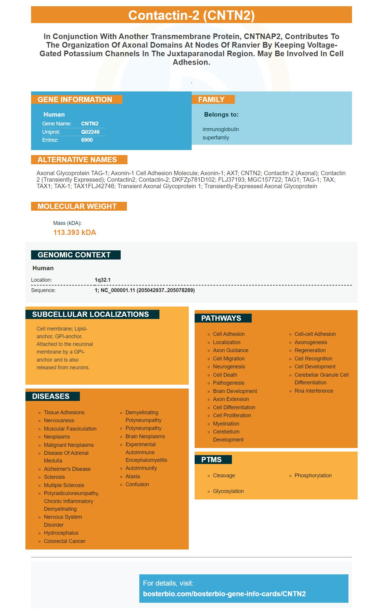
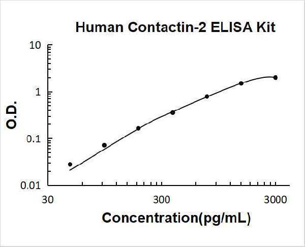
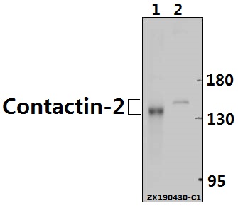
Facts about Contactin-2.
.
Human | |
---|---|
Gene Name: | CNTN2 |
Uniprot: | Q02246 |
Entrez: | 6900 |
Belongs to: |
---|
immunoglobulin superfamily |
Axonal glycoprotein TAG-1; axonin-1 cell adhesion molecule; Axonin-1; AXT; CNTN2; contactin 2 (axonal); contactin 2 (transiently expressed); Contactin2; Contactin-2; DKFZp781D102; FLJ37193; MGC157722; TAG1; TAG-1; TAX; TAX1; TAX-1; TAX1FLJ42746; Transient axonal glycoprotein 1; transiently-expressed axonal glycoprotein
Mass (kDA):
113.393 kDA
Human | |
---|---|
Location: | 1q32.1 |
Sequence: | 1; NC_000001.11 (205042937..205078289) |
Cell membrane; Lipid-anchor, GPI-anchor. Attached to the neuronal membrane by a GPI-anchor and is also released from neurons.
This Boster Bio article is for you, whether you're an immunologist looking for a diagnosis or a scientist researching a disease. Learn about its Clinical significance, Specificity, Application, and more. This Boster Bio product can be used for a variety of purposes, including immunohistochemistry, Western Blotting, and ELISA.
The CNTN2 protein marker is a distinct protein with a unique structure. Although it isn't known how it folds 3D, its structure indicates that the N terminal portion folds back onto the C-terminal. The N-terminal part of the protein's headpiece looks like a horseshoe. In a crystalline environment, the major conformation of the protein is determinable by the contacts between the headpieces.
Structure studies were performed on CNTN2, a protein with multi-domain flexibility, and the CNTN2 molecule was successfully purified from human cells by using a recombinant DNA technique. Traditional methods for determining the structure of proteins, such X-ray Crystallography, reveal only one conformation. Single particle 3D EM however, reveals millions protein particles and their conformations.
Ten domains are present in the CNTN2 molecule. Each one represents an Ig and FNIII domain. The extracellular domain in CNTN2 is difficult for cryo-EM to image because it is made of a small molecular structure. It also has a lasso-shaped terminus. Categor 1 particles also have a horseshoe shape.
The headpiece of CNTN2 molecule looks horseshoe-like. The molecule's conformation has been fully elongated. Windowed 2D data shows 150 representative class averages per 200 classes. The number and types of particles in each class vary from six to 96. The termini are indicated by the yellow arrows. There are four CNTN2 molecules in the mouse brain.
CNTNs contain six Ig-like domains. GPI anchors hold the ectodomain (CNTN2) to the cell membrane. The crystal structure reveals a horseshoe shaped structure for CNTN2 proteins. Its Ig1 domain and Ig4 domains make a U-shaped arrangement, with six residue linkers.
The CNTN2 gene product is a member of the immunoglobulin superfamily and is a membrane-associated glycosylphosphatidylinositol-anchored neuronal protein. It functions as a cell adhesion molecule and may play a role in axon connections during development of the nervous system. Other roles CNTN2 could play include glial tumourgenesis.
The expression patterns and function of the CNTN2 marker were examined in various cell lines from the peripheral nervous systems. These two genes were expressed in U-118MG cells and LN-18 human astrocytes. Both CNTN2 and RACK1 were not expressed in DTmesV neuronal cells, the latter being a known marker of terminally differentiated neurons. The results show that CNTN2 is a good marker to detect glioma.
A new Cntn2-dependent transcriptional regulator has been developed and tested in murine cardiac tissues. This marker may provide a powerful new tool to study Purkinje cellular function. The regulation of Cntn2 expression may provide a mechanistic base for patterning the Purkinje cell conduction system network. Its role in human cardiac tissue is still unclear.
The CNTN2 proteins interact with RACK1. Antibodies to this receptor inhibit glioma cells differentiation and increase CNTN2 expression within cancerous cells. RACK1 also binds to CNTN2, so knockdown of this gene abolishes these effects. The protein is crucial for the differentiation of glioma cells and their growth. RACK1 appears not to be necessary for human glioma development.
Researchers used western blot to examine the protein expression of CNTN2 in human glioma cells to determine if it was associated with glioma growth. RACK1 as well as CNTN2 protein levels in glioma cells were significantly higher than in normal brain tissue. High-grade glioma cells also had higher levels of RACK1 (CNTN2) and CNTN2.
RACK1 has been shown to interact well with the CNTN2 marker. It has also been shown to increase cell proliferation, which could be an indicator of glioma development. RACK1 had its effect reversed after knockdown of the gene. Moreover, RACK1-mediated glioma growth was completely abated by knockdown of CNTN2. These results suggest that CNTN2 may play a part in glioma development.
CNTN2 protein is an overexpressed protein in many types brain tumors. Rickman and collaborators reported in 2010 CNTN2 being frequently overexpressed within gliomas. It plays a critical function in the formation of oligodendroglioma. The CNTN2 protein also interacts with other proteins, possibly mediating its role in tumor development. Researchers did a western blot to determine if CNTN2 had been overexpressed within glioma tissue.
Knockdown of CNTN2 reduced the effects of RACK1 upon cell differentiation in SW1783 MG cells and U118 MG cells. Knockdown of CNTN2 abrogated both RACK1 and CNTN2 expression. These results suggest that knockdown CNTN2 might inhibit RACK1's effect on CNTN2 transcription. There are several other ways that the CNTN2 genes can be knocked down.
CNTN2 in the mouse's spinal cord is a marker for postmitotic neurons. Although its function is unknown, extensive studies on mice and zebrafish suggest that it is involved facial branchiomotor neuron movement. Cntn2 expression was also found in a subset of peripheral neurons during adulthood. This gene is associated to the formation of and maintenance of myelinated fibrils.
The expression pattern of Chga, Tagln3, and Cntn2 was investigated by whole-mount in situ hybridization. Expression patterns of Cntn2 were similar to those of Nhlh2, a pan-neuronal marker. In addition, CNTN2 was found to be restricted to the MZ and floor plate of the trigeminal nerve. Despite these differences, the study indicated that Cntn2 is a neuronal marker that is specifically expressed in the trigeminal nerve.
In situ hybridization was used and HuC/D stained to determine the expression of CNTN2 in flat-mounted rhombencephalon preparations. Cntn2, Tagln3, Chga showed limited expression. Spotted expression in the neural tubes corresponds to the development of somatic motor neurons. Nhlh2 (Chga) and Tagln2 (ventral part of neural tube) were detected.
Expression of CNTN2 first became apparent at Hh22 in Rathke’s pouch. It was a small lateral stripe that is distal to the optic vesicles. Nhlh2, Tagln3, Tagln3 & Tagln3 markers had been detected in the trigeminal Placode cell lineage at Hh25. These markers were discovered in all cranial glands by HH20. Similar to Chga or Stmn2, these markers were not found in the gV or mx lobes. This indicates that CNTN2 has a greater specificity.
The CNTN2 protein marker is a protein that is elevated in many human glioma cell line. The protein expression was also studied in normal human astrocytes. Both RACK1 as well as CNTN2 were upregulated in cell lines SW1783 and U118 MG, according to the researchers. In this study, the CNTN2 gene was shown to predict the likelihood of developing glioma in these cell types.
CNTN2 acts as a cell adhesion molecule in cells and is involved in the development of a variety of types of cancers. Recent studies show that CNTN2 protein expression is high in gliomas. This protein is a hub in the protein protein interaction network. It may also play a key role in tumorigenesis.
PMID: 8425542 by Hasler T.H., et al. cDNA cloning, structural features, and eucaryotic expression of human TAG-1/axonin-1.
PMID: 8307567 by Tsiotra C.P., et al. Isolation of the cDNA and chromosomal localization of the gene (TAX1) encoding the human axonal glycoprotein TAG-1.