This website uses cookies to ensure you get the best experience on our website.


- Table of Contents
Plan COX5A chromogenic IHC in paraffin sections using its granular cytoplasmic tissue pattern (HPA tissue IHC) and mitochondrial inner membrane location (UniProt). Compare cell types with documented high and undetected staining when selecting controls (HPA tissue IHC).

Expected localisation, validated protocols, controls and antibodies — the at-a-glance facts below, then the full design guide.
| Expected localisation | Granular cytoplasm (HPA tissue IHC); mitochondrial inner membrane (UniProt) | |
| Staining pattern | Granular cytoplasmic staining in glandular and epithelial cells (HPA tissue IHC) | |
| Antigen retrieval | EDTA pH 8.0 HIER, heat-mediated (datasheet A07895-2) | |
| Positive control | Appendix+4 more · see all | |
| Negative control | Adipose tissue+2 more · see all |
| Fixation | Keep fixation consistent across sections. (standard IHC practice; not target-specific) | |
| Caveat | Staining varies by cell type: colon glands high, adipocytes undetected (HPA tissue IHC) | |
| Regulation | No specific regulator annotated (UniProt) | |
| Isoform / epitope | No isoforms; processing removes residues 1–41 from the mature chain (UniProt) |
The catalog antibody’s IHC-P protocol (datasheet: A07895-2) is accompanied by published COX5A IHC methods for rat brain, renal carcinoma, and gastric tissues (PMC7191708; PMC2783948; PMC12582873).
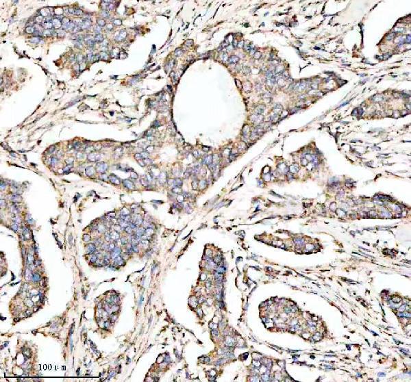
| Sample | Paraffin-embedded human breast cancer tissue; fixative not specified (datasheet A07895-2) |
| Fixation | Image fixative and duration unreported (datasheet A07895-2); verify before use. |
| Sectioning | 4–5 µm sections on charged slides (standard) |
| Deparaffinisation | Xylene, graded ethanol series to water (standard) |
| Antigen retrieval | Heat retrieval: EDTA pH 8.0 (datasheet A07895-2); 20 min, 95–100 °C (standard) |
| Peroxidase block | 3% H2O2, 10 min, room temperature (standard) |
| Blocking | 10% goat serum (datasheet A07895-2) |
| Primary antibody | Rabbit anti-COX5A, 1:50 recommended; image 1:100 (datasheet A07895-2) |
| Primary incubation | Overnight at 4 °C (datasheet A07895-2) |
| Detection | HRP-conjugated secondary, DAB chromogen (datasheet A07895-2) |
| Counterstain | Hematoxylin, blue, dehydrate and mount (standard) |
| Expected result | COX5A-positive staining in glandular cells of appendix (HPA tissue IHC: High). HPA tissue profile: General cytoplasmic expression with a granular pattern. No signal in the no-primary control. |
COX5A is a component of respiratory complex IV in the mitochondrial inner membrane and has no annotated transmembrane segment (UniProt P20674). In paraffin section IHC, expect granular cytoplasmic staining, including in glandular cells of colon and appendix and respiratory epithelial cells of bronchus (HPA tissue IHC). HPA rates the tissue pattern Supported, with medium consistency between antibody staining and RNA expression (HPA tissue IHC).
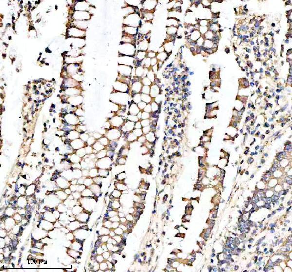
| Granular cytoplasmic stain in colon glandular cells, with nuclei remaining defined by counterstain. | This matches HPA's general granular cytoplasmic pattern and High staining in colon glandular cells (HPA tissue IHC). It is consistent with mitochondrial localization (UniProt P20674), although a chromogenic pattern alone cannot resolve the inner membrane (standard IHC interpretation). |
| Predominantly nuclear, sharply membranous, or extracellular stain replaces the granular cytoplasmic pattern. | These compartments conflict with the annotated mitochondrial inner membrane location and HPA tissue pattern (UniProt P20674; HPA tissue IHC). Treat the result as suspect; review morphology and detection controls before calling it COX5A signal (standard IHC practice). |
| Strong stain appears in adipocytes, ovarian stroma cells, or soft tissue fibroblasts. | HPA reports COX5A as Not detected in those sampled cell populations (HPA tissue IHC). Consider nonspecific antibody binding or endogenous detection activity; confirm the cell identity and compare controls before interpreting the stain (standard IHC practice). |
| Broad, smooth color covers tissue or surrounding spaces and obscures cell boundaries. | This differs from HPA's granular cytoplasmic profile (HPA tissue IHC). Uneven reagent coverage, residual detection activity, or nonspecific background are possible workflow causes; the appearance alone does not identify which one occurred (standard IHC practice). |
| No stain is visible in colon glandular cells or bronchial respiratory epithelial cells. | Both populations are High in the supplied HPA tissue IHC observations (HPA tissue IHC). A blank result warrants review of section integrity, reagent performance, retrieval conditions, and detection controls; it does not by itself establish biological absence (standard IHC practice). |
| Compartment and topology | COX5A is annotated in the mitochondrial inner membrane, with no transmembrane segment (UniProt P20674). Evaluate the stain as a cytoplasmic granular pattern at light-microscope resolution (HPA tissue IHC); topology does not identify an antibody epitope. |
| Protein processing | UniProt annotates the mature chain as residues 42–150 of a 150-residue precursor (UniProt P20674). With no epitope location supplied, the record cannot predict whether the antibody recognizes precursor, mature protein, or both. |
| Tissue and cell context | HPA records High stain in several epithelial and glandular populations, but Low stain in skeletal myocytes and Not detected in adipocytes (HPA tissue IHC). Its RNA tissue enhancement in skeletal muscle is a different measurement and does not override the Low protein stain observation (HPA tissue IHC). |
| Strength of IHC evidence | HPA rates the overall tissue IHC profile Supported and lists antibody HPA027526 as IHC Supported (HPA tissue IHC; HPA antibodies). Medium staining–RNA consistency leaves room for discordant samples; use the reported cell pattern as a reference, not an absolute threshold (HPA tissue IHC). |
| IF/ICC evidence: can it independently confirm the compartment? | HPA supplies no main subcellular location or ICC-IF image set for COX5A (HPA subcellular). The IHC pattern therefore has no supplied HPA cell-image confirmation; consult the separate IF/ICC guide for that application. |
| Situation | Likely cause | Next action |
|---|---|---|
| Positive tissue is blank. | The slide may have a failed staining step; HPA reports High signal in colon glandular and bronchial respiratory epithelial cells (HPA tissue IHC). | Check tissue preservation and the run's positive control, then review retrieval, primary antibody, and chromogen steps against the validated IHC workflow (standard IHC practice). No COX5A-specific retrieval condition is supplied. |
| Only nuclei or tissue edges stain. | The distribution conflicts with HPA's granular cytoplasmic profile and UniProt's mitochondrial location (HPA tissue IHC; UniProt P20674). | Compare with a no-primary control, inspect section edges and morphology, and repeat detection if the pattern persists (standard IHC practice). Do not score this distribution as COX5A-positive. |
| Adipocytes or fibroblasts appear strongly positive. | HPA records Not detected in adipocytes and soft tissue fibroblasts; nonspecific binding or detection background is possible (HPA tissue IHC; standard IHC practice). | Verify the cells on the counterstained section and compare a no-primary control with a HPA High-staining reference tissue (standard IHC practice; HPA tissue IHC). |
| Stain is diffuse across cells and empty spaces. | This differs from the HPA granular cytoplasmic profile; background from the staining workflow is possible (HPA tissue IHC; standard IHC practice). | Check reagent coverage and washing, then compare a no-primary control and review blocking of endogenous detection activity where applicable (standard IHC practice). |
| Skeletal myocytes look weaker than expected from the RNA profile. | HPA reports Low IHC staining in skeletal myocytes despite tissue-enhanced RNA in skeletal muscle (HPA tissue IHC). | Score the observed protein stain separately from RNA expression; use cell-resolved IHC references and the same run's controls when judging whether the assay worked (HPA tissue IHC; standard IHC practice). |
| A proposed negative tissue shows scattered granular positive cells. | HPA's Not detected calls apply to named cell populations, such as ovarian stroma cells, rather than every cell in an entire organ (HPA tissue IHC). | Identify the stained cell population before scoring. Record cell-level location and intensity, and compare it with the relevant HPA cell-level observation (HPA tissue IHC; standard IHC practice). |
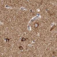


Comprehensive Human Protein Atlas IHC scoring per tissue (reliability: Supported — Medium consistency between antibody staining and RNA expression data.). Rows are taken directly from the HPA tissue chart — click any row's HPA link to view the source.
| Tissue | Cell type | Level | Evidence | Source |
|---|---|---|---|---|
| Appendix | Glandular cells | High | Protein (IHC) | HPA → |
| Bronchus | Respiratory epithelial cells | High | Protein (IHC) | HPA → |
| Cerebellum | Cells in molecular layer | High | Protein (IHC) | HPA → |
| Cerebral cortex | Neuropil | High | Protein (IHC) | HPA → |
| Colon | Glandular cells | High | Protein (IHC) | HPA → |
Troubleshoot COX5A staining in paraffin sections by checking retrieval, tissue processing, mitochondrial localisation, controls, and scoring.
Both antibodies have IHC images from human paraffin sections (catalog IHC captions). Both list Human, Mouse and Rat reactivity; A07895-2 also lists IF/ICC, although no IF image is supplied (catalog).
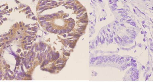
A07895-1 shows IHC staining in human paraffin-embedded colon carcinoma at 1:50 (A07895-1 IHC caption). A07895-2 shows IHC staining in human paraffin-embedded breast cancer, colon tissue and colon cancer; its application list also includes IF/ICC (A07895-2 IHC captions; catalog applications).
Which to pick: For tissue IHC, A07895-2 has a documented EDTA pH 8.0 retrieval and DAB workflow in human paraffin sections; A07895-1 is another IHC option with a human paraffin-section image (respective IHC captions). For IF/ICC, choose A07895-2 because those applications are listed, while A07895-1 lists IHC without IF/ICC; no IF image is supplied for A07895-2 (catalog applications; catalog IF images). Both are described as polyclonal and list Human, Mouse and Rat reactivity, but their IHC images show human samples only; neither caption reports the fixative (A07895-1 IHC caption; A07895-2 dilution record and IHC captions; catalog reactivity).