This website uses cookies to ensure you get the best experience on our website.


- Table of Contents
Plan chromogenic CPLX1 IHC in paraffin sections using cytoplasmic staining in neural and neuroendocrine tissues and synaptic staining in brain as expected readouts (HPA tissue IHC). The guide uses the catalog antibody’s documented IHC workflow and flags the possibility of signal from more than one gene product (datasheet A05254; HPA tissue IHC).

Expected localisation, validated protocols, controls and antibodies — the at-a-glance facts below, then the full design guide.
| Expected localisation | Cytoplasmic in neural and neuroendocrine tissues; synaptic in brain (HPA tissue IHC) | |
| Staining pattern | Neuronal and neuroendocrine cytoplasm; brain synapses (HPA tissue IHC) | |
| Antigen retrieval | EDTA pH 8.0 HIER, heat-mediated (datasheet A05254) | |
| Positive control | Adrenal gland+4 more · see all | |
| Negative control | Adipose tissue+4 more · see all |
| Fixation | Keep fixation conditions consistent across sections. (standard IHC practice; not target-specific) | |
| Caveat | Signal may include products of more than one gene (HPA tissue IHC) | |
| Regulation | Increased in Parkinson substantia nigra (UniProt) | |
| Isoform / epitope | No isoforms; single cytosolic chain, no ectodomain (UniProt) |
The catalog antibody’s IHC-P protocol (datasheet A05254) is followed by two published CPLX1 protocols for chromogenic staining of paraffin sections (PMC6944231; PMC12283636).
| Sample | Paraffin-embedded rat brain tissue; fixative not specified (datasheet A05254) |
| Fixation | Image fixative and duration unreported (datasheet A05254); verify before use. |
| Sectioning | 4–5 µm sections on charged slides (standard) |
| Deparaffinisation | Xylene, graded ethanol series to water (standard) |
| Antigen retrieval | Heat retrieval: EDTA pH 8.0 (datasheet A05254); 20 min, 95–100 °C (standard) |
| Peroxidase block | 3% H2O2, 10 min, room temperature (standard) |
| Blocking | 10% goat serum (datasheet A05254) |
| Primary antibody | Rabbit anti-CPLX1, 1:50 (datasheet A05254) |
| Primary incubation | Overnight at 4 °C (datasheet A05254) |
| Detection | HRP-conjugated secondary, DAB chromogen (datasheet A05254) |
| Counterstain | Hematoxylin, blue, dehydrate and mount (standard) |
| Expected result | CPLX1-positive staining in medullary cells of adrenal gland (HPA tissue IHC: High). HPA tissue profile: Cytoplasmic expression in nervous and neuroendocrine tissues. Synaptic positivity in brain. No signal in the no-primary control. |
CPLX1 is a cytosolic protein enriched at presynaptic release sites in mature neurons; it has no transmembrane segment (UniProt O14810). In paraffin IHC, expect cytoplasmic staining in neuronal and neuroendocrine cells, with synaptic positivity in brain (HPA tissue IHC). HPA rates the tissue pattern Supported, with medium agreement between staining and RNA data, and cautions that the profile includes antibodies targeting proteins from more than one gene (HPA tissue IHC).
| Cytoplasmic staining in cerebral cortical neurons and cerebellar granular-layer cells, with staining in synaptic regions. | This fits HPA's High staining in both cell groups and its reported synaptic positivity in brain (HPA tissue IHC). Presynaptic enrichment provides a localization rationale, but an individual positive cell cannot be identified as inhibitory from staining alone (UniProt O14810). |
| Cytoplasmic staining in adrenal medullary cells or pancreatic endocrine cells. | These are useful comparisons outside brain: HPA reports High staining in adrenal medullary cells and Medium staining in pancreatic endocrine cells (HPA tissue IHC). Judge intensity within the identified cell population; other cells on the section need not match it. |
| Strong, predominantly nuclear staining in cells expected to be positive. | Nuclear dominance conflicts with the reported cytosolic and presynaptic localization (UniProt O14810; HPA tissue IHC). Treat it as a localization warning, then compare the same compartment in a known-positive tissue and a primary-antibody omission control before interpreting it as CPLX1. |
| Prominent staining in an HPA-listed negative cell population, such as adipocytes or bronchial respiratory epithelium. | HPA reports CPLX1 as Not detected in those cell populations (HPA tissue IHC). Unexpected staining warrants checks for background, endogenous detection activity or antibody cross-reactivity; the multi-gene antibody caution makes target attribution particularly uncertain (HPA tissue IHC). |
| Diffuse stain across the section, or no signal in cerebral cortical neurons. | Neither result gives a convincing cell-restricted positive pattern: HPA reports High cortical neuronal staining (HPA tissue IHC). Use an omission control to assess diffuse signal, and check section and detection controls before treating absent cortical staining as biological absence (general IHC practice). |
| Cell and tissue selection | HPA reports High staining in cortical neurons, cerebellar granular-layer cells and adrenal medullary cells; hippocampal neurons and pancreatic endocrine cells are Medium (HPA tissue IHC). Use those distinctions when choosing a comparison section and judging intensity. |
| Compartment and protein topology | CPLX1 is cytosolic, occurs in the perikaryon and presynapse, and lacks a transmembrane segment (UniProt O14810). Score cytoplasmic and synaptic staining separately; a crisp cell-surface rim is not the localization predicted by this record. |
| Strength of target attribution | HPA calls its tissue IHC reliability Supported but notes medium agreement with RNA and antibodies targeting proteins from more than one gene (HPA tissue IHC). The reported pattern supports interpretation, yet an unexpected positive population needs independent validation. |
| IF/ICC: What pattern can be used for comparison? | HPA describes mainly vesicular localization, marked uncertain, from antibodies targeting proteins from multiple genes (HPA subcellular ICC-IF). Treat it as a cautious comparison only; the IHC tissue profile reports cytoplasmic and synaptic staining (HPA tissue IHC). |
| Situation | Likely cause | Next action |
|---|---|---|
| No stain in a cerebral cortex positive-control section | Cortical neurons are reported High, so a blank result may reflect the section, staining workflow or detection system rather than expected tissue distribution (HPA tissue IHC; general IHC practice). | Confirm neuronal morphology and section integrity; check primary incubation, antigen retrieval and chromogen controls using the validated IHC workflow (general IHC practice). Target-specific fixation sensitivity is unreported in the supplied sources. |
| Diffuse chromogen across tissue and empty spaces | A field-wide deposit lacks the cell restriction in HPA's neuronal and neuroendocrine profile; nonspecific detection or excess background is possible (HPA tissue IHC; general IHC practice). | Compare a primary-antibody omission control, then review blocking, wash steps and detection reagent exposure (general IHC practice). Do not score diffuse deposit as CPLX1 solely because a positive cell group is present. |
| Strong signal in adipocytes or bronchial respiratory epithelium | HPA lists these cell populations as Not detected, and flags a multi-gene antibody limitation for its tissue profile (HPA tissue IHC). The stain therefore needs a specificity check. | Compare a known-positive section and omission control in the same run; review whether signal follows cells, edges or deposits (general IHC practice). Report the unexpected population separately rather than folding it into the expected CPLX1 pattern. |
| Nuclear staining dominates while neuronal cytoplasm is faint | That distribution differs from the cytosolic, perikaryal and presynaptic locations recorded for CPLX1 (UniProt O14810). A nuclear pattern alone is insufficient evidence of the expected IHC result. | Recheck compartment boundaries against the counterstain and compare with a cortical neuronal positive control (HPA tissue IHC; general IHC practice). If nuclear dominance persists, flag localization as discordant and seek independent target validation. |
| A pancreatic section appears weaker than an adrenal section | HPA reports Medium staining in pancreatic endocrine cells and High staining in adrenal medullary cells (HPA tissue IHC). A weaker pancreatic signal can therefore fit the observed tissue profile. | Identify the endocrine or medullary cells before comparing intensity, and score each cell population within its own section (HPA tissue IHC; general IHC practice). Avoid calling the pancreatic section failed solely because it is lighter. |
| Cerebellar staining is present, but its cell identity is unclear | HPA's High cerebellar call concerns cells in the granular layer; UniProt says CPLX1 is expressed mainly by inhibitory neurons in cerebellum (HPA tissue IHC; UniProt O14810). These statements do not identify every stained cell. | Use anatomy and counterstain to record the stained layer and compartment (general IHC practice). Describe the observed cells without assigning inhibitory identity from CPLX1 staining alone; investigate a different distribution with appropriate controls. |


Comprehensive Human Protein Atlas IHC scoring per tissue (reliability: Supported — Medium consistency between antibody staining and RNA expression data. Caution, targets protein from more than one gene.). Rows are taken directly from the HPA tissue chart — click any row's HPA link to view the source.
| Tissue | Cell type | Level | Evidence | Source |
|---|---|---|---|---|
| Adrenal gland | Medullary cells | High | Protein (IHC) | HPA → |
| Cerebellum | Cells in granular layer | High | Protein (IHC) | HPA → |
| Cerebral cortex | Neuronal cells | High | Protein (IHC) | HPA → |
| Caudate | Neuronal cells | Medium | Protein (IHC) | HPA → |
| Hippocampus | Neuronal cells | Medium | Protein (IHC) | HPA → |
| Tissue | Cell type | Level | Evidence | Source |
|---|---|---|---|---|
| Adipose tissue | Adipocytes | Not detected | Protein (IHC) | HPA → |
| Appendix | Endocrine cells | Not detected | Protein (IHC) | HPA → |
| Bone marrow | Hematopoietic cells | Not detected | Protein (IHC) | HPA → |
| Breast | Adipocytes | Not detected | Protein (IHC) | HPA → |
| Bronchus | Respiratory epithelial cells | Not detected | Protein (IHC) | HPA → |
Troubleshoot CPLX1 staining in paraffin sections by checking retrieval, expected cellular patterns, controls, and scoring before interpreting chromogenic signal.
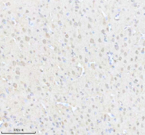
A05254 has real IHC data from paraffin-embedded rat brain (image caption); its catalog lists IF/ICC applications and Human, Mouse, and Rat reactivity (catalog: applications and reactivity).
A05254 is the only SKU shown and has an IHC image from a paraffin-embedded rat brain section (image caption). IF/ICC are listed applications, but no IF image is supplied (catalog: applications and IF image alts).
Which to pick: For tissue IHC, choose A05254: its rat brain image documents heat retrieval in EDTA at pH 8.0 and primary antibody at 1:200 overnight at 4°C (A05254 image caption); the fixative is unreported (A05254 image caption). For IF/ICC or work across Human, Mouse, and Rat, A05254 is the listed option because it is polyclonal and includes those applications and species (catalog: dilution_raw, applications, reactivity); the supplied figure documents rat brain IHC only (A05254 image caption).