CREB3L2 / Cyclic AMP-responsive element-binding protein 3-like protein 2 · IHC design guide

Design Immunohistochemistry for CREB3L2

Plan chromogenic CREB3L2 IHC in paraffin sections with the catalog antibody at 2–5 μg/ml (datasheet A04591-1). Compare cytoplasmic and membranous staining, using the strong signal in bronchial ciliary rootlets as a reference (HPA tissue IHC).

Evidence assembled Oct 2026 · For research use; verify linked source records and product datasheet before use
Immunohistochemistry protocol sheet for CREB3L2 (IHC for CREB3L2): expected localisation Mainly cytoplasmic, with membranous staining (HPA tissue IHC), antibody A04591-1, validated IHC image, and IHC protocol steps
Printable CREB3L2 IHC protocol sheet — expected localisation Mainly cytoplasmic, with membranous staining (HPA tissue IHC), antibody A04591-1, controls and protocol steps. Open the full CREB3L2 IHC guide →

CREB3L2 Immunohistochemistry Experimental Design Guide

Expected localisation, validated protocols, controls and antibodies — the at-a-glance facts below, then the full design guide.

Must know before staining
Expected localisation Mainly cytoplasmic, with membranous staining (HPA tissue IHC)
Staining pattern High ciliary-rootlet staining in bronchial ciliated cells (HPA tissue IHC)
Antigen retrieval EDTA pH 8.0 HIER, heat-mediated (datasheet A04591-1)
Positive control ⓘ Bronchus+4 more · see all
Negative control ⓘ Adipose tissue+4 more · see all
Important caveats
Reasons your staining may differ from the expected pattern.
Fixation Keep fixation consistent across sections. (standard IHC practice; not target-specific)
Caveat ER-stress cleavage can shift staining toward nuclei (UniProt)
Regulation ER stress triggers proteolytic activation (UniProt)
Isoform / epitope 3 isoforms; cleavage separates N- and C-terminal epitopes (UniProt)
Section 1

Recommended CREB3L2 IHC & IF Protocols

The catalog antibody’s IHC-P protocol is accompanied by published CREB3L2 staining methods for cartilage (PMC4952234) and breast cancer tissue (PMC11721608).

Recommended immunohistochemistry (IHC-P) protocol parameters
SampleParaffin-embedded human thyroid cancer tissue; fixative not specified (datasheet A04591-1)
FixationImage fixative and duration unreported (datasheet A04591-1); verify before use.
Sectioning4–5 µm sections on charged slides (standard)
DeparaffinisationXylene, graded ethanol series to water (standard)
Antigen retrievalHeat retrieval: EDTA pH 8.0 (datasheet A04591-1); 20 min, 95–100 °C (standard)
Peroxidase block3% H2O2, 10 min, room temperature (standard)
Blocking10% goat serum (datasheet A04591-1)
Primary antibodyRabbit anti-CREB3L2, 2-5 μg/ml (datasheet A04591-1)
Primary incubationOvernight at 4 °C (datasheet A04591-1)
DetectionHRP-conjugated secondary, DAB chromogen (datasheet A04591-1)
CounterstainHematoxylin, blue, dehydrate and mount (standard)
Expected resultCREB3L2-positive staining in ciliated cells (ciliary rootlets) of bronchus (HPA tissue IHC: High). HPA tissue profile: Mainly cytoplasmic expression with additional membranous expression. No signal in the no-primary control.
💡Decision noteStart with EDTA pH 8.0 heat retrieval (datasheet A04591-1). For matching samples, consider the published cartilage proteinase K treatment (PMC4952234) or breast tissue pH 9.0 retrieval (PMC11721608).
Section 2

What Is the Expected CREB3L2 Staining Pattern?

CREB3L2 is an ER membrane protein with a cytosolic N terminus; after stress related cleavage, its released N terminal fragment can enter the nucleus (UniProt Q70SY1). In tissue IHC, expect mainly cytoplasmic staining with some membranous staining, especially in bronchial and fallopian tube ciliated cells at the ciliary rootlets (HPA tissue IHC: High). HPA rates the tissue pattern Enhanced, with medium consistency between staining and RNA data (HPA tissue IHC).

What am I looking at on my slide?
Strong staining at ciliary rootlets in bronchial or fallopian tube ciliated cells.This matches the clearest reported tissue positives (HPA tissue IHC: High in both cell populations). Assess staining within the named cells; an adjacent unstained cell population does not make the positive result inconsistent.
Predominantly cytoplasmic staining, with some membranous staining; nuclear signal may also appear.The tissue pattern is mainly cytoplasmic with additional membranous expression (HPA tissue IHC). Nuclear localisation is biologically plausible after cleavage (UniProt Q70SY1); nucleoplasm and ER localisation are reported by ICC-IF (HPA subcellular). Nuclear staining alone does not establish ER stress in that section.
A strong, uniform stain sits outside cells or lacks a discernible cellular compartment.That distribution does not match the reported cellular pattern (HPA tissue IHC; HPA subcellular). Treat it as possible artefact and inspect the negative control and tissue morphology before assigning CREB3L2 positivity (general IHC practice).
Unexpected staining dominates a cell population reported as undetected, such as adipocytes.HPA reports CREB3L2 as not detected in adipocytes, specifically, rather than declaring all adipose tissue negative (HPA tissue IHC). Check for cross-reactivity or endogenous detection activity with appropriate controls (general IHC practice); a positive stain alone cannot distinguish those causes.
No signal appears in bronchial or fallopian tube ciliated cells at the ciliary rootlets.These are reported High staining populations (HPA tissue IHC). Review whether the relevant cells are present and intact, then compare a positive control and the antibody's validated IHC conditions (general IHC practice). Absence in one section does not overturn the HPA tissue observation.
💡Expected CREB3L2 appearanceCall a result consistent when ciliary rootlets of bronchial or fallopian tube ciliated cells stain strongly (HPA tissue IHC: High), within a mainly cytoplasmic, sometimes membranous tissue pattern (HPA tissue IHC); broad, compartmentless staining is suspect (general IHC interpretation).
How each factor affects the staining
Subcellular processing and topologyThe membrane segment spans residues 380–400, with the N terminus on the cytosolic side (UniProt Q70SY1 topology). ER localisation and nuclear localisation can therefore reflect different protein states (UniProt Q70SY1 processing); staining alone does not identify which state is present.
Antibody epitopeCleavage releases an N terminal fragment for nuclear transport (UniProt Q70SY1). Epitope position would affect which forms an antibody can recognise, but the supplied antibody records do not locate either epitope; do not infer fragment specificity.
Cell population and tissue choiceBronchial and fallopian tube ciliated cells are High, while kidney tubule cells are Medium and adipocytes are Not detected (HPA tissue IHC). Select and score the specified cell population, since a whole tissue label hides these differences.
Evidence for tissue and cell localisationThe tissue IHC profile is Enhanced despite medium staining–RNA consistency (HPA tissue IHC). Nucleoplasm is supported and ER is approved in ICC-IF (HPA subcellular). These are evidence summaries for different assays, so interpret a paraffin section against its tissue IHC pattern.
Isoforms and modificationUniProt lists 3 isoforms and glycosylation sites at 480, 504 and 517 (UniProt Q70SY1). Their effect on staining by the catalog antibody is unreported in the supplied evidence; avoid assigning a staining difference to them.
Why is my staining missing, weak or wrong?
SituationLikely causeNext action
A known High staining ciliated cell population is blank.The target cells may be absent or poorly preserved, or the staining run may have failed (general IHC practice); the expected population is High (HPA tissue IHC).Confirm the ciliated cells and their rootlets on the section, then check a positive control and the catalog antibody's validated IHC procedure (general IHC practice).
Every compartment and nearby tissue stain diffusely.Background from detection or insufficient blocking is possible (general IHC practice); diffuse staining is unlike the mainly cytoplasmic tissue profile (HPA tissue IHC).Inspect the no-primary control, blocking and wash steps, and whether the signal follows cell boundaries (general IHC practice).
A purported negative control tissue contains positive cells.A tissue-level negative assignment may overlook cell-specific HPA results: colon endothelial cells, for example, are Not detected, rather than every colon cell (HPA tissue IHC).Score the exact HPA-listed cell population and compare its morphology with the stain; investigate unexpected signal with assay controls (HPA tissue IHC; general IHC practice).
Nuclear staining appears alongside cytoplasmic staining.Nuclear transport of the processed N terminal domain is described (UniProt Q70SY1), and nucleoplasmic staining is supported in ICC-IF (HPA subcellular).Record nuclear and cytoplasmic staining separately; do not infer ER stress or cleavage solely from the IHC pattern (UniProt Q70SY1 processing; general IHC interpretation).
A different cell population stains more strongly than the expected ciliated cells.Cell-specific expression differs across tissues (HPA tissue IHC); cross-reactivity or endogenous detection activity may also produce an unexpected signal (general IHC practice).Verify cell identity, compare the positive and no-primary controls, and score the expected and unexpected populations separately (general IHC practice).
Q: IF/ICC shows ER and nuclear signal; should I apply an IF protocol to this IHC result?A: ER and nucleoplasm are reported ICC-IF locations (HPA subcellular), while the paraffin tissue IHC profile is mainly cytoplasmic with additional membranous expression (HPA tissue IHC).Interpret this chromogenic section using its IHC controls and cell-specific tissue pattern (HPA tissue IHC; general IHC practice); use the separate IF/ICC guide for that assay.

Sample controls for CREB3L2 IHC & IF

🧪Run bronchus first and score staining in ciliated cells at the ciliary rootlets (HPA: High in bronchial ciliated cells). Use adipose tissue adipocytes as the tissue negative (HPA: Not detected in adipocytes); on the bronchus slide, treat unstained neighboring cells as background references without assuming every non-ciliated cell is CREB3L2-negative.
Positive control tissue: Bronchus (Ciliated cells (ciliary rootlets), HPA High)
Negative control tissue: Adipose tissue (HPA Not detected)
ICC-IF cell lines (HPA subcellular resource): HPA ICC-IF images show CREB3L2 in BJ [Human fibroblast], Hep-G2, U2OS, with annotated localisation: Nucleoplasm (supported), Endoplasmic reticulum (approved) (HPA subcellular).
Technical controls: Include a no-primary (secondary-only) slide, a concentration-matched rabbit IgG isotype control, and CREB3L2-knockout material as a biological negative (caption: rabbit primary antibody; standard IHC practice). Quench endogenous peroxidase for DAB detection and inspect bronchial luminal material for trapped chromogen before scoring ciliary-rootlet staining (caption: HRP/DAB detection; standard IHC practice).
⚠️Feasibility: A target-specific fixation window or fixation effect is unreported, and the selected A04591-1 tissue-IHC caption does not report the fixative (selected-SKU caption: fixative not stated). The paraffin-section example used heat-mediated EDTA retrieval at pH 8.0, but it does not establish that retrieval is required under other conditions (selected-SKU caption: EDTA pH 8.0). The supplied evidence does not establish whether frozen sections or IF are easier; IF can assess the reported nucleoplasmic and ER locations, while bronchial ciliary-rootlet staining requires careful separation from luminal staining artefact (HPA: nucleoplasm and ER; HPA: High in bronchial ciliary rootlets; standard IHC practice).

HPA tissue IHC evidence for CREB3L2

Comprehensive Human Protein Atlas IHC scoring per tissue (reliability: Enhanced — Medium consistency between antibody staining and RNA expression data.). Rows are taken directly from the HPA tissue chart — click any row's HPA link to view the source.

Positive expression · recommended positive controls

TissueCell typeLevelEvidenceSource
Bronchus Ciliated cells (ciliary rootlets) High Protein (IHC) HPA →
Fallopian tube Ciliated cells (ciliary rootlets) High Protein (IHC) HPA →
Adrenal gland Glandular cells Medium Protein (IHC) HPA →
Bone marrow Hematopoietic cells Medium Protein (IHC) HPA →
Cerebral cortex Neuronal cells Medium Protein (IHC) HPA →

Undetected expression · recommended negative controls

TissueCell typeLevelEvidenceSource
Adipose tissue Adipocytes Not detected Protein (IHC) HPA →
Appendix Glandular cells Not detected Protein (IHC) HPA →
Colon Endothelial cells Not detected Protein (IHC) HPA →
Duodenum Glandular cells Not detected Protein (IHC) HPA →
Endometrium Cells in endometrial stroma Not detected Protein (IHC) HPA →
Section 3

Advanced CREB3L2 IHC Tips

Use the catalog antibody’s paraffin section result as the starting point, then assess retrieval, compartment, cell type and controls when CREB3L2 staining differs.

Which retrieval conditions should I use when CREB3L2 staining is weak?
Start with heat mediated retrieval in EDTA at pH 8.0 for paraffin sections (datasheet A04591-1). The selected image used that retrieval before overnight incubation at 4°C with 2 μg/ml catalog antibody, so keep those conditions together for the first comparison (caption A04591-1). If staining remains weak, compare a second retrieval condition on adjacent sections while holding antibody concentration, detection and development constant (standard IHC practice). Include a known staining compartment in each run, because CREB3L2 can appear in the endoplasmic reticulum and nucleus, while tissue IHC is mainly cytoplasmic with additional membranous staining (UniProt Q70SY1; HPA tissue IHC).
Could fixation explain inconsistent CREB3L2 staining across paraffin sections?
The selected paraffin section caption does not state a fixative, so target specific fixation sensitivity is unknown for this antibody (caption A04591-1). Record each specimen’s fixative, fixation duration and processing history before comparing staining intensity; variation in those steps can change antigen accessibility in IHC (standard IHC practice). Use adjacent sections and hold EDTA pH 8.0 retrieval, 2 μg/ml primary antibody and DAB development consistent when investigating the discrepancy (datasheet A04591-1; standard IHC practice). A weak section should be interpreted alongside morphology and a run control, since staining differences alone cannot establish a CREB3L2 specific fixation effect (standard IHC practice).
How should I assess cytoplasmic versus nuclear CREB3L2 staining?
Score nuclear, cytoplasmic and membranous staining separately rather than treating every stained cell as equivalent (standard IHC practice). CREB3L2 is an ER membrane protein whose released N terminal domain can enter the nucleus after stress dependent cleavage (UniProt Q70SY1). HPA tissue IHC describes mainly cytoplasmic staining with additional membranous staining, while its subcellular data support nucleoplasmic and ER localisation (HPA tissue IHC; HPA subcellular). On the same EDTA pH 8.0 retrieved run, compare staining with intact cellular morphology and record the fraction of nuclei stained within each cell population (datasheet A04591-1; standard IHC practice).
Can the antibody’s epitope change how I interpret nuclear staining?
Yes: the epitope must be mapped before assigning a nuclear signal to a particular CREB3L2 form (standard IHC interpretation). CREB3L2 has 3 reported isoforms, a cytoplasmic region spanning residues 1–379, a transmembrane segment at 380–400 and a lumenal region at 401–520 (UniProt Q70SY1). Stress dependent cleavage releases an N terminal domain that can enter the nucleus, so an antibody recognizing only a C terminal epitope would answer a different localisation question (UniProt Q70SY1; standard epitope interpretation). If the catalog antibody’s epitope is unspecified, report compartment patterns without claiming which isoform or cleavage product produced the staining (standard IHC practice).
How can IF help check the cell type and compartment seen by IHC?
Use IF as a separate localisation check and multiplex CREB3L2 with a marker for the expected cell population; ciliated cells in bronchus and fallopian tube show high tissue IHC staining (HPA tissue IHC; standard IF practice). Choose fluorophores after checking tissue autofluorescence in an unstained section, and use spectral separation and single stain controls for the multiplex image (standard IF practice). Because residues 1–379 face the cytoplasm whereas 401–520 are lumenal, select and compare permeabilisation conditions only after establishing which side contains the antibody epitope (UniProt Q70SY1; standard IF practice). Compare nuclear and ER associated signals with the matched chromogenic section without treating IF intensity as a direct DAB score (HPA subcellular; standard IF practice).
What should I change when DAB background obscures CREB3L2?
First inspect a section processed without primary antibody and a matched stained section to distinguish detection background from candidate CREB3L2 staining (standard IHC practice). The selected paraffin section used 10% goat serum blocking, 2 μg/ml rabbit primary overnight at 4°C, a peroxidase conjugated secondary and DAB development (caption A04591-1). Check peroxidase blocking, reagent washes and DAB development time as general chromogenic IHC controls, changing one step at a time (standard IHC practice). Retain EDTA pH 8.0 retrieval during that comparison so a background reduction is not confused with loss of antigen exposure (datasheet A04591-1; standard IHC practice).
How should I quantify CREB3L2 across sections with mixed cell populations? ⚠ ANSWER MARKED FOR VERIFICATION
Define the cell population and compartment before scoring, then record the percentage of positive cells and staining intensity from 0–3 in each compartment (standard IHC scoring practice). An H score combines intensity with the percentage of cells at each intensity and spans 0–300; report it alongside the number of evaluable cells or tissue area (standard IHC scoring practice). Normalise comparisons to the same eligible cell type, tissue area and staining run, because tissue IHC shows cell dependent patterns including high ciliated cell staining (HPA tissue IHC; standard IHC practice). Keep retrieval at EDTA pH 8.0 and development conditions consistent, and report nuclear and cytoplasmic scores separately (datasheet A04591-1; standard IHC practice).
Which CREB3L2 staining patterns warrant an artefact check?
Treat staining concentrated at section edges, necrotic regions or sites positive in the no primary control as suspect before assigning it to CREB3L2 (standard IHC practice). Check endogenous peroxidase when DAB signal persists without primary antibody, and compare suspect areas with intact cells on the same slide (standard IHC practice). ER associated cytoplasmic staining and some nuclear signal are biologically plausible, while HPA tissue IHC reports mainly cytoplasmic with additional membranous staining (UniProt Q70SY1; HPA tissue IHC). For a disputed positive, repeat the EDTA pH 8.0 run and verify that signal follows the expected cell population and compartment rather than tissue damage or detection background (datasheet A04591-1; standard IHC practice).
Boster reagents

Best CREB3L2 / Cyclic AMP-responsive element-binding protein 3-like protein 2 IHC Antibodies

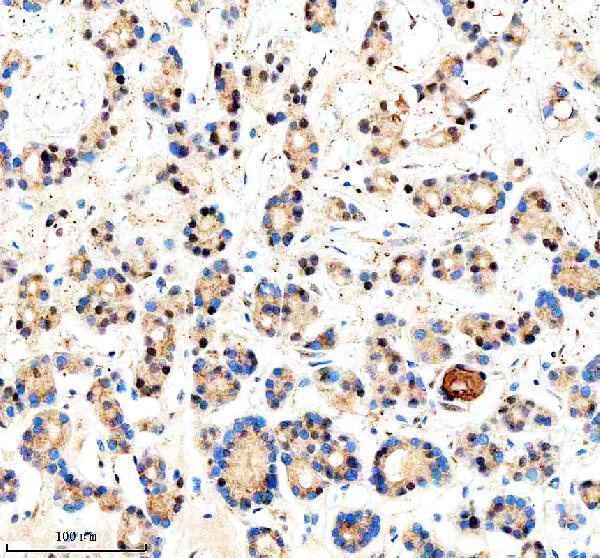
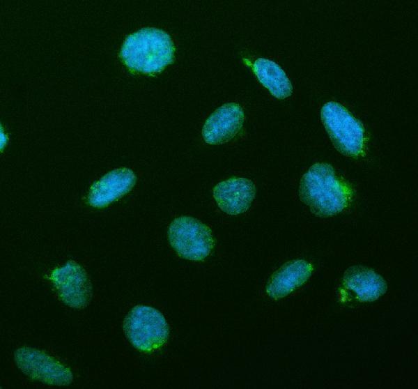
A04591-1 has IHC data from paraffin-embedded human thyroid cancer tissue and IF/ICC data from U2OS cells (catalog IHC and IF captions); reported reactivity is human (catalog: reactivity).

Real IHC data IHC analysis of CREB3L2 using anti-CREB3L2 antibody (A04591-1). CREB3L2 was detected in a paraffin-embedded section of human thyroid cancer tissue. Heat mediated antigen retrieval was performed in EDTA buffer (pH 8.0, epitope retrieval solution). The tissue section was blocked with 10% goat serum. The tissue section was then incubated with 2 μg/ml rabbit anti-CREB3L2 Antibody (A04591-1) overnight at 4°C. Peroxidase Conjugated Goat Anti-rabbit IgG was used as secondary antibody and incubated for 30 minutes at 37°C. The tissue section was developed using HRP Conjugated Rabbit IgG Super Vision Assay Kit (Catalog # SV0002) with DAB as the chromogen.
Anti-CREB3L2 Antibody ®
Cat # A04591-1

A04591-1 is listed for IHC and shown on a paraffin-embedded human thyroid cancer section (catalog: applications; IHC caption). A04591-1 is also listed for IF/ICC and shown in U2OS cells (catalog: applications; IF caption).

Which to pick: Choose A04591-1 for paraffin-section IHC; its IHC caption documents that preparation, but does not report the fixative (catalog: IHC caption). Choose the same SKU for IF/ICC in U2OS cells (catalog: IF caption). No cross-species option is documented: A04591-1 lists human reactivity, and its clonality is unreported (catalog: reactivity; clone field).

Each figure is that product's own IHC / IF validation image from its datasheet.

References

  1. UniProt Consortium. UniProt entry Q70SY1 (CR3L2_HUMAN, Cyclic AMP-responsive element-binding protein 3-like protein 2).
  2. Human Protein Atlas. CREB3L2 tissue IHC expression (reliability: Enhanced).
  3. Human Protein Atlas. CREB3L2 subcellular location (ICC-IF): Localized to the nucleoplasm and endoplasmic reticulum..
  4. Human Protein Atlas. CREB3L2 antibody validation summary (2 antibodies).
  5. The unfolded protein response genes in human osteoarthritic chondrocytes: PERK emerges as a potential therapeutic target. Arthritis research & therapy 2016 — PMC4952234.
  6. Low-Grade Fibromyxoid Sarcoma and Related Subtypes: A Systematic Review and Pooled Analysis of 773 Cases. Cancers 2026 — PMC12896792.
  7. Clinical and molecular characterization of primary sclerosing epithelioid fibrosarcoma of bone and review of the literature. Genes, chromosomes & cancer 2020 — PMC7082133.
  8. Noncanonical UPR factor CREB3L2 drives immune evasion of triple-negative breast cancer through Hedgehog pathway modulation in T cells. Science advances 2025 — PMC11721608.
  9. PubMed PMID:12915480 — UniProt-cited evidence.
  10. PubMed PMID:14702039 — UniProt-cited evidence.
  11. PubMed PMID:12690205 — UniProt-cited evidence.