This website uses cookies to ensure you get the best experience on our website.


- Table of Contents
Plan CRTC3 staining in paraffin sections using high-staining adrenal glandular cells and undetected adipocytes as controls (HPA tissue IHC). Score nuclear and cytoplasmic staining separately, since nuclear expression predominates across tissues and activation can promote nuclear translocation (HPA tissue IHC; UniProt).

Expected localisation, validated protocols, controls and antibodies — the at-a-glance facts below, then the full design guide.
| Expected localisation | Mainly nuclear; cytoplasmic staining also occurs (HPA tissue IHC) | |
| Staining pattern | Nuclear in most tissues; cytoplasmic in several (HPA tissue IHC) | |
| Antigen retrieval | EDTA pH 8.0 HIER, heat-mediated (datasheet A04568-2) | |
| Positive control | Adrenal gland+4 more · see all | |
| Negative control | Adipose tissue+4 more · see all |
| Fixation | Matched tissue-IHC evidence does not establish the fixation claim. Validate the specimen-specific method before use. (selected-SKU IHC image A04568-2) | |
| Caveat | Antibody staining and RNA expression show low consistency (HPA tissue IHC) | |
| Regulation | Activation promotes nuclear translocation (UniProt) | |
| Isoform / epitope | 2 isoforms; confirm antibody epitope coverage (UniProt) |
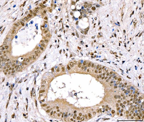
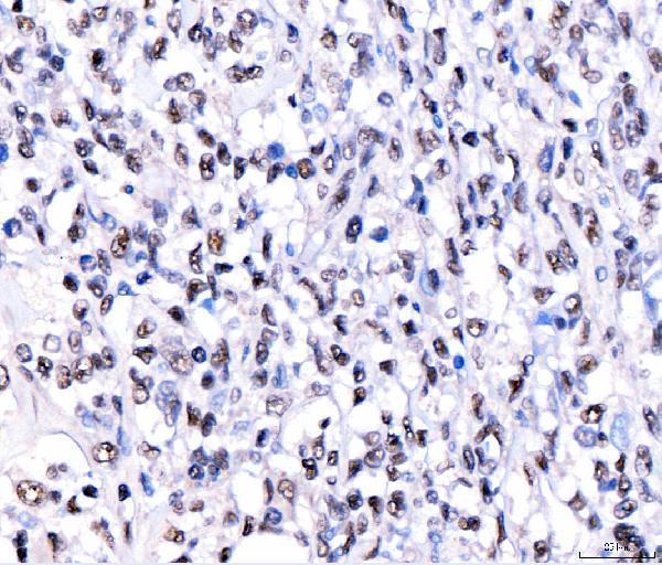
The catalog antibody protocol uses EDTA pH 8.0 heat retrieval (datasheet: A04568-2). The published IHC reports below provide additional tissue and staining details (PMC10477178; PMC8581419).
| Sample | Paraffin-embedded human gallbladder carcinoma tissue; fixative not specified (datasheet A04568-2) |
| Fixation | Image fixative and duration unreported (datasheet A04568-2); verify before use. |
| Sectioning | 4–5 µm sections on charged slides (standard) |
| Deparaffinisation | Xylene, graded ethanol series to water (standard) |
| Antigen retrieval | Heat retrieval: EDTA pH 8.0 (datasheet A04568-2); 20 min, 95–100 °C (standard) |
| Peroxidase block | 3% H2O2, 10 min, room temperature (standard) |
| Blocking | 10% goat serum (datasheet A04568-2) |
| Primary antibody | Rabbit anti-CRTC3, 2-5 μg/ml (datasheet A04568-2) |
| Primary incubation | Overnight at 4 °C (datasheet A04568-2) |
| Detection | Streptavidin-biotin complex (SABC), DAB chromogen (datasheet A04568-2) |
| Counterstain | Hematoxylin, blue, dehydrate and mount (standard) |
| Expected result | CRTC3-positive staining in glandular cells of adrenal gland (HPA tissue IHC: High). HPA tissue profile: Nuclear expression in most tissues. Additional cytoplasmic expression in several tissues. No signal in the no-primary control. |
CRTC3 should stain mainly in nuclei, with cytoplasmic staining possible (HPA: nuclear expression in most tissues; UniProt Q6UUV7: mainly nuclear, also cytoplasmic). Look for signal in the specific cells HPA rates High, including adrenal glandular cells, ovarian follicle cells and skin fibroblasts (HPA: High). CRTC3 has no transmembrane segment (UniProt Q6UUV7 topology). HPA rates its tissue IHC profile Approved but reports low consistency with RNA expression (HPA: reliability description).
| Distinct nuclear staining in adrenal glandular cells or ovarian follicle cells, with some cytoplasmic signal. | This fits the reported compartment and cell pattern (HPA: nuclear expression in most tissues, additional cytoplasmic expression in several; High in these cells). Score the named cell population and nuclear signal separately; a High HPA rating does not mean every cell on the section must stain. |
| Strong membrane rim or extracellular staining dominates, while nuclei are unstained. | This conflicts with the reported nuclear and cytoplasmic locations and the absence of a transmembrane segment (HPA: tissue IHC profile; UniProt Q6UUV7 topology). Treat the pattern as suspect and compare a control section before assigning it to CRTC3 (general IHC practice). |
| Signal appears mainly in an HPA Not detected cell population, such as adipocytes. | HPA reports CRTC3 as Not detected in adipocytes, so this pattern warrants a specificity check (HPA: adipose tissue IHC). Cross-reactivity or endogenous detection activity is possible (general IHC practice); the HPA rating applies to adipocytes, not automatically to every cell in adipose tissue. |
| Uniform chromogen haze covers cells and surrounding tissue without clear nuclear boundaries. | The distribution is hard to reconcile with the reported nuclear emphasis (HPA: tissue IHC profile). Nonspecific primary or detection reagent binding, or endogenous detection activity, can produce background (general IHC practice); do not score the haze as positive nuclei. |
| No discernible signal appears in a section containing HPA High cells. | Check that the relevant cells are present: HPA rates salivary glandular cells, skin fibroblasts and smooth muscle cells High (HPA: tissue IHC). A blank result could reflect assay failure or a specimen difference (general IHC practice). HPA's Approved profile has low consistency with RNA expression, so one blank section is not conclusive (HPA: reliability description). |
| Compartment and signaling state | CRTC3 is mainly nuclear and can also occupy the cytoplasm (UniProt Q6UUV7: subcellular location); UniProt reports nuclear translocation after adenylyl cyclase or MAP kinase activation (UniProt Q6UUV7). Interpret a nuclear-to-cytoplasmic difference in context rather than requiring one fixed ratio in every specimen. |
| Choice of positive and low-signal cells | Use the named cell population when judging a control: adrenal glandular cells and ovarian follicle cells are High, whereas adipocytes and duodenal glandular cells are Not detected (HPA: tissue IHC). A tissue name alone does not specify which cells should stain. |
| Strength of IHC validation | The tissue profile is Approved and has low consistency between antibody staining and RNA expression (HPA: reliability description). HPA063691 is IHC Approved; its Enhanced rating applies to ICC, while HPA043735 has no listed IHC rating (HPA: antibody validation). |
| Isoforms and antibody coverage | UniProt lists isoforms 1 and 3 (UniProt Q6UUV7: isoforms). The supplied record does not locate an antibody epitope or establish coverage of either isoform, so an isoform-specific explanation for weak staining remains unverified. |
| Topology and processing | CRTC3 has no transmembrane segment or signal peptide, and its annotated chain spans residues 1–619 (UniProt Q6UUV7: topology and processing). These annotations support an intracellular reading of the stain; they do not establish fixation sensitivity or an antigen retrieval setting. |
| Situation | Likely cause | Next action |
|---|---|---|
| An HPA High control shows no nuclear stain. | The expected cell population may be absent from the field, or the IHC run may have failed (HPA: cell-specific High ratings; general IHC practice). | Confirm the named cells on a counterstained section, then review the IHC-validated antibody's documented retrieval, dilution and detection conditions (general IHC practice). Interpret a persistent blank result alongside HPA's low staining–RNA consistency (HPA: reliability description). |
| Membrane-like or extracellular chromogen is stronger than nuclear signal. | That distribution conflicts with HPA's nuclear emphasis and UniProt's lack of a transmembrane segment (HPA: tissue IHC profile; UniProt Q6UUV7 topology). | Compare a named HPA High cell population and a primary-antibody omission control; review blocking and detection background before scoring (general IHC practice). |
| Adipocytes or another HPA Not detected cell population appears strongly positive. | Off-target binding or endogenous detection activity may account for unexpected staining (HPA: Not detected in adipocytes; general IHC practice). | Check a reagent control and the relevant endogenous-activity block for the detection chemistry; judge the result by the identified cell type, not the tissue label (general IHC practice). |
| The whole section has diffuse brown background. | Nonspecific binding or endogenous activity can obscure nuclear boundaries in chromogenic IHC (general IHC practice). | Review blocking, primary-antibody dilution and detection controls, then score only distinguishable cellular signal (general IHC practice; HPA: predominantly nuclear tissue profile). |
| Cytoplasmic signal is visible alongside nuclear stain. | Additional cytoplasmic tissue staining is reported, and CRTC3 can occur in the cytoplasm (HPA: tissue IHC profile; UniProt Q6UUV7: subcellular location). | Record nuclear and cytoplasmic compartments separately and compare the named positive cells; do not discard an otherwise convincing result solely for cytoplasmic signal (HPA: tissue IHC profile). |
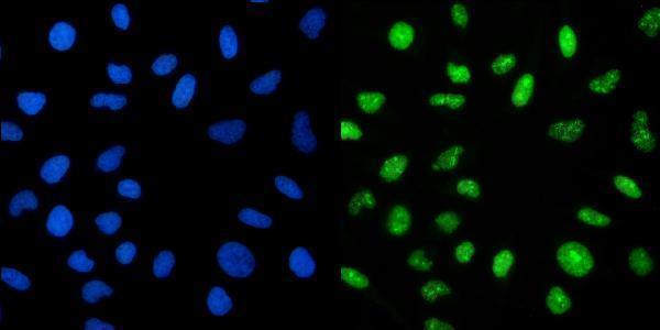
| IF/ICC Q: What distribution should a fluorescent image show? | A: Mainly nucleoplasmic signal with additional cytosolic signal (HPA: enhanced ICC-IF subcellular localization). | Compare nuclear and cytosolic channels with the HPA subcellular pattern; keep IF/ICC protocol choices on its own guide page (HPA: ICC-IF subcellular record). |


Comprehensive Human Protein Atlas IHC scoring per tissue (reliability: Approved — Low consistency between antibody staining and RNA expression data.). Rows are taken directly from the HPA tissue chart — click any row's HPA link to view the source.
| Tissue | Cell type | Level | Evidence | Source |
|---|---|---|---|---|
| Adrenal gland | Glandular cells | High | Protein (IHC) | HPA → |
| Appendix | Lymphoid tissue | High | Protein (IHC) | HPA → |
| Endometrium | Cells in endometrial stroma | High | Protein (IHC) | HPA → |
| Ovary | Follicle cells | High | Protein (IHC) | HPA → |
| Salivary gland | Glandular cells | High | Protein (IHC) | HPA → |
| Tissue | Cell type | Level | Evidence | Source |
|---|---|---|---|---|
| Adipose tissue | Adipocytes | Not detected | Protein (IHC) | HPA → |
| Cerebellum | Cells in granular layer | Not detected | Protein (IHC) | HPA → |
| Cervix | Glandular cells | Not detected | Protein (IHC) | HPA → |
| Colon | Endothelial cells | Not detected | Protein (IHC) | HPA → |
| Duodenum | Glandular cells | Not detected | Protein (IHC) | HPA → |
Use compartment-aware controls and matched processing to troubleshoot CRTC3 staining in paraffin sections; assess IF on its separate guide page.
A04568-2 has human paraffin-section IHC images and a HeLa IF image (A04568-2 image captions). M04568 lists IHC and ICC/IF applications with human, mouse, and rat reactivity (M04568 catalog).
A04568-2 has IHC images from human gallbladder carcinoma, lymphoma, breast cancer, and colonic adenocarcinoma paraffin sections, plus a HeLa IF image (A04568-2 image captions). M04568 lists IHC and ICC/IF applications and human, mouse, and rat reactivity; no image is supplied (M04568 catalog).
Which to pick: Choose A04568-2 for human tissue IHC because its own paraffin-section captions document EDTA retrieval and chromogenic detection (A04568-2 IHC captions). For IF/ICC, A04568-2 has a HeLa IF image; choose M04568 when mouse or rat reactivity is needed, based on its listed reactivity and applications (A04568-2 IF caption; M04568 catalog). The A04568-2 IHC captions do not report a fixative, so fixation is unconfirmed (A04568-2 IHC captions). The selected A04568-2 tissue-IHC caption documents paraffin sections, but does not specify the fixative (selected-SKU IHC image A04568-2).