CSNK2A1 / Casein kinase II subunit alpha · IHC design guide

Design Immunohistochemistry for CSNK2A1

Plan chromogenic CSNK2A1 IHC in paraffin sections using the reported nuclear and cytoplasmic tissue pattern (HPA tissue IHC). Use colon glandular cells as a positive reference and adipocytes as a negative reference (HPA tissue IHC); start antibody titration at 1:50 (datasheet A02398-2).

Evidence assembled Oct 2026 · For research use; verify linked source records and product datasheet before use
Immunohistochemistry protocol sheet for CSNK2A1 (IHC for CSNK2A1): expected localisation Nuclear and cytoplasmic tissue staining (HPA tissue IHC), antibody A02398-2, validated IHC image, and IHC protocol steps
Printable CSNK2A1 IHC protocol sheet — expected localisation Nuclear and cytoplasmic tissue staining (HPA tissue IHC), antibody A02398-2, controls and protocol steps. Open the full CSNK2A1 IHC guide →

CSNK2A1 Immunohistochemistry Experimental Design Guide

Expected localisation, validated protocols, controls and antibodies — the at-a-glance facts below, then the full design guide.

Must know before staining
Expected localisation Nuclear and cytoplasmic tissue staining (HPA tissue IHC)
Staining pattern Nuclear and cytoplasmic staining in most tissues (HPA tissue IHC)
Antigen retrieval EDTA pH 8.0 HIER, heat-mediated (datasheet A02398-2)
Positive control ⓘ Adrenal gland+4 more · see all
Negative control ⓘ Adipose tissue
Important caveats
Reasons your staining may differ from the expected pattern.
Fixation Keep fixation consistent across sections (standard IHC practice; not target-specific)
Caveat Antibody staining may reflect more than one gene (HPA tissue IHC)
Regulation Rises with gastric carcinoma progression (UniProt)
Isoform / epitope Two isoforms; check whether the epitope is shared (UniProt)
Section 1

Recommended CSNK2A1 IHC & IF Protocols

The catalog antibody’s IHC-P protocol (datasheet A02398-2) is accompanied by 4 published CSNK2A1 IHC protocols (PMC8303481; PMC6945564; PMC8487869; PMC7395738).

Recommended immunohistochemistry (IHC-P) protocol parameters
SampleParaffin-embedded human stomach cancer tissue; fixative not specified (datasheet A02398-2)
FixationImage fixative and duration unreported (datasheet A02398-2); verify before use.
Sectioning4–5 µm sections on charged slides (standard)
DeparaffinisationXylene, graded ethanol series to water (standard)
Antigen retrievalHeat retrieval: EDTA pH 8.0 (datasheet A02398-2); 20 min, 95–100 °C (standard)
Peroxidase block3% H2O2, 10 min, room temperature (standard)
Blocking10% goat serum (datasheet A02398-2)
Primary antibodyRabbit anti-CSNK2A1, 1:50 recommended; image 2 μg/ml (datasheet A02398-2)
Primary incubationOvernight at 4 °C (datasheet A02398-2)
DetectionHRP-conjugated secondary, DAB chromogen (datasheet A02398-2)
CounterstainHematoxylin, blue, dehydrate and mount (standard)
Expected resultCSNK2A1-positive staining in glandular cells of adrenal gland (HPA tissue IHC: High). HPA tissue profile: Nuclear and cytoplasmic expression in most tissues. No signal in the no-primary control.
💡Decision noteStart with EDTA pH 8.0 heat retrieval for the catalog antibody (datasheet A02398-2). The published retrieval conditions belong to their respective antibodies (PMC8303481; PMC6945564; PMC7395738).
Section 2

What Is the Expected CSNK2A1 Staining Pattern?

CSNK2A1 is annotated in the nucleus (UniProt P68400) and shows nuclear and cytoplasmic staining across most tissues by IHC (HPA tissue IHC: Supported). Expect signal in glandular cells of colon, breast, cervix, duodenum, and adrenal gland, among other listed cell types (HPA tissue IHC: High). Interpret tissue staining cautiously: HPA reports medium agreement with RNA data and cautions that the staining may detect protein from more than one gene (HPA tissue IHC: reliability description).

What am I looking at on my slide?
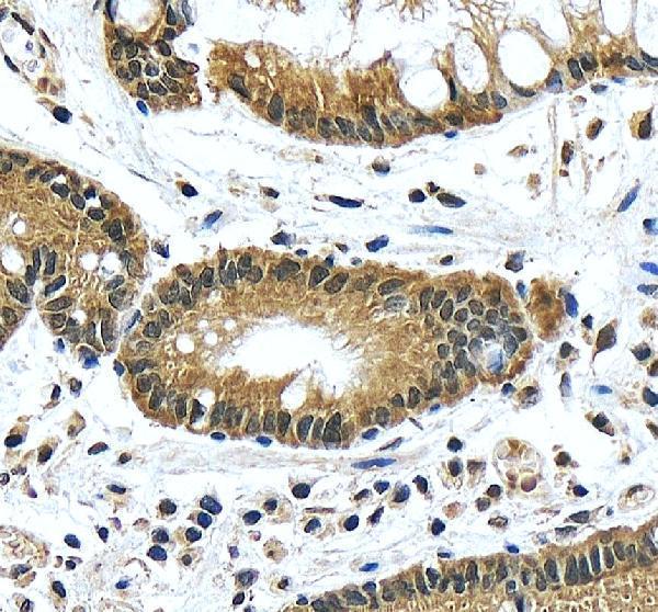
Nuclear and cytoplasmic stain in colon glandular cells.This matches the broad tissue pattern and a listed High cell population (HPA tissue IHC: nuclear and cytoplasmic; colon glandular cells High). Judge the cellular pattern alongside a negative control, because HPA cautions that staining may detect protein from more than one gene (HPA tissue IHC: reliability description).
Staining appears only at cell borders or as a sharply membranous rim.That compartment is inconsistent with the reported nuclear and cytoplasmic IHC pattern (HPA tissue IHC: profile) and with the absence of a transmembrane segment (UniProt P68400 topology). Treat it as a possible artefact; review morphology and controls before assigning it to CSNK2A1 (general IHC practice).
Adipocytes stain strongly while nearby expected positive cells are weak.Adipocytes are listed as Not detected (HPA tissue IHC: adipose tissue). Strong signal there raises concern for cross-reactivity or endogenous detection activity; neither cause can be established from the image alone. Compare a matched negative control and a listed High cell population (general IHC practice; HPA tissue IHC: positive cells).
A uniform haze obscures nuclei, cytoplasm, and tissue boundaries.Diffuse background prevents a reliable compartment call (general IHC practice). Check whether the haze persists without primary antibody, then assess blocking, washing, and detection conditions (general IHC practice). Do not score diffuse staining as evidence of the nuclear and cytoplasmic pattern reported by HPA (HPA tissue IHC: profile).
No signal appears in colon glandular cells.Colon glandular cells are listed as High (HPA tissue IHC: colon). A blank result therefore warrants a workflow check, but one field cannot establish target absence (general IHC practice). Examine section integrity, primary antibody use, and detection controls before interpreting the specimen (general IHC practice).
💡Expected CSNK2A1 appearanceA convincing positive shows discernible nuclear and cytoplasmic stain in a listed High population, such as colon glandular cells (HPA tissue IHC: profile; colon High); a uniform haze or adipocyte dominated signal is suspect (general IHC practice; HPA tissue IHC: adipocytes Not detected).
How each factor affects the staining
Tissue and cell selectionHPA lists High staining in several glandular and other cell populations, Low staining in stomach glandular cells, and Not detected in adipocytes (HPA tissue IHC: tissue entries). Select comparison fields by cell type; a low staining field is a less useful positive benchmark than a listed High field (general IHC practice).
Compartment expectationNuclear localisation is annotated by UniProt, while HPA describes nuclear and cytoplasmic tissue staining (UniProt P68400: subcellular location; HPA tissue IHC: profile). Score the observed compartment without requiring exclusively nuclear IHC staining; the supplied sources do not establish a required nuclear to cytoplasmic intensity ratio.
Antibody validationThe IHC entries for HPA061698, CAB020680, and CAB069395 are Supported; HPA059206 has no IHC status in this payload (HPA antibodies: IHC status). A Supported entry aids interpretation but does not remove HPA's caution that staining may detect protein from more than one gene (HPA tissue IHC: reliability description).
Isoforms and epitope coverageUniProt lists two isoforms and a 1–391 chain without a signal peptide or propeptide (UniProt P68400: isoforms; processing). Antibody epitopes and isoform coverage are not supplied, so the record cannot predict whether both isoforms contribute equally to IHC staining. Avoid inferring specificity from the processing annotation alone.
IF/ICC question: what localisation should I compare?HPA reports mainly nucleoplasmic IF/ICC localisation; primary cilium localisation is additional but uncertain (HPA subcellular: supported nucleoplasm; uncertain primary cilium). Use that as an IF/ICC interpretation reference, while judging paraffin section IHC against HPA's nuclear and cytoplasmic tissue pattern (HPA tissue IHC: profile).
Why is my staining missing, weak or wrong?
SituationLikely causeNext action
Colon glandular cells remain unstained.A listed High cell population should provide an interpretable comparison (HPA tissue IHC: colon High); a blank field alone does not identify the failed step (general IHC practice).Check section morphology, primary antibody application, detection reagents, and counterstain; compare another listed High population if available (general IHC practice; HPA tissue IHC: positive entries).
The entire section has a brown haze.Nonspecific binding or detection background can obscure cellular detail (general IHC practice); the haze cannot be equated with HPA's nuclear and cytoplasmic pattern (HPA tissue IHC: profile).Review the no primary control, blocking, washing, and detection conditions, then rescore only clearly resolved cells (general IHC practice).
Adipocytes show prominent stain.This conflicts with their Not detected designation (HPA tissue IHC: adipocytes) and may reflect cross-reactivity or endogenous detection activity (general IHC practice).Compare matched controls and nearby cell types; assess endogenous detection activity using the controls appropriate to the chromogenic system (general IHC practice).
Only a membranous outline is visible.A membrane only pattern does not match reported tissue staining (HPA tissue IHC: nuclear and cytoplasmic profile); CSNK2A1 lacks a transmembrane segment (UniProt P68400 topology).Confirm the outline follows intact cells rather than edges or deposits, and compare control sections before scoring it (general IHC practice).
Stomach glandular cells look faint.Low staining is reported in that population (HPA tissue IHC: stomach glandular cells Low), so faint signal there does not by itself establish a failed assay.Check a listed High population, such as colon glandular cells, on a suitable comparison section before changing detection conditions (HPA tissue IHC: colon High; general IHC practice).
Staining varies between tissues or antibody preparations.HPA reports low RNA tissue specificity but only medium agreement between antibody staining and RNA data, with a multi gene detection caution (HPA tissue IHC: RNA specificity; reliability description).Record the exact antibody and cell type for each comparison; interpret disagreement with matched controls and the relevant antibody's IHC status (general IHC practice; HPA antibodies: IHC status).

Sample controls for CSNK2A1 IHC & IF

🧪Run bone marrow first: hematopoietic cells should stain (HPA: High in bone marrow hematopoietic cells). Run adipose tissue as the negative: adipocytes are not detected there (HPA: Not detected in adipose tissue adipocytes); adipocytes within the marrow section, if present, should lack specific brown DAB staining, but confirm their status on that section (HPA: adipocytes Not detected; standard IHC practice).
Positive control tissue: Adrenal gland (Glandular cells, HPA High)
Negative control tissue: Adipose tissue (HPA Not detected)
ICC-IF cell lines (HPA subcellular resource): HPA ICC-IF images show CSNK2A1 in A-431, U-251MG, Hep-G2, U2OS, KOLF2.1J, with annotated localisation: Nucleoplasm (supported) (HPA subcellular).
Technical controls: Include a no-primary, secondary-only control; use nonimmune rabbit IgG matched to the primary antibody format; and compare with CSNK2A1-knockout material if available (caption: rabbit primary; standard IHC practice). Quench endogenous peroxidase and inspect the no-primary slide because marrow myeloid cells can produce background with peroxidase detection (standard IHC practice).
⚠️Feasibility: No target-specific fixation window or fixation effect is reported, and the selected paraffin-section caption does not state a fixative (caption: fixative not stated). Heat retrieval in EDTA at pH 8.0 was used, but retrieval dependence was not tested in the supplied evidence (caption: EDTA pH 8.0 retrieval). The evidence does not establish whether frozen sections or IF are easier; decalcification of a marrow core, if used, may affect epitope preservation (standard IHC practice).

HPA tissue IHC evidence for CSNK2A1

Comprehensive Human Protein Atlas IHC scoring per tissue (reliability: Supported — Medium consistency between antibody staining and RNA expression data. Caution, targets protein from more than one gene.). Rows are taken directly from the HPA tissue chart — click any row's HPA link to view the source.

Positive expression · recommended positive controls

TissueCell typeLevelEvidenceSource
Adrenal gland Glandular cells High Protein (IHC) HPA →
Bone marrow Hematopoietic cells High Protein (IHC) HPA →
Breast Glandular cells High Protein (IHC) HPA →
Cerebellum Cells in molecular layer High Protein (IHC) HPA →
Cerebral cortex Neuronal cells High Protein (IHC) HPA →

Undetected expression · recommended negative controls

TissueCell typeLevelEvidenceSource
Adipose tissue Adipocytes Not detected Protein (IHC) HPA →
Section 3

Advanced CSNK2A1 IHC Tips

Troubleshoot CSNK2A1 staining in paraffin sections by checking retrieval, compartment, controls, and scoring before interpreting differences between samples.

How should I optimize retrieval when CSNK2A1 staining is weak in paraffin sections?
Start with heat-mediated retrieval in EDTA pH 8.0 for paraffin sections (datasheet A02398-2). Keep section thickness, heating device, and cooling interval constant while comparing retrieval durations (standard IHC practice). If nuclear staining remains weak, check a matched positive section before trying another buffer or pH (HPA: nucleoplasm supported; standard IHC practice). Excess heating can damage morphology or increase diffuse DAB, so inspect tissue integrity and a no-primary control alongside signal (standard IHC practice). Use 2 μg/ml antibody overnight at 4°C as the documented starting condition, changing one variable per comparison (datasheet A02398-2; standard IHC practice).
Could fixation explain weak or patchy CSNK2A1 staining?
The selected paraffin-section caption does not report a fixative, so CSNK2A1-specific fixation sensitivity is unknown (datasheet A02398-2). Record the fixative, fixation duration, tissue thickness, and delay before fixation for each specimen because inconsistent processing can change IHC staining (standard IHC practice). Compare sections processed together using the same EDTA pH 8.0 retrieval and antibody conditions before attributing a difference to biology (datasheet A02398-2; standard IHC practice). If patchiness follows poorly preserved areas, assess morphology and repeat staining on a better preserved block when available (standard IHC practice). Do not infer a fixation effect from nuclear localisation or phosphorylation sites alone (UniProt P68400: nucleus and modified residues).
Should CSNK2A1 staining appear only in nuclei?
Evaluate nuclear staining first: UniProt assigns CSNK2A1 to the nucleus, and HPA supports a predominantly nucleoplasmic location (UniProt P68400: subcellular location; HPA: nucleoplasm supported). Cytoplasmic staining can also be compatible with the HPA tissue profile, which reports nuclear and cytoplasmic expression in most tissues (HPA: tissue IHC profile). Score the nuclear and cytoplasmic compartments separately rather than merging their intensities (standard IHC practice). Compare signal with a no-primary section and intact tissue morphology, especially if DAB spreads uniformly across compartments (standard IHC practice). Treat an apparent membrane-only pattern cautiously because the recorded protein has no transmembrane segment (UniProt P68400: topology).
How can epitope choice affect interpretation across CSNK2A1 isoforms?
CSNK2A1 has 2 recorded isoforms, so establish which sequence region the antibody recognizes before interpreting unequal staining as isoform abundance (UniProt P68400: isoforms; standard IHC practice). The kinase domain spans residues 39–324, while recorded CDK1-related phosphorylation sites include residues 344, 360, 362, and 370 (UniProt P68400: domain and modified residues). Those sites warrant particular attention if the documented epitope overlaps them, because epitope accessibility can affect immunostaining (UniProt P68400: modified residues; standard IHC practice). Neither the supplied caption nor the protein record maps this antibody’s epitope, so these data cannot establish isoform specificity (datasheet A02398-2; UniProt P68400: isoforms). Compare any alternative antibody only after documenting its epitope and IHC validation (standard IHC practice).
How should I check a CSNK2A1 IHC pattern with multiplex IF?
Use IF as a separate validation experiment and pair CSNK2A1 with a marker for the cell type being evaluated in the section (standard IF practice). Select a fluorophore channel with low tissue autofluorescence, and include single-stain and no-primary controls to assess bleed-through and background (standard IF practice). CSNK2A1 has no transmembrane segment and is mainly nucleoplasmic, so optimize permeabilisation for intracellular, including nuclear, epitope access (UniProt P68400: topology; HPA: nucleoplasm supported; standard IF practice). Check whether CSNK2A1 signal overlaps a nuclear counterstain and the chosen cell marker, while assessing cytoplasmic signal separately (HPA: tissue IHC profile; standard IF practice). The supplied paraffin-section caption reports chromogenic detection, not an IF fixation or permeabilisation condition (datasheet A02398-2).
What should I change when CSNK2A1 DAB staining is diffuse?
First inspect a no-primary control and assess whether diffuse DAB follows endogenous peroxidase activity, tissue edges, or damaged areas (standard IHC practice). Apply an appropriate peroxidase block before HRP detection, then rinse thoroughly and develop matched sections for the same DAB interval (standard IHC practice). The documented section used 10% goat serum blocking, 2 μg/ml primary antibody overnight at 4°C, and an HRP-based detection system (datasheet A02398-2). If background persists, titrate primary antibody concentration and review blocking and wash conditions one variable at a time (standard IHC practice). Judge improvement against preserved nuclear detail and a positive section, since nucleoplasmic staining is supported (HPA: nucleoplasm supported; standard IHC practice).
How should I score CSNK2A1 staining across paraffin sections? ⚠ ANSWER MARKED FOR VERIFICATION
Define the cell population and score nuclear and cytoplasmic DAB separately, because the HPA tissue profile reports both compartments (HPA: tissue IHC profile; standard IHC practice). For each compartment, record the percentage of positive cells and intensity, then calculate an H-score with the same thresholds across sections (standard IHC practice). If spatial abundance is the question, count positive cells per mm² of viable, evaluable tissue and report the sampled area (standard IHC practice). Normalize comparisons to the same cell population, viable tissue area, staining run, and exposure to DAB rather than total section area alone (standard IHC practice). Exclude folds, necrosis, and tissue edges using rules set before scoring (standard IHC practice).
How can I distinguish genuine CSNK2A1 staining from artefact?
Give greatest weight to reproducible nuclear staining in intact cells, while allowing a separately assessed cytoplasmic component (UniProt P68400: nucleus; HPA: tissue IHC profile; standard IHC practice). Compare the candidate-positive cell population with tissue architecture and a matched no-primary control; staining limited to edges, necrosis, or peroxidase-rich areas is suspect (standard IHC practice). A membrane-only pattern warrants further validation because CSNK2A1 has no recorded transmembrane segment (UniProt P68400: topology). Interpret apparent tissue differences cautiously: HPA reports low RNA tissue specificity and warns that its staining may detect proteins from more than one gene (HPA: tissue IHC reliability and RNA specificity). Confirm any disease-associated difference with matched processing and an independently validated reagent before assigning it to CSNK2A1 (standard IHC practice).
Boster reagents

Best CSNK2A1 / Casein kinase II subunit alpha IHC Antibodies

Both catalog antibodies have human paraffin-section IHC images; A02398-2 also lists IF/ICC, and both list Human, Mouse and Rat reactivity (catalog applications/reactivity; IHC image captions).

Real IHC data IHC analysis of CSNK2A1 using anti-CSNK2A1 antibody (A02398-2). CSNK2A1 was detected in a paraffin-embedded section of human stomach cancer tissue. Heat mediated antigen retrieval was performed in EDTA buffer (pH 8.0, epitope retrieval solution). The tissue section was blocked with 10% goat serum. The tissue section was then incubated with 2 μg/ml rabbit anti-CSNK2A1 Antibody (A02398-2) overnight at 4°C. Peroxidase Conjugated Goat Anti-rabbit IgG was used as secondary antibody and incubated for 30 minutes at 37°C. The tissue section was developed using HRP Conjugated Rabbit IgG Super Vision Assay Kit (Catalog # SV0002) with DAB as the chromogen.
Anti-CSNK2A1 Antibody
Cat # A02398-2

A02398-2 has an IHC image from a paraffin-embedded human stomach cancer section and lists IF/ICC applications (A02398-2 image caption; catalog applications). M02398 has an IHC image from a paraffin-embedded human colon section and lists IHC, but no IF/ICC application (M02398 image caption; catalog applications).

Which to pick: For tissue IHC, choose the rabbit polyclonal A02398-2 for its documented stomach cancer section or the rabbit monoclonal M02398 for its documented colon section (catalog antibody descriptions; respective IHC image captions). For IF/ICC, choose A02398-2 because those applications are listed, although the payload provides no IF image (catalog applications; IF image alts). Both list Human, Mouse and Rat reactivity, but their IHC captions show human tissue only; neither caption reports the fixative (catalog reactivity; respective IHC image captions).

Each figure is that product's own IHC / IF validation image from its datasheet.

References

  1. UniProt Consortium. UniProt entry P68400 (CSK21_HUMAN, Casein kinase II subunit alpha).
  2. Human Protein Atlas. CSNK2A1 tissue IHC expression (reliability: Supported).
  3. Human Protein Atlas. CSNK2A1 subcellular location (ICC-IF): Mainly localized to the nucleoplasm. In addition localized to the primary cilium..
  4. Human Protein Atlas. CSNK2A1 antibody validation summary (4 antibodies).
  5. CK2α/CSNK2A1 Induces Resistance to Doxorubicin through SIRT6-Mediated Activation of the DNA Damage Repair Pathway. Cells 2021 — PMC8303481.
  6. Effects of Casein Kinase 2 Alpha 1 Gene Expression on Mice Liver Susceptible to Type 2 Diabetes Mellitus and Obesity. International journal of medical sciences 2020 — PMC6945564.
  7. An Integrative Pan-Cancer Analysis of the Prognostic and Immunological Role of Casein Kinase 2 Alpha Protein 1 (CSNK2A1) in Human Cancers: A Study Based on Bioinformatics and Immunohistochemical Analysis. International journal of general medicine 2021 — PMC8487869.
  8. Signatures of co-deregulated genes and their transcriptional regulators in colorectal cancer. NPJ systems biology and applications 2020 — PMC7395738.
  9. PubMed PMID:2752008 — UniProt-cited evidence.
  10. PubMed PMID:2174700 — UniProt-cited evidence.
  11. PubMed PMID:8420794 — UniProt-cited evidence.