This website uses cookies to ensure you get the best experience on our website.


- Table of Contents
Plan paraffin-section CUBN IHC using kidney tubules as a positive reference and cytoplasmic and membranous staining as the expected tissue pattern (HPA tissue IHC). Keep fixation consistent and optimise the catalog antibody within its 1:50–1:200 IHC dilution range (datasheet).

Expected localisation, validated protocols, controls and antibodies — the at-a-glance facts below, then the full design guide.
| Expected localisation | Cytoplasmic and membranous tissue staining (HPA tissue IHC) | |
| Staining pattern | Proximal renal tubules and small intestinal epithelia: cytoplasmic/membranous (HPA tissue IHC) | |
| Antigen retrieval | Citrate pH 6.0 HIER, 95–98 °C, 20 min (rule: cytoplasmic / membrane antigen) | |
| Positive control | Kidney | |
| Negative control | Adipose tissue+4 more · see all |
| Fixation | Keep fixation consistent across paraffin sections. (standard IHC practice; not target-specific) | |
| Caveat | Plasma-membrane localisation depends on AMN interaction (UniProt) | |
| Regulation | Kidney-enriched RNA expression (HPA tissue RNA) | |
| Isoform / epitope | No isoforms listed; mature chain aa 36–3623; no transmembrane segment (UniProt) |
The catalog antibody protocol is followed by published CUBN IHC methods for renal tissue (PMC6247592) and human renal cell carcinoma tissue microarrays (PMC5215231).
| Sample | Paraffin-embedded human kidney tissue; fixative not specified (datasheet M03670) |
| Fixation | Image fixative and duration unreported (datasheet M03670); verify before use. |
| Sectioning | 4–5 µm sections on charged slides (standard) |
| Deparaffinisation | Xylene, graded ethanol series to water (standard) |
| Antigen retrieval | Heat-induced epitope retrieval in citrate buffer, pH 6.0, 20 min at 95–98 °C (standard rule: cytoplasmic / membrane antigen) |
| Peroxidase block | 3% H2O2, 10 min, room temperature (standard) |
| Blocking | 10% normal serum of the secondary host, 30 min, room temperature (standard) |
| Primary antibody | Rabbit monoclonal (clone GA-3) anti-CUBN, 1:50-1:200 (datasheet M03670) |
| Primary incubation | Overnight at 4 °C (standard) |
| Detection | HRP-polymer secondary, DAB chromogen 5–10 min (standard) |
| Counterstain | Hematoxylin, blue, dehydrate and mount (standard) |
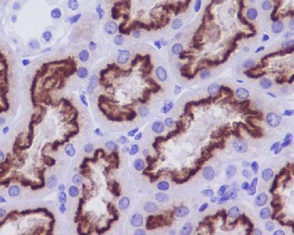
| Expected result | CUBN-positive staining in cells in tubules of kidney (HPA tissue IHC: High). HPA tissue profile: Cytoplasmic and membranous expression in the proximal renal tubules and small intestine epithelia. No signal in the no-primary control. |
CUBN should stain proximal renal tubule cells and small intestinal epithelium with an apical membranous and cytoplasmic pattern (HPA: tissue IHC; UniProt O60494: apical membrane, endosomes). Kidney tubular staining is High, and HPA rates its tissue IHC profile Enhanced for consistency with RNA expression (HPA: kidney; HPA: reliability). CUBN lacks a transmembrane segment and depends on AMN for plasma membrane localisation (UniProt O60494: topology and subunit).
| Strong staining outlines the luminal edge of renal tubule cells, with some cytoplasmic staining. | This fits the expected renal pattern: HPA reports High staining in kidney tubule cells and describes cytoplasmic and membranous expression in proximal tubules (HPA: tissue IHC). An apical emphasis is consistent with CUBN localisation (UniProt O60494: subcellular location). Judge the pattern within tubules, since the supplied HPA level applies to tubular cells (HPA: kidney). |
| Small intestinal epithelial cells show membranous and cytoplasmic staining. | This is an expected tissue and cell type (HPA: tissue IHC; UniProt O60494: tissue specificity). The supplied HPA profile does not assign these cells a High staining level, so compare their compartment pattern without requiring renal intensity (HPA: tissue IHC). |
| Staining is predominantly nuclear or distributed uniformly across unrelated compartments. | That differs from HPA's cytoplasmic and membranous tissue pattern and UniProt's membrane and endocytic locations (HPA: tissue IHC; UniProt O60494: subcellular location). Check morphology and controls before scoring it as CUBN; nonspecific detection or interpretation of counterstain is possible (general IHC practice). |
| Strong staining appears in adipocytes or adrenal glandular cells. | These cell types are listed as Not detected in the supplied tissue IHC data (HPA: adipose tissue and adrenal gland). Treat the result as discordant and assess cross-reactivity and endogenous detection activity with appropriate controls (general IHC practice). A discordant stain alone does not establish either cause. |
| Kidney tubules show no staining despite a visible detection reaction elsewhere. | The result conflicts with High kidney tubular staining in HPA's reference profile (HPA: kidney). Review section quality, tissue identity and morphology, then check antibody and detection controls (general IHC practice). A failed positive control limits interpretation of negative study sections (general IHC practice). |
| Tissue and cell selection | Kidney proximal tubules provide a supported positive reference: HPA reports High tubular staining, and UniProt reports CUBN in kidney cortex at protein level (HPA: kidney; UniProt O60494: tissue specificity). Small intestinal epithelium is another expected site, while the listed adipocytes and adrenal glandular cells are Not detected comparators (HPA: tissue IHC). |
| Apical organisation and AMN | CUBN has no transmembrane segment and depends on AMN for plasma membrane localisation (UniProt O60494: topology and subunit). Interpret an apical rim in the context of epithelial polarity and morphology (UniProt O60494: apical cell membrane; general IHC practice). |
| Intracellular signal | Cytoplasmic staining can fit the tissue profile (HPA: tissue IHC). UniProt also places CUBN in coated pits, endosomes and lysosome membranes, supporting intracellular signal alongside membrane staining; those annotations do not identify individual puncta on a chromogenic section (UniProt O60494: subcellular location). |
| Processing and antibody epitope | UniProt annotates a signal peptide at residues 1–23, a propeptide at 24–35 and a mature chain at 36–3623 (UniProt O60494: processing). No epitope location is supplied for the staining antibody, so these annotations cannot predict which processed form it detects or explain a missing tissue signal. |
| IHC validation | Both listed antibodies, HPA004133 and HPA043854, have Enhanced IHC status, and HPA rates the tissue profile Enhanced for agreement with RNA expression (HPA: antibodies; HPA: reliability). This supports the reference distribution but does not establish that every antibody or detection run will reproduce it. |
| IF/ICC applicability? | HPA supplies no CUBN ICC-IF images or assigned main subcellular location (HPA: subcellular). Use the IHC tissue pattern and UniProt localisation as expectations, while treating IF/ICC staining performance as unverified by the supplied HPA record (HPA: tissue IHC; UniProt O60494: subcellular location). |
| Situation | Likely cause | Next action |
|---|---|---|
| No staining in the kidney positive reference | The run or tissue may be unsuitable; a CUBN-specific failure mechanism cannot be assigned from these records (HPA: High kidney tubular staining). | Verify proximal tubule morphology and section integrity, then review antibody, detection and control performance before scoring other sections negative (general IHC practice). |
| Tubules stain, but the luminal edge is hard to distinguish | Cytoplasmic staining is part of the reported pattern, and section orientation can obscure epithelial polarity (HPA: tissue IHC; general IHC practice). | Examine well-oriented tubules and compare the luminal rim with cytoplasm; record both compartments instead of calling all tubular colour apical (general IHC practice). |
| Colour spreads broadly across tissue or obscures cell borders | Diffuse signal may reflect detection background; the reference pattern is cell and compartment specific (general IHC practice; HPA: tissue IHC). | Inspect the negative reagent control, blocking and wash steps, then adjust detection conditions according to the assay instructions (general IHC practice). |
| Unexpected staining appears in HPA Not detected cells | Cross-reactivity or endogenous chromogenic detection activity may contribute; the image alone cannot distinguish them (HPA: listed negative cell types; general IHC practice). | Compare a negative reagent control and an appropriate endogenous activity control, then recheck tissue and cell identity (general IHC practice). |
| Signal is mainly nuclear | Nuclear localisation is outside the supplied HPA tissue pattern and UniProt location list (HPA: tissue IHC; UniProt O60494: subcellular location). | Check counterstain appearance and controls, then score CUBN only where cell type and compartment agree with the reference pattern (general IHC practice; HPA: tissue IHC). |
| Signal remains weak after a run with working controls | The supplied sources do not establish CUBN-specific antigen retrieval or fixation sensitivity; weak signal has several possible assay causes. | Review tissue preservation and the antibody's validated IHC conditions; if changing antigen retrieval, compare conditions with the kidney positive reference in the same run (general IHC practice; HPA: High kidney tubular staining). |
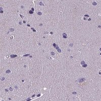

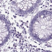

Comprehensive Human Protein Atlas IHC scoring per tissue (reliability: Enhanced — High consistency between antibody staining and RNA expression data.). Rows are taken directly from the HPA tissue chart — click any row's HPA link to view the source.
| Tissue | Cell type | Level | Evidence | Source |
|---|---|---|---|---|
| Kidney | Cells in tubules | High | Protein (IHC) | HPA → |
| Tissue | Cell type | Level | Evidence | Source |
|---|---|---|---|---|
| Adipose tissue | Adipocytes | Not detected | Protein (IHC) | HPA → |
| Adrenal gland | Glandular cells | Not detected | Protein (IHC) | HPA → |
| Appendix | Endocrine cells | Not detected | Protein (IHC) | HPA → |
| Bone marrow | Hematopoietic cells | Not detected | Protein (IHC) | HPA → |
| Breast | Adipocytes | Not detected | Protein (IHC) | HPA → |
Troubleshoot CUBN staining by checking retrieval, compartment, epithelial cell identity and controls before interpreting changes in chromogenic signal.
M03670 has real IHC data from paraffin-embedded human kidney (M03670 IHC image caption) and lists human reactivity (catalog reactivity). No IF/ICC data are supplied (catalog applications and IF image alts).
M03670 is listed for IHC with human reactivity (catalog applications and reactivity). Its own IHC figure shows paraffin-embedded human kidney stained for Cubilin (M03670 IHC image caption).
Which to pick: For tissue IHC, choose M03670: it is a rabbit monoclonal (catalog host and clone) with an IHC image from paraffin-embedded human kidney (M03670 IHC image caption); the fixative is unreported (M03670 IHC image caption). There is no supported IF/ICC pick (catalog applications and IF image alts). There is no supported cross-species pick because M03670 lists human reactivity only (catalog reactivity).