This website uses cookies to ensure you get the best experience on our website.


- Table of Contents
Plan DBN1 chromogenic IHC in paraffin sections using strongly stained cerebellar processes as a positive reference (HPA tissue IHC). Assess cytoplasmic and membranous staining (HPA tissue IHC) with consistent fixation across samples.

Expected localisation, validated protocols, controls and antibodies — the at-a-glance facts below, then the full design guide.
| Expected localisation | Cytoplasmic and membranous tissue staining (HPA tissue IHC) | |
| Staining pattern | High cerebellar process signal; cytoplasmic/membranous overall (HPA tissue IHC) | |
| Antigen retrieval | EDTA pH 8.0 HIER, heat-mediated (datasheet A05530-1) | |
| Positive control | Cerebellum+4 more · see all | |
| Negative control | Adipose tissue+4 more · see all |
| Fixation | Keep paraffin-section fixation consistent across samples. (standard IHC practice; not target-specific) | |
| Caveat | Hippocampal neurons stain weakly as a positive control (HPA tissue IHC) | |
| Regulation | Expression regulation is not reported (UniProt) | |
| Isoform / epitope | 3 isoforms; epitope coverage is unspecified (UniProt) |
The catalog antibody’s IHC-P protocol is followed by published DBN1 staining conditions from three PMC articles (datasheet; PMC6456690; PMC12262305; PMC7352383).
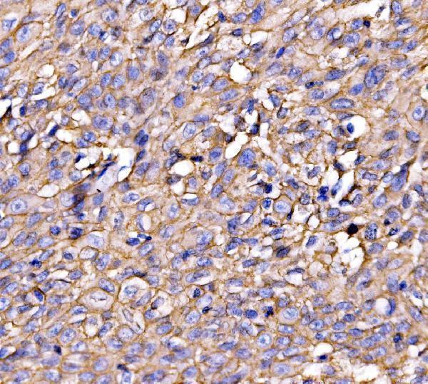
| Sample | Paraffin-embedded human laryngeal carcinoma tissue; fixative not specified (datasheet A05530-1) |
| Fixation | Image fixative and duration unreported (datasheet A05530-1); verify before use. |
| Sectioning | 4–5 µm sections on charged slides (standard) |
| Deparaffinisation | Xylene, graded ethanol series to water (standard) |
| Antigen retrieval | Heat retrieval: EDTA pH 8.0 (datasheet A05530-1); 20 min, 95–100 °C (standard) |
| Peroxidase block | 3% H2O2, 10 min, room temperature (standard) |
| Blocking | 10% goat serum (datasheet A05530-1) |
| Primary antibody | Rabbit anti-DBN1, 2-5 μg/ml (datasheet A05530-1) |
| Primary incubation | Overnight at 4 °C (datasheet A05530-1) |
| Detection | Streptavidin-biotin complex (SABC), DAB chromogen (datasheet A05530-1) |
| Counterstain | Hematoxylin, blue, dehydrate and mount (standard) |
| Expected result | DBN1-positive staining in processes in granular layer of cerebellum (HPA tissue IHC: High). HPA tissue profile: Cytoplasmic and membranous expression in several tissues, most abundant in CNS. No signal in the no-primary control. |
DBN1 staining is expected in the cytoplasm, cell cortex and projections, with membranous staining in some tissues (UniProt Q16643 subcellular location; HPA tissue IHC). Cerebellar granular-layer processes and testicular spermatogonia provide high-staining examples; cortical neuropil shows medium staining (HPA tissue IHC). HPA rates the tissue pattern Enhanced, citing consistency between antibody staining and RNA expression (HPA tissue IHC reliability). DBN1 has no transmembrane segment, so a membrane-associated pattern does not imply a membrane-spanning protein (UniProt Q16643 topology).
| Strong staining outlines processes in the cerebellar granular layer; cortical neuropil stains more moderately (HPA tissue IHC). | This supports the expected process-rich CNS pattern. Assess the stained structures as well as intensity: HPA reports High in cerebellar processes and Medium in cortical neuropil, while UniProt places DBN1 in dendrites and the cell cortex (HPA tissue IHC; UniProt Q16643 subcellular location). |
| Cytoplasmic or peripheral staining appears in spermatogonia, stromal cells or glandular cells (HPA tissue IHC). | Interpret intensity against the named cell population: spermatogonia are High, endometrial stromal cells and epididymal or gallbladder glandular cells are Medium (HPA tissue IHC). Cytoplasmic and membranous expression across several tissues is compatible with the HPA profile (HPA tissue IHC). |
| Predominantly nuclear staining appears without convincing cytoplasmic, peripheral or process staining. | Treat this as an unexpected compartment pattern requiring review, because UniProt lists cytoplasm, cell cortex, junctions and projections, while HPA describes cytoplasmic and membranous tissue staining (UniProt Q16643 subcellular location; HPA tissue IHC). Compare a known-positive section and detection controls before assigning it to DBN1 (general IHC practice). |
| Strong staining appears in adipocytes or cardiomyocytes, which HPA records as Not detected (HPA tissue IHC). | Check whether the signal follows the named cells or surrounding structures. Persistent cell-associated staining raises cross-reactivity or endogenous detection activity as possibilities (general IHC practice); HPA's Not detected calls apply to those specific cell types, not every cell in the tissue (HPA tissue IHC). |
| No signal appears in cerebellar granular-layer processes or testicular spermatogonia. | A blank known-positive population makes the run difficult to interpret: both populations are High in HPA tissue IHC (HPA tissue IHC). Check section quality, retrieval, antibody application and detection with appropriate controls (general IHC practice) before treating another tissue as DBN1-negative. |
| Anatomical and cell-type context | HPA reports abundant CNS staining but different levels by structure: cerebellar granular-layer processes are High, cortical neuropil is Medium, and hippocampal neuronal cells are Low (HPA tissue IHC). UniProt documents expression in specific hippocampal layers and pyramidal-cell regions; that record does not assign those structures a uniform IHC intensity (UniProt Q16643 tissue specificity). |
| Peripheral versus cytoplasmic signal | DBN1 is an actin cytoskeleton-organizing protein associated with projections, the cortex and junctions (UniProt Q16643 function and subcellular location). HPA describes cytoplasmic and membranous tissue staining (HPA tissue IHC). With no transmembrane segment, a peripheral signal should be read as localisation, not evidence of membrane-spanning topology (UniProt Q16643 topology). |
| Antibody validation and scope | HPA lists 2 rabbit polyclonal antibodies, HPA051452 and HPA056940, with Enhanced IHC status (HPA antibodies). That supports the reported tissue pattern; it does not establish that every chromogenic protocol, section or unexpected stained cell will perform alike (HPA antibodies; general IHC practice). |
| Isoforms and epitope information | UniProt lists 3 DBN1 isoforms (UniProt Q16643 isoforms). No antibody epitope or isoform coverage is supplied here, so an apparent difference between sections cannot be assigned to a particular isoform from this evidence alone (UniProt Q16643 isoforms; supplied antibody record). |
| IF/ICC Q&A | Q: Should IF/ICC show the same pattern as tissue IHC? A: HPA places the main ICC-IF signal at the plasma membrane, with additional actin-filament localisation, in its imaged cell lines (HPA subcellular ICC-IF). Treat that as cell-culture localisation evidence; tissue intensity and cell-type expectations come from HPA tissue IHC. |
| Situation | Likely cause | Next action |
|---|---|---|
| Known-positive cerebellar processes show little or no chromogenic signal (HPA tissue IHC). | The expected High population is missing; a failed run or unsuitable section is possible (HPA tissue IHC; general IHC practice). | Inspect tissue preservation and compare a positive-control section; verify retrieval, primary-antibody application and the detection reagents before interpreting low-staining samples (general IHC practice). No DBN1-specific retrieval condition is supplied. |
| Brown precipitate covers many compartments or extends beyond cell boundaries. | Diffuse background can arise from incomplete blocking, excess antibody or insufficient washing (general IHC practice); it cannot be assigned to DBN1 localisation from appearance alone. | Review the no-primary control, blocking and washes; adjust antibody concentration within a validated workflow, then reassess whether the signal follows HPA-described cells and structures (general IHC practice; HPA tissue IHC). |
| A strong, predominantly nuclear pattern replaces cytoplasmic or process staining. | That compartment is outside the listed DBN1 locations and HPA tissue profile (UniProt Q16643 subcellular location; HPA tissue IHC). | Compare with a known-positive section and a no-primary control; confirm that the cytoplasmic or peripheral signal is reproducible before scoring nuclear staining as specific (general IHC practice; HPA tissue IHC). |
| Adipocytes, cardiomyocytes or adrenal glandular cells stain strongly despite HPA Not detected calls (HPA tissue IHC). | Cross-reactivity or endogenous detection activity may explain unexpected cell-associated chromogen (general IHC practice); an HPA call concerns the specified cell population (HPA tissue IHC). | Check no-primary and detection controls, inspect which cells contain signal, and compare staining with an HPA High population in the same run (general IHC practice; HPA tissue IHC). |
| Hippocampal neuronal cells look weak beside strongly stained cerebellar processes. | This contrast can match HPA's Low hippocampal neuronal-cell and High cerebellar-process calls (HPA tissue IHC). UniProt's hippocampal expression record does not require equal IHC intensity (UniProt Q16643 tissue specificity). | Score each anatomical population separately, using its own expected HPA level; avoid calling the run failed solely because hippocampal neuronal staining is weak (HPA tissue IHC). |
| A peripheral rim is interpreted as proof that DBN1 spans the plasma membrane. | UniProt lists no transmembrane segment, despite peripheral and junctional localisation (UniProt Q16643 topology and subcellular location). | Report the observed cytoplasmic or membranous staining pattern without inferring membrane-spanning topology; use the named positive cell population and compartment to support the IHC interpretation (HPA tissue IHC; UniProt Q16643 topology). |


Comprehensive Human Protein Atlas IHC scoring per tissue (reliability: Enhanced — High consistency between antibody staining and RNA expression data.). Rows are taken directly from the HPA tissue chart — click any row's HPA link to view the source.
| Tissue | Cell type | Level | Evidence | Source |
|---|---|---|---|---|
| Cerebellum | Processes in granular layer | High | Protein (IHC) | HPA → |
| Testis | Spermatogonia cells | High | Protein (IHC) | HPA → |
| Cerebral cortex | Neuropil | Medium | Protein (IHC) | HPA → |
| Endometrium | Cells in endometrial stroma | Medium | Protein (IHC) | HPA → |
| Epididymis | Glandular cells | Medium | Protein (IHC) | HPA → |
| Tissue | Cell type | Level | Evidence | Source |
|---|---|---|---|---|
| Adipose tissue | Adipocytes | Not detected | Protein (IHC) | HPA → |
| Adrenal gland | Glandular cells | Not detected | Protein (IHC) | HPA → |
| Appendix | Endocrine cells | Not detected | Protein (IHC) | HPA → |
| Bronchus | Respiratory epithelial cells | Not detected | Protein (IHC) | HPA → |
| Cervix | Glandular cells | Not detected | Protein (IHC) | HPA → |
Troubleshoot DBN1 staining in paraffin sections by checking retrieval, cellular pattern, controls, and scoring before interpreting differences between samples.
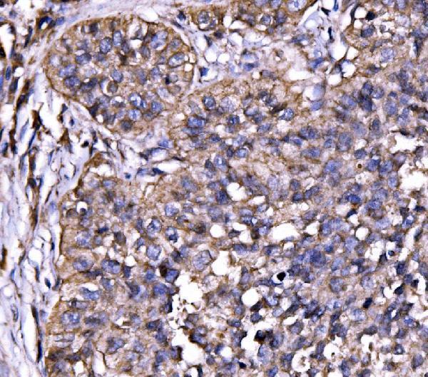
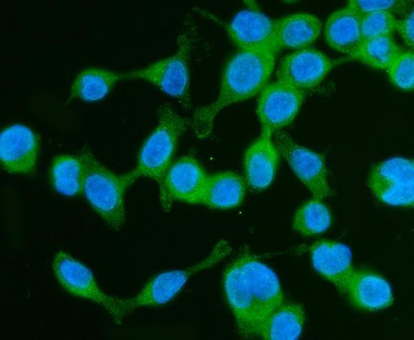
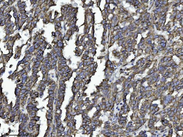
Two antibodies have paraffin-section IHC images from human tissues, and one also has ICC/IF imagery from CACO-2 cells (catalog image captions).
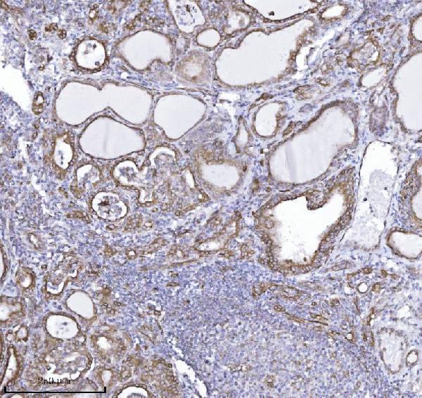
A05530-1 has IHC images from human laryngeal carcinoma, lung cancer, thyroid papillary carcinoma, and cerebellum, plus an ICC/IF image from CACO-2 cells (catalog image captions). M05530-4 has IHC images from human ovarian serous carcinoma, human thyroid cancer, and rat brain (catalog IHC image captions).
Which to pick: For paraffin-section IHC, choose A05530-1 for its human tissue examples (A05530-1 IHC image captions), or M05530-4 for its monoclonal clone 4F6E7 and human and rat tissue examples (catalog clone field; M05530-4 IHC image captions). For IF/ICC, choose A05530-1: both applications are listed and its CACO-2 image documents ICC/IF staining (A05530-1 applications; A05530-1 IF image caption). For mouse-reactive tissue IHC, M05530-4 lists mouse reactivity, although its IHC images show only human and rat sections; the fixative is unreported for both antibodies’ paraffin-section images (M05530-4 reactivity; catalog IHC image captions).