DGAT1 / Diacylglycerol O-acyltransferase 1 · IHC design guide

Design Immunohistochemistry for DGAT1

Plan chromogenic DGAT1 IHC on paraffin sections with a 1:50 starting dilution (datasheet M02522). Assess cytoplasmic staining in intestinal endocrine cells and include adipocytes as a tissue comparison where staining was not detected (HPA tissue IHC).

Evidence assembled Oct 2026 · For research use; verify linked source records and product datasheet before use
Immunohistochemistry protocol sheet for DGAT1 (IHC for DGAT1): expected localisation Cytoplasmic staining (HPA tissue IHC); ER membrane (UniProt), antibody M02522, validated IHC image, and IHC protocol steps
Printable DGAT1 IHC protocol sheet — expected localisation Cytoplasmic staining (HPA tissue IHC); ER membrane (UniProt), antibody M02522, controls and protocol steps. Open the full DGAT1 IHC guide →

DGAT1 Immunohistochemistry Experimental Design Guide

Expected localisation, validated protocols, controls and antibodies — the at-a-glance facts below, then the full design guide.

Must know before staining
Expected localisation Cytoplasmic staining (HPA tissue IHC); ER membrane (UniProt)
Staining pattern Cytoplasmic, high in small-intestinal endocrine cells (HPA tissue IHC)
Antigen retrieval EDTA pH 8.0 HIER, heat-mediated (datasheet M02522)
Positive control ⓘ Adrenal gland+4 more · see all
Negative control ⓘ Adipose tissue+4 more · see all
Important caveats
Reasons your staining may differ from the expected pattern.
Fixation Matched tissue-IHC evidence does not establish the fixation claim. Validate the specimen-specific method before use. (selected-SKU IHC image M02522)
Caveat Check specificity: datasheet includes unrelated GluR1 text (datasheet M02522)
Regulation Intestine-enhanced expression (HPA tissue RNA)
Isoform / epitope No isoforms annotated; epitope access depends on membrane side (UniProt)
Section 1

Recommended DGAT1 IHC & IF Protocols

The catalog antibody’s IHC-P protocol (datasheet: M02522) is accompanied by published DGAT1 IHC methods for patient biopsies (PMC6058035) and rat brain (PMC12082899).

Recommended immunohistochemistry (IHC-P) protocol parameters
SampleParaffin-embedded human stomach cancer tissue; fixative not specified (datasheet M02522)
FixationImage fixative and duration unreported (datasheet M02522); verify before use.
Sectioning4–5 µm sections on charged slides (standard)
DeparaffinisationXylene, graded ethanol series to water (standard)
Antigen retrievalHeat retrieval: EDTA pH 8.0 (datasheet M02522); 20 min, 95–100 °C (standard)
Peroxidase block3% H2O2, 10 min, room temperature (standard)
Blocking10% goat serum (datasheet M02522)
Primary antibodyRabbit monoclonal (clone 23D11) anti-DGAT1, 1:50 (datasheet M02522)
Primary incubationOvernight at 4 °C (datasheet M02522)
DetectionHRP-conjugated secondary, DAB chromogen (datasheet M02522)
CounterstainHematoxylin, blue, dehydrate and mount (standard)
Expected resultDGAT1-positive staining in glandular cells of adrenal gland (HPA tissue IHC: High). HPA tissue profile: General cytoplasmic expression, most abundant in small intestine and duodenum. No signal in the no-primary control.
💡Decision noteStart with heat-mediated EDTA pH 8.0 retrieval (datasheet: M02522); the rat-brain IHC method reports EDTA pH 9.0 or citrate pH 6.0 (PMC12082899: IHC methods).
Section 2

What Is the Expected DGAT1 Staining Pattern?

DGAT1 is an endoplasmic reticulum membrane protein with 9 transmembrane segments (UniProt O75907 topology). In paraffin IHC, expect predominantly cytoplasmic staining, especially in intestinal cells; HPA describes general cytoplasmic expression most abundant in small intestine and duodenum (HPA tissue IHC). HPA rates its tissue pattern Enhanced for consistency with RNA expression, pending external verification (HPA tissue IHC).

What am I looking at on my slide?
Cytoplasmic staining in small intestine or duodenum, strongest in identifiable endocrine cells.This fits the reported high endocrine-cell staining in both tissues and the general cytoplasmic pattern (HPA tissue IHC). DGAT1 resides in the endoplasmic reticulum membrane, so a cytoplasmic distribution is plausible (UniProt O75907 subcellular location). Assess signal in the named cells against neighboring cells on the same section; a tissue-wide intensity label does not mean every cell must stain equally (HPA tissue IHC; general IHC practice).
Predominantly nuclear or nucleolar chromogen, with little cytoplasmic staining.Question this as an IHC result because HPA tissue sections show a general cytoplasmic pattern and UniProt places DGAT1 in the endoplasmic reticulum membrane (HPA tissue IHC; UniProt O75907 subcellular location). HPA also reports additional nucleolar and nucleolar-rim localization by ICC-IF, so nucleolar signal alone cannot be called impossible (HPA subcellular ICC-IF). Check the IHC controls and the full cellular pattern before interpreting it as DGAT1 (general IHC practice).
Strong signal in adipocytes or another cell population reported as undetected.HPA reports adipocytes as not detected, making strong adipocyte staining discordant with its tissue IHC reference (HPA tissue IHC). Cross-reactivity or endogenous chromogenic activity are possibilities, rather than a diagnosis from appearance alone (general IHC practice). Compare an adjacent no-primary control and a known-positive section processed together (general IHC practice).
Diffuse color across cells, extracellular spaces, or the whole section.A widespread haze does not preserve the reported cytoplasmic and cell-type pattern (HPA tissue IHC). Nonspecific binding, endogenous detection activity, or incomplete washing can produce background in chromogenic IHC (general IHC practice). Judge the section against a no-primary control before assigning the haze to DGAT1 (general IHC practice).
No convincing signal in small-intestine or duodenum endocrine cells.These are high-staining cell populations in HPA tissue IHC, so their absence makes a negative run difficult to interpret (HPA tissue IHC). Check that the relevant cells are present, then review the antibody, antigen retrieval, detection reagents, and a concurrently stained positive control (general IHC practice). An HPA high rating is a reference observation, not a guarantee for every specimen or run (HPA tissue IHC; general IHC practice).
💡Expected DGAT1 appearanceCall a section positive when identifiable intestinal endocrine cells show clear cytoplasmic chromogen above local background, consistent with HPA’s High ratings in small intestine and duodenum (HPA tissue IHC); uniform haze or dominant signal in HPA-undetected adipocytes is a warning to check controls, not sufficient evidence of a DGAT1-positive result (HPA tissue IHC; general IHC practice).
How each factor affects the staining
Cell and tissue choiceHPA rates duodenal and small-intestinal endocrine cells, rectal enterocytes, and adrenal glandular cells High (HPA tissue IHC). It rates lung macrophages Low and adipocytes Not detected (HPA tissue IHC). Use the named cell population when choosing and scoring a reference section; do not turn a tissue label into an expectation for every cell (HPA tissue IHC; general IHC practice).
Membrane topology and epitopeDGAT1 has 9 annotated transmembrane segments and both cytoplasmic and lumenal regions (UniProt O75907 topology). The supplied antibody record does not locate its epitope, so topology alone cannot identify which retrieval condition will expose it or predict its staining intensity (UniProt O75907 topology; HPA antibody record).
IHC antibody evidenceThe supplied rabbit polyclonal antibody, CAB032853, has an Enhanced IHC status; HPA’s tissue assessment reports high staining–RNA consistency while external verification remains pending (HPA antibody record; HPA tissue IHC). Treat the reported pattern as a reference and evaluate controls for the actual staining run (general IHC practice).
What should IF/ICC show?Predominantly endoplasmic reticulum localization is expected; HPA also approves nucleolar and nucleolar-rim localization in ICC-IF (HPA subcellular ICC-IF). This IF observation adds context when judging compartment differences, but it does not establish that nucleolar chromogen alone is a valid paraffin IHC result (HPA subcellular ICC-IF; HPA tissue IHC).
Why is my staining missing, weak or wrong?
SituationLikely causeNext action
No staining in the expected intestinal cells.The positive cell population may be absent from the section, or a staining step may have failed (HPA tissue IHC; general IHC practice).Confirm the cell population on the counterstained section; run a small-intestine or duodenum reference alongside it, then check retrieval, antibody incubation, and detection controls (HPA tissue IHC; general IHC practice).
All cells show a faint, even brown haze.Nonspecific binding, endogenous detection activity, or incomplete washing can obscure a cellular pattern (general IHC practice).Inspect a no-primary control; review blocking, washing, and chromogen development before scoring cytoplasmic signal (general IHC practice).
Only nuclei or nucleoli appear positive.The IHC distribution conflicts with the general cytoplasmic tissue pattern, although nucleoli are an additional ICC-IF location (HPA tissue IHC; HPA subcellular ICC-IF).Compare positive and no-primary controls and look for cytoplasmic signal in the expected cells before accepting the result (HPA tissue IHC; general IHC practice).
Adipocytes stain strongly.That result differs from HPA’s Not detected adipocyte entry; cross-reactivity or endogenous detection activity may contribute (HPA tissue IHC; general IHC practice).Compare no-primary and known-positive sections from the same run; assess whether staining follows the expected cell-type pattern (HPA tissue IHC; general IHC practice).
The positive reference stains, but the study section is negative.DGAT1 staining varies by cell population: HPA reports High intestinal endocrine-cell staining but Not detected staining in several other named populations (HPA tissue IHC).Identify and score the cell population actually present, and report the negative finding for that population rather than the whole tissue (HPA tissue IHC; general IHC practice).

Sample controls for DGAT1 IHC & IF

🧪Run small intestine first and score its endocrine cells, where DGAT1 staining is High (HPA: High in small-intestine endocrine cells). Use lymph-node germinal-center cells as the negative tissue (HPA: Not detected in lymph-node germinal-center cells); on the positive slide, use neighboring cells without specific staining as local background comparators, without assuming every non-endocrine cell is DGAT1-negative.
Positive control tissue: Adrenal gland (Glandular cells, HPA High)
Negative control tissue: Adipose tissue (HPA Not detected)
ICC-IF cell lines (HPA subcellular resource): HPA ICC-IF images show DGAT1 in A-431, U-251MG, U2OS, with annotated localisation: Endoplasmic reticulum (approved) (HPA subcellular).
Technical controls: Include a no-primary, secondary-only control and an isotype control matched to the primary antibody’s host and clonality; M02522 uses a rabbit primary (selected IHC caption: rabbit anti-DGAT1). Confirm specificity with matched DGAT1-knockout material or a cognate-peptide block if available, and block endogenous peroxidase before chromogenic detection in small-intestine sections (HPA: High in small-intestine endocrine cells).
⚠️Feasibility: A target-specific fixation window or fixation effect is unreported, and the selected M02522 paraffin-section caption does not state the fixative (selected IHC caption: fixative not stated). Heat-mediated retrieval in EDTA at pH 8.0 was used for the captioned stomach-cancer section, but retrieval dependence has not been established (selected IHC caption: EDTA retrieval, pH 8.0). Relative ease of frozen-section IHC versus IF is unreported; ICC-IF images and approved endoplasmic-reticulum localization support an IF localization check (HPA subcellular), while intestinal endogenous peroxidase can create chromogenic background and should be assessed with the no-primary control (standard IHC practice).

HPA tissue IHC evidence for DGAT1

Comprehensive Human Protein Atlas IHC scoring per tissue (reliability: Enhanced — High consistency between antibody staining and RNA expression data. Pending external verification.). Rows are taken directly from the HPA tissue chart — click any row's HPA link to view the source.

Positive expression · recommended positive controls

TissueCell typeLevelEvidenceSource
Adrenal gland Glandular cells High Protein (IHC) HPA →
Duodenum Endocrine cells High Protein (IHC) HPA →
Rectum Enterocytes High Protein (IHC) HPA →
Small intestine Endocrine cells High Protein (IHC) HPA →
Appendix Endocrine cells Medium Protein (IHC) HPA →

Undetected expression · recommended negative controls

TissueCell typeLevelEvidenceSource
Adipose tissue Adipocytes Not detected Protein (IHC) HPA →
Bone marrow Hematopoietic cells Not detected Protein (IHC) HPA →
Cervix Glandular cells Not detected Protein (IHC) HPA →
Epididymis Glandular cells Not detected Protein (IHC) HPA →
Lymph node Germinal center cells Not detected Protein (IHC) HPA →
Section 3

Advanced DGAT1 IHC Tips

Troubleshoot DGAT1 staining in paraffin sections by checking retrieval, compartment, cell type and controls before comparing chromogenic signal across samples.

What retrieval should I start with for DGAT1 in paraffin sections?
Start with heat-mediated retrieval in EDTA at pH 8.0 for paraffin sections (datasheet M02522). The selected tissue image used this retrieval, followed by 10% goat serum blocking and an overnight incubation with the catalog antibody at 1:50 and 4°C (datasheet M02522). If staining is weak, compare retrieval duration on adjacent sections while holding antibody concentration and chromogen development constant (standard IHC practice). Inspect morphology alongside signal, because excessive heating can damage sections and make diffuse staining difficult to interpret (standard IHC practice). An intestinal control can help assess expected signal, but its staining does not establish performance in every tissue (HPA: intestine-enhanced RNA and abundant small-intestinal staining; standard IHC practice).
Could fixation explain weak DGAT1 staining in my paraffin sections?
DGAT1-specific fixation sensitivity is unknown from the supplied evidence, and the selected paraffin-section caption does not state a fixative (datasheet M02522). Record the fixative, fixation duration and processing history for each specimen before comparing staining intensity across cases (standard IHC practice). If weak staining tracks with processing history, run the same EDTA pH 8.0 retrieval and antibody conditions on a control processed alongside the affected sections (datasheet M02522; standard IHC practice). Examine tissue preservation and compare serial sections before changing retrieval, since damage or uneven processing can confound a chromogenic score (standard IHC practice). HPA tissue patterns provide expression context, not a measurement of fixation sensitivity (HPA: tissue IHC; standard IHC practice).
Where should convincing DGAT1 staining appear in tissue sections?
Prioritise cytoplasmic staining with a distribution compatible with the endoplasmic reticulum, because DGAT1 is an ER membrane protein and HPA describes general cytoplasmic tissue expression (UniProt O75907 localisation; HPA: tissue IHC). Its 9 transmembrane segments support a membrane-associated pattern, although chromogenic tissue sections may not resolve individual ER membranes (UniProt O75907 topology; standard IHC practice). Compare stained cells with local morphology and an appropriate positive-control section, especially when assessing intestinal epithelium (UniProt O75907 function; standard IHC practice). Predominantly nuclear signal warrants review of counterstain, background and controls before assignment to DGAT1; HPA reports additional nucleolar localisation in ICC/IF images, which does not establish the same pattern in this IHC assay (HPA: subcellular; standard IHC practice).
How should DGAT1 topology affect epitope troubleshooting?
DGAT1 has 9 annotated transmembrane segments and alternating cytoplasmic and lumenal regions, so epitope access depends on where the antibody binds (UniProt O75907 topology; standard IHC practice). The supplied product caption does not identify the antibody epitope, so do not assign its staining behaviour to a particular membrane face (datasheet M02522). UniProt lists 0 annotated isoforms and phosphoserines at residues 17 and 18; these annotations do not establish whether this antibody recognises a modified epitope (UniProt O75907 isoforms and modified residues). If retrieval changes the signal, compare adjacent sections and a consistent positive control, then seek epitope information before attributing the change to topology or phosphorylation (standard IHC practice).
How can IF help assess an ambiguous DGAT1 IHC pattern?
Use IF as a separate localisation check and multiplex DGAT1 with a marker for the expected cell type, such as an appropriate intestinal epithelial marker when investigating intestinal IHC staining (UniProt O75907 function; standard IF practice). Choose spectrally separated fluorophores, favouring a far-red channel when tissue autofluorescence obscures shorter-wavelength signal, and inspect single-channel controls (standard IF practice). Because DGAT1 spans the ER membrane 9 times, select permeabilisation according to whether the antibody epitope faces cytoplasm or ER lumen; the supplied caption does not identify that epitope (UniProt O75907 topology; datasheet M02522; standard IF practice). HPA reports ER localisation in ICC/IF images, providing a pattern to compare with tissue IHC without treating the assays as interchangeable (HPA: subcellular; standard IHC/IF practice).
What should I check when DGAT1 DAB staining is diffuse?
First compare the suspect section with a no-primary control and inspect whether colour follows tissue edges, folds or damaged regions (standard IHC practice). The selected paraffin-section method used 10% goat serum blocking, a 1:50 primary incubation overnight at 4°C, a peroxidase-conjugated secondary and DAB development (datasheet M02522). Include a peroxidase block and control chromogen development consistently, because endogenous enzyme activity or overdevelopment can produce misleading colour (standard IHC practice). If background persists, assess washing, blocking and primary concentration on serial sections while preserving the EDTA pH 8.0 retrieval as the reference condition (datasheet M02522; standard IHC practice). Compare the resulting pattern with the expected cytoplasmic distribution before scoring (HPA: tissue IHC; standard IHC practice).
How should I score DGAT1 staining across sections? ⚠ ANSWER MARKED FOR VERIFICATION
Define the cell population and tissue region before scoring, then record either the percentage of positive cells, an intensity-weighted H-score, or positive-cell density per mm² as appropriate to the study question (standard IHC practice). For an H-score, multiply each intensity category by its percentage of cells to obtain a scale from 0 to 300 (standard IHC practice). Normalise positive-cell counts to the area or number of eligible cells, and keep retrieval, exposure to DAB and scoring thresholds consistent across sections (standard IHC practice). Score relevant compartments separately when mixed cell types are present, since HPA reports differing DGAT1 levels among named tissue cell populations (HPA: tissue IHC; standard IHC practice).
When is a DGAT1-positive IHC pattern credible?
A credible pattern is reproducible in well-preserved cells, compatible with cytoplasmic ER-associated staining, and supported by controls processed under the same conditions (UniProt O75907 localisation; HPA: tissue IHC; standard IHC practice). HPA reports abundant small-intestinal expression and high staining in duodenal endocrine cells, making cell identity important when interpreting a positive intestinal field (HPA: tissue IHC). Recheck staining restricted to cut edges, necrotic areas or folds, and use a no-primary control to assess nonspecific or endogenous-enzyme signal (standard IHC practice). Strong nuclear-only colour should prompt review before assigning it to DGAT1, while HPA's additional nucleolar ICC/IF localisation should be interpreted in its own assay context (HPA: subcellular; standard IHC/IF practice).
Boster reagents

Best DGAT1 / Diacylglycerol O-acyltransferase 1 IHC Antibodies

M02522 has a human paraffin-section IHC image (catalog IHC caption); IF/ICC is listed for human samples, without an IF image (catalog applications, reactivity and image records).

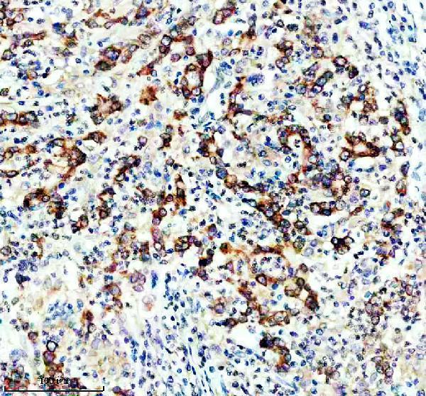
Real IHC data IHC analysis of DGAT1 using anti-DGAT1 antibody (M02522). DGAT1 was detected in a paraffin-embedded section of human stomach cancer tissue. Heat mediated antigen retrieval was performed in EDTA buffer (pH 8.0, epitope retrieval solution). The tissue section was blocked with 10% goat serum. The tissue section was then incubated with 1:50 rabbit anti-DGAT1 Antibody (M02522) overnight at 4°C. Peroxidase Conjugated Goat Anti-rabbit IgG was used as secondary antibody and incubated for 30 minutes at 37°C. The tissue section was developed using HRP Conjugated Rabbit IgG Super Vision Assay Kit (Catalog # SV0002) with DAB as the chromogen.
Anti-DGAT1 Rabbit Monoclonal Antibody
Cat # M02522

M02522 will render with an IHC image of a paraffin-embedded human stomach cancer section (catalog IHC caption). IF/ICC is listed for M02522, but no IF image is supplied (catalog applications and image records).

Which to pick: Choose M02522 for paraffin-section IHC: its own image documents that use, though the fixative is unreported (catalog IHC caption). M02522 is also listed for human IF/ICC at 1:50, without an IF image (catalog applications, reactivity, dilution and image records). Cross-species use is unsupported because only human reactivity is listed (catalog reactivity).

Each figure is that product's own IHC / IF validation image from its datasheet.

References

  1. UniProt Consortium. UniProt entry O75907 (DGAT1_HUMAN, Diacylglycerol O-acyltransferase 1).
  2. Human Protein Atlas. DGAT1 tissue IHC expression (reliability: Enhanced).
  3. Human Protein Atlas. DGAT1 subcellular location (ICC-IF): Mainly localized to the endoplasmic reticulum. In addition localized to the nucleoli and nucleoli rim..
  4. Human Protein Atlas. DGAT1 antibody validation summary (1 antibodies).
  5. Intestinal Failure and Aberrant Lipid Metabolism in Patients With DGAT1 Deficiency. Gastroenterology 2018 — PMC6058035.
  6. Lazy neutrophils - a lack of DGAT1 reduces the chemotactic activity of mouse neutrophils. Inflammation research : official journal of the European Histamine Research Society ... [et al.] 2024 — PMC11445369.
  7. Inhibition of diacylglycerol O-acyltransferase 1 provides neuroprotection by inhibiting ferroptosis in ischemic stroke. Molecular medicine (Cambridge, Mass.) 2025 — PMC12082899.
  8. Targeting DGAT1 Ameliorates Glioblastoma by Increasing Fat Catabolism and Oxidative Stress. Cell metabolism 2020 — PMC7415721.
  9. PubMed PMID:9756920 — UniProt-cited evidence.
  10. PubMed PMID:15489334 — UniProt-cited evidence.
  11. PubMed PMID:16214399 — UniProt-cited evidence.