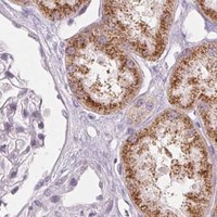
EHHADH / Peroxisomal bifunctional enzyme · IHC design guide

Design Immunohistochemistry for EHHADH

Plan chromogenic IHC in paraffin sections with catalog antibody A05757-3 at 1:10–1:50 (datasheet: A05757-3). Use hepatocytes as a high-staining reference and assess granular cytoplasmic staining in renal proximal tubules (HPA tissue IHC).

Evidence assembled Oct 2026 · For research use; verify linked source records and product datasheet before use
Immunohistochemistry protocol sheet for EHHADH (IHC for EHHADH): expected localisation Observed: granular cytoplasm (HPA tissue IHC); expected: peroxisomes (UniProt), antibody A05757-1, validated IHC image, and IHC protocol steps
Printable EHHADH IHC protocol sheet — expected localisation Observed: granular cytoplasm (HPA tissue IHC); expected: peroxisomes (UniProt), antibody A05757-1, controls and protocol steps. Open the full EHHADH IHC guide →

EHHADH Immunohistochemistry Experimental Design Guide

Expected localisation, validated protocols, controls and antibodies — the at-a-glance facts below, then the full design guide.

Must know before staining
Expected localisation Observed: granular cytoplasm (HPA tissue IHC); expected: peroxisomes (UniProt)
Staining pattern Granular cytoplasm in hepatocytes and proximal tubules (HPA tissue IHC)
Antigen retrieval Citrate pH 6.0 HIER, 95–98 °C, 20 min (rule: cytoplasmic / membrane antigen)
Positive control ⓘ Liver+1 more · see all
Negative control ⓘ Adipose tissue+4 more · see all
Important caveats
Reasons your staining may differ from the expected pattern.
Fixation Matched tissue-IHC evidence does not establish the fixation claim. Validate the specimen-specific method before use. (selected-SKU IHC image A05757-1)
Caveat Presumed off-target binding may confound staining (HPA tissue IHC)
Regulation Liver high; kidney medium (HPA tissue IHC)
Isoform / epitope 2 isoforms; epitope differences unspecified (UniProt)
Section 1

Recommended EHHADH IHC & IF Protocols

The catalog antibody’s IHC-P protocol is accompanied by published EHHADH IHC methods for mouse liver and tumor, adjacent, and distant normal tissues (PMC7672977; PMC13503599).

Recommended immunohistochemistry (IHC-P) protocol parameters
SampleParaffin-embedded human breast carcinoma tissue; fixative not specified (datasheet A05757-1)
FixationImage fixative and duration unreported (datasheet A05757-1); verify before use.
Sectioning4–5 µm sections on charged slides (standard)
DeparaffinisationXylene, graded ethanol series to water (standard)
Antigen retrievalHeat-induced epitope retrieval in citrate buffer, pH 6.0, 20 min at 95–98 °C (standard rule: cytoplasmic / membrane antigen)
Peroxidase block3% H2O2, 10 min, room temperature (standard)
Blocking10% normal serum of the secondary host, 30 min, room temperature (standard)
Primary antibodyRabbit anti-EHHADH, 1:100-1:300 (datasheet A05757-1)
Primary incubationOvernight at 4 °C (standard)
DetectionHRP-polymer secondary, DAB chromogen 5–10 min (standard)
CounterstainHematoxylin, blue, dehydrate and mount (standard)
Expected resultEHHADH-positive staining in hepatocytes of liver (HPA tissue IHC: High). HPA tissue profile: Cytoplasmic granular expression in hepatocytes and renal proximal tubules. No signal in the no-primary control.
💡Decision noteStart with citrate pH 6.0 retrieval at 95–98 °C for 20 min for this peroxisomal target (page retrieval; UniProt Q08426).
Section 2

What Is the Expected EHHADH Staining Pattern?

EHHADH is a peroxisomal protein with no transmembrane segment (UniProt Q08426: subcellular location and topology). In paraffin section IHC, expect granular cytoplasmic staining in hepatocytes and renal proximal tubules, with higher staining in hepatocytes (HPA: high in hepatocytes; medium in proximal tubules). HPA rates its tissue IHC evidence Enhanced but reports medium agreement with RNA data and presumed off target binding that was disregarded (HPA: tissue IHC reliability).

What am I looking at on my slide?
Distinct cytoplasmic granules in hepatocytes; weaker granules in proximal tubule cell bodies.This matches the observed liver and kidney pattern (HPA: granular cytoplasm; high in hepatocytes, medium in proximal tubules). Compare cell types within each section rather than requiring identical staining intensity across tissues (general IHC practice).
Predominantly nuclear, membrane rim, or smooth diffuse staining replaces cytoplasmic granules.Treat this as a localization mismatch, not confirmed EHHADH: UniProt places EHHADH in peroxisomes and reports no transmembrane segment (UniProt Q08426: location and topology). Check morphology and the detection controls before scoring (general IHC practice).
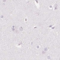
Strong staining appears in adipocytes or other cells recorded as not detected.Suspect nonspecific antibody binding or detection activity, especially if the same signal persists in a negative control (HPA: adipocytes not detected; presumed off target binding observed). A negative HPA entry is a reference pattern, not proof that every specimen must remain unstained.
A broad haze obscures cell borders and granular detail throughout the section.The expected HPA pattern is cell associated and granular (HPA: tissue IHC profile). Widespread haze is background until controls establish otherwise; review blocking, antibody concentration, wash steps, and chromogen development (general IHC practice).
Hepatocytes show no convincing cytoplasmic granules.This conflicts with the HPA high hepatocyte result (HPA: liver IHC). Check the positive control and section quality, then review retrieval, primary antibody use, and detection steps (general IHC practice). Absence alone cannot distinguish assay failure from specimen variation.
💡Expected EHHADH appearanceCall a result positive when hepatocytes show clear cytoplasmic granules and proximal tubules show weaker granular staining (HPA: high in hepatocytes; medium in proximal tubules); nuclear staining or diffuse haze alone does not match the reported pattern (UniProt Q08426: peroxisome; HPA: granular cytoplasm).
How each factor affects the staining
Tissue and cell typeUse hepatocytes as the stronger pattern reference and proximal tubules as a second reference (HPA: high in hepatocytes; medium in proximal tubules). HPA lists adipocytes as not detected, useful for checking unexpected widespread staining (HPA: adipose tissue IHC).
Compartment and protein topologyPeroxisomal localization supports cytoplasmic granules; no transmembrane segment supports caution with a membrane rim pattern (UniProt Q08426: location and topology). Light microscopy does not identify individual granules as peroxisomes without further evidence (general IHC interpretation).
Strength of tissue IHC evidenceBoth listed rabbit polyclonal antibodies have Enhanced IHC status (HPA: HPA036401 and HPA042021). HPA also reports medium agreement between staining and RNA and disregarded presumed off target binding; interpret a novel pattern with controls, not validation status alone (HPA: reliability description).
Isoforms and processingUniProt lists two isoforms and a full length 1–723 chain, with no signal peptide or propeptide (UniProt Q08426: isoforms and processing). Without an epitope map in this record, the staining contribution of each isoform cannot be assigned.
IF/ICC Q&A: Is a matching fluorescent cell pattern established?No ICC-IF image bearing cell lines or main subcellular location are available in this HPA payload (HPA: subcellular summary). The liver and kidney observations above are tissue IHC findings (HPA: tissue IHC), so do not present them as an observed IF/ICC result.
Why is my staining missing, weak or wrong?
SituationLikely causeNext action
No signal in the liver positive referenceThe assay may have failed, or this specimen may differ from the HPA reference; HPA reports high hepatocyte staining (HPA: liver IHC).Confirm that the section contains intact hepatocytes, then check the positive control, retrieval, primary incubation, and detection reagents (general IHC practice). Do not infer a target specific fixation effect from this result.
Kidney looks negative while liver stains correctlyProximal tubule staining is reported at medium level, below the high hepatocyte level (HPA: kidney and liver IHC). A weak signal may be hard to resolve against background.Identify proximal tubules by morphology and inspect cell bodies for granules; compare with the liver reference under the same scoring criteria (HPA: granular proximal tubule profile; general IHC practice).
Nuclei or cell borders dominate the stainThis compartment disagrees with peroxisomal localization and the observed granular cytoplasmic pattern (UniProt Q08426: location and topology; HPA: tissue IHC profile).Review counterstain and cell boundaries, then assess the negative control and repeat staining with adjusted detection conditions if needed (general IHC practice). Avoid scoring the mismatched compartment as EHHADH.
Adipocytes stain stronglyHPA records adipocytes as not detected and notes disregarded presumed off target binding in its tissue assessment (HPA: adipose tissue IHC; reliability description).Compare the stain with a control lacking primary antibody. If that control is clear, reassess antibody concentration and pattern specificity; if stained, investigate detection background (general IHC practice).
Diffuse chromogen appears across many cell typesThis is inconsistent with the reported cell specific granular pattern (HPA: tissue IHC profile). Excess detection signal or endogenous enzyme activity is possible (general chromogenic IHC practice).Check the negative control, endogenous enzyme blocking where relevant, wash steps, and development time (general IHC practice). Judge EHHADH only after distinct cellular granules can be resolved.
Two antibodies give different tissue patternsEnhanced IHC status for each antibody does not establish that every observed signal is specific; HPA reports presumed off target binding in its assessment (HPA: antibody status and reliability description).Compare cell type and compartment against the HPA liver and kidney references, then review controls for each antibody (HPA: tissue IHC profile; general IHC practice). Report the disagreement rather than merging the patterns.

Sample controls for EHHADH IHC & IF

🧪Run liver first: hepatocytes should stain (HPA: High in hepatocytes). Use adipose tissue as the negative tissue (HPA: Not detected in adipocytes); on the liver slide, assess non-hepatocyte cells as candidate internal negatives and expect only background staining where EHHADH is absent (HPA: High in hepatocytes).
Positive control tissue: Liver (Hepatocytes, HPA High)
Negative control tissue: Adipose tissue (HPA Not detected)
ICC-IF cell lines (HPA subcellular resource): HPA carries no ICC-IF cell line for EHHADH; derive a cell-line control from the positive tissue's cell type (Hepatocytes) and confirm it by RNA or western blot first.
Technical controls: Include a no-primary, secondary-only control and an isotype control matched to the primary antibody’s host species and immunoglobulin class; use an EHHADH knockout sample or peptide-blocked primary as a biological specificity control (selected A05757-1 caption: peptide block shown). For liver chromogenic IHC, block endogenous peroxidase and account for endogenous biotin if using biotin-based detection (standard IHC practice).
⚠️Feasibility: A target-specific fixation window and retrieval dependence are unreported in the supplied evidence; the fixative is also unreported in the selected A05757-1 paraffin-section caption (selected A05757-1 caption). The evidence does not establish that frozen sections or IF/ICC are easier than paraffin IHC (HPA subcellular: no ICC-IF image cell lines). Endogenous liver peroxidase or biotin can complicate chromogenic interpretation (standard IHC practice).

HPA tissue IHC evidence for EHHADH

Comprehensive Human Protein Atlas IHC scoring per tissue (reliability: Enhanced — Medium consistency between antibody staining and RNA expression data. Presumed off target binding observed and disregarded.). Rows are taken directly from the HPA tissue chart — click any row's HPA link to view the source.

Positive expression · recommended positive controls

TissueCell typeLevelEvidenceSource
Liver Hepatocytes High Protein (IHC) HPA →
Kidney Proximal tubules (cell body) Medium Protein (IHC) HPA →

Undetected expression · recommended negative controls

TissueCell typeLevelEvidenceSource
Adipose tissue Adipocytes Not detected Protein (IHC) HPA →
Adrenal gland Glandular cells Not detected Protein (IHC) HPA →
Appendix Glandular cells Not detected Protein (IHC) HPA →
Bone marrow Hematopoietic cells Not detected Protein (IHC) HPA →
Breast Adipocytes Not detected Protein (IHC) HPA →
Section 3

Advanced EHHADH IHC Tips

Troubleshoot EHHADH staining in paraffin sections by checking retrieval, cellular distribution, controls and scoring before interpreting chromogenic signal.

How should I retrieve EHHADH antigen in paraffin sections?
Start with citrate buffer at pH 6.0 for heat-induced retrieval at 95–98 °C for 20 min (page retrieval setting). Cool sections gradually, then compare staining in liver hepatocytes or kidney proximal tubules with a matched no-primary control (HPA: high in hepatocytes; medium in proximal tubules; standard IHC practice). If staining remains weak, vary heating time in small increments while keeping the buffer and detection conditions fixed (standard IHC practice). Judge improvement by sharper cytoplasmic granules and a stable control background, since granular cytoplasmic staining is the reported tissue pattern (HPA: tissue IHC profile).
Could fixation explain weak or uneven EHHADH staining?
Target-specific fixation sensitivity is unknown because the selected antibody’s paraffin-section caption does not state a fixative (caption: A05757-1). Record the actual fixative and fixation interval for each specimen, and compare sections processed together before changing retrieval or antibody concentration (standard IHC practice). If staining varies between blocks, examine section thickness, tissue preservation and retrieval uniformity alongside the fixation record (standard IHC practice). Use hepatocytes or proximal tubules as tissue-pattern references, but do not treat their reported staining as evidence that EHHADH tolerates a particular fixative (HPA: tissue IHC profile; UniProt Q08426: tissue specificity).
Where should convincing EHHADH staining appear in tissue?
Expect punctate or granular cytoplasmic staining consistent with a peroxisomal protein, rather than a dominant nuclear or cell-surface pattern (UniProt Q08426: peroxisome, no transmembrane segment; HPA: cytoplasmic granular expression). In liver, assess hepatocytes; in kidney, assess proximal tubules, including the terminal proximal segments highlighted in the protein record (HPA: tissue IHC profile; UniProt Q08426: tissue specificity). Compare the distribution within intact cells at the same magnification used for the negative control (standard IHC practice). Diffuse color alone is insufficient: check whether discrete granules remain visible and whether the expected cells stain more strongly than surrounding structures (HPA: tissue IHC profile; standard IHC practice).
Can isoforms or epitope accessibility alter EHHADH staining?
EHHADH has 2 annotated isoforms, but the supplied antibody caption does not identify its epitope or establish isoform specificity (UniProt Q08426: isoforms; caption: A05757-1). Avoid assigning a staining difference to a particular isoform without an epitope map and independent isoform evidence (standard IHC interpretation). The record lists modified residues, including alternative modifications at residue 165, yet it does not show that these changes affect this antibody’s binding (UniProt Q08426: modified residues). Use consistent retrieval and processing when comparing samples, and interpret peptide-blocked staining only as support for competition by the supplied peptide, not proof of isoform selectivity (caption: A05757-1; standard IHC practice).
How can I check the EHHADH pattern with multiplex immunofluorescence?
For a separate IF experiment, pair EHHADH with a validated marker of hepatocytes or renal proximal tubules, the expected expressing cells (HPA: tissue IHC profile; standard IF practice). Choose a fluorophore whose signal can be separated from the specimen’s autofluorescence, and include single-label and no-primary controls to inspect bleed-through and background (standard IF practice). Because EHHADH is peroxisomal and has no transmembrane segment, permeabilise cells sufficiently to expose an intracellular epitope while preserving puncta (UniProt Q08426: localisation and topology; standard IF practice). Optimise fixation and detergent empirically for IF, since the supplied evidence provides no IF-specific fixation or antibody-validation result (HPA: no ICC/IF images; caption: A05757-1).
What should I check when brown background obscures EHHADH granules?
Run no-primary and detection-only controls, then inspect whether diffuse brown signal persists outside intact cells (standard chromogenic IHC practice). Block endogenous peroxidase before DAB development and check for incomplete blocking in the tissue under study (standard chromogenic IHC practice). Titrate the catalog antibody and shorten detection or chromogen development if the control background rises with signal (standard IHC practice). Keep the expected pattern in view: granular cytoplasmic staining in hepatocytes and proximal tubules supports interpretation more than uniform staining across unrelated structures (HPA: tissue IHC profile). The selected image includes peptide blocking, but that comparison alone does not exclude every off-target signal (caption: A05757-1; HPA: presumed off-target binding disregarded).
How should I score EHHADH staining across sections? ⚠ ANSWER MARKED FOR VERIFICATION
Define hepatocytes or proximal tubule cells before scoring, and use the same region-selection rules across cases (HPA: tissue IHC profile; standard IHC practice). Report the percentage of positive cells and an intensity-based H-score, documenting the threshold and scorer agreement; if counting puncta, report density per mm² of viable tissue (standard IHC quantification). Normalise comparisons to the number or area of the relevant cells and account for section thickness and staining batch (standard IHC practice). Keep liver and kidney cell populations separate because their reported staining levels differ, and exclude necrotic or damaged areas from the denominator (HPA: high in hepatocytes, medium in proximal tubules; standard IHC practice).
How do I distinguish true EHHADH signal from artefact?
Prioritise reproducible cytoplasmic granules in hepatocytes or proximal tubules, matching EHHADH’s peroxisomal location and the reported tissue pattern (UniProt Q08426: peroxisome; HPA: tissue IHC profile). Question predominantly nuclear, surface-like or widespread staining in cells outside those expected populations, particularly when no-primary controls show similar color (UniProt Q08426: no transmembrane segment; HPA: tissue IHC profile; standard IHC practice). Exclude section-edge deposits, necrotic regions and residual endogenous peroxidase signal before scoring (standard chromogenic IHC practice). Peptide blocking in the selected paraffin-tissue image offers a useful comparison, while the HPA record also notes presumed off-target binding; interpret both alongside morphology and controls (caption: A05757-1; HPA: reliability description).
Boster reagents

Best EHHADH / Peroxisomal bifunctional enzyme IHC Antibodies

Two anti-EHHADH antibodies have human paraffin-section IHC figures; A05757-1 also lists IF for human and rat, though no IF figure is supplied (catalog: image captions, applications, reactivity).

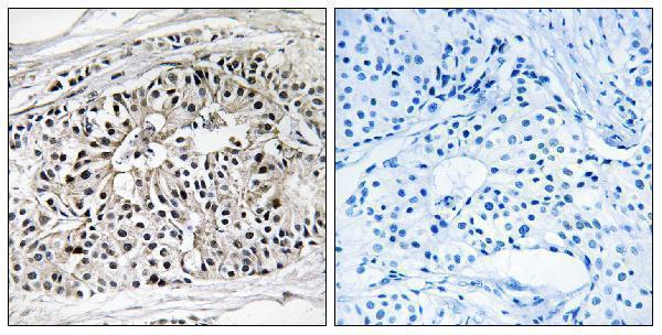
Real IHC data Immunohistochemistry analysis of paraffin-embedded human breast carcinoma tissue, using EHHADH Antibody. The picture on the right is blocked with the synthesized peptide.
Anti-PBFE EHHADH Antibody
Cat # A05757-1
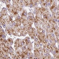
Real IHC data Formalin-fixed and paraffin-embedded human hepatocarcinoma with EHHADH Antibody (C-term), which was peroxidase-conjugated to the secondary antibody, followed by DAB staining. This data demonstrates the use of this antibody for immunohistochemistry; clinical relevance has not been evaluated.
Anti-EHHADH Antibody (C-term)
Cat # A05757-3

A05757-1 renders with IHC of paraffin-embedded human breast carcinoma and a peptide-blocking comparison (A05757-1 image caption). A05757-3 renders with peroxidase/DAB IHC of formalin-fixed, paraffin-embedded human hepatocarcinoma (A05757-3 image caption).

Which to pick: For tissue IHC, A05757-1 offers a peptide-blocking comparison in human paraffin sections; its caption does not report the fixative (A05757-1 image caption). For IF, choose A05757-1 at its listed 1:50 dilution; IF is listed, but no IF figure or ICC application is supplied (A05757-1 applications, dilution, image alts). For IHC across human, mouse, and rat, A05757-3 lists all three species and IHC-P; its figure shows human tissue only, and both pictured antibodies are rabbit polyclonals (A05757-3 reactivity, applications, image caption; catalog: hosts, dilution_raw).

Each figure is that product's own IHC / IF validation image from its datasheet.

References

  1. UniProt Consortium. UniProt entry Q08426 (ECHP_HUMAN, Peroxisomal bifunctional enzyme).
  2. Human Protein Atlas. EHHADH tissue IHC expression (reliability: Enhanced).
  3. Human Protein Atlas. EHHADH subcellular location (ICC-IF): Highest expression in Hep-G2: 27.8 nTPM.
  4. Human Protein Atlas. EHHADH antibody validation summary (2 antibodies).
  5. Tandem Mass Tag-based quantitative proteomics analysis of metabolic associated fatty liver disease induced by high fat diet in mice. Nutrition & metabolism 2020 — PMC7672977.
  6. Linoleic acid metabolic reprogramming is linked to immunometabolic remodeling and post-transplant recurrence risk in hepatocellular carcinoma. Frontiers in immunology 2026 — PMC13503599.
  7. VHL-dependence of EHHADH Expression in a Human Renal Cell Carcinoma Cell Line. Journal of kidney cancer and VHL 2024 — PMC10834178.
  8. Telomerase-related gene EHHADH drives lung cancer progression and shapes the immunosuppressive tumor microenvironment. Translational oncology 2026 — PMC13272562.
  9. PubMed PMID:8188243 — UniProt-cited evidence.
  10. PubMed PMID:14702039 — UniProt-cited evidence.
  11. PubMed PMID:16641997 — UniProt-cited evidence.