This website uses cookies to ensure you get the best experience on our website.
- Table of Contents
3 Q&As
Facts about Echinoderm microtubule-associated protein-like 4.

.
| Human | |
|---|---|
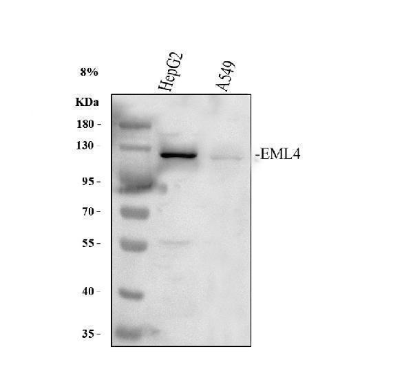
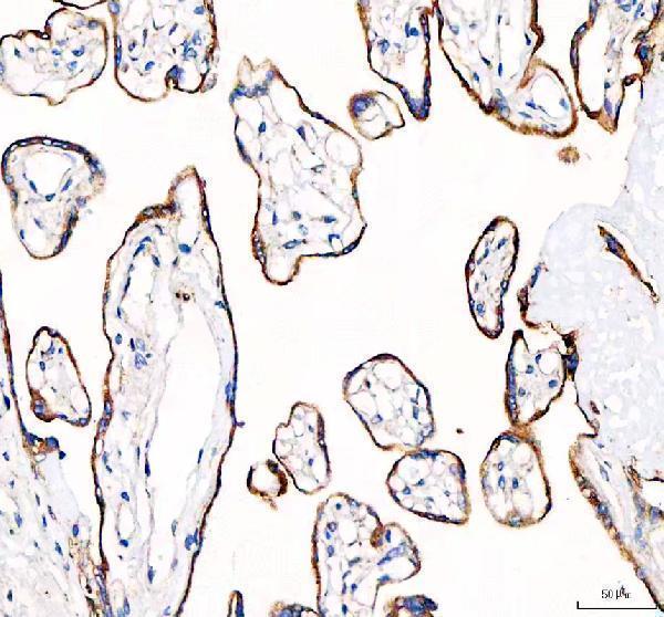
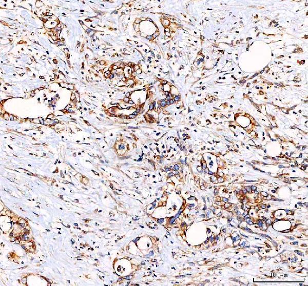
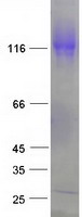
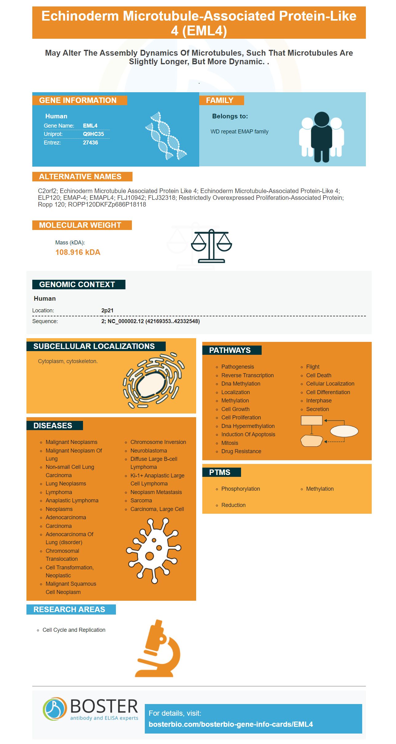
| Gene Name: | EML4 |
| Uniprot: | Q9HC35 |
| Entrez: | 27436 |

| Belongs to: |
|---|
| WD repeat EMAP family |

C2orf2; echinoderm microtubule associated protein like 4; echinoderm microtubule-associated protein-like 4; ELP120; EMAP-4; EMAPL4; FLJ10942; FLJ32318; Restrictedly overexpressed proliferation-associated protein; Ropp 120; ROPP120DKFZp686P18118
Mass (kDA):
108.916 kDA

| Human | |
|---|---|
| Location: | 2p21 |
| Sequence: | 2; NC_000002.12 (42169353..42332548) |
Cytoplasm, cytoskeleton.


PMID: 10995578 by Heidebrecht H.J., et al. Cloning and localization of C2orf2(ropp120), a previously unknown WD repeat protein.