This website uses cookies to ensure you get the best experience on our website.


- Table of Contents
Plan paraffin-section EPN2 IHC around medium neuronal staining in caudate, cerebral cortex and hippocampus (HPA tissue IHC). This guide covers a 1:200 starting dilution (datasheet A08001-1), expected cytoplasmic vesicle localisation (UniProt), and specificity controls prompted by presumed off-target staining (HPA tissue IHC).

Expected localisation, validated protocols, controls and antibodies — the at-a-glance facts below, then the full design guide.
| Expected localisation | Cytoplasmic vesicles expected (UniProt); IHC compartment unreported (HPA tissue IHC) | |
| Staining pattern | Medium neuronal staining in CNS; compartment and texture unreported (HPA tissue IHC) | |
| Antigen retrieval | Citrate pH 6.0 HIER, heat-mediated (datasheet A08001-1) | |
| Positive control | Caudate+2 more · see all | |
| Negative control | Adipose tissue+4 more · see all |
| Fixation | Keep fixation consistent across sections (standard IHC practice; not target-specific). Selected-image fixative and duration unreported (datasheet A08001-1); verify before use. | |
| Caveat | Presumed off-target staining may affect interpretation (HPA tissue IHC) | |
| Regulation | Highest expression in brain (UniProt) | |
| Isoform / epitope | 4 isoforms; no extracellular segment; epitope coverage unknown (UniProt) |
The catalog antibody’s IHC-P protocol (datasheet A08001-1) is accompanied by a published EPN2 protocol for paraffin-embedded bladder cancer tissue (PMC12573180).
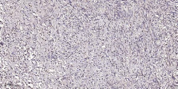
| Sample | Paraffin-embedded human small intestinal carcinoma tissue; fixative not specified (datasheet A08001-1) |
| Fixation | Image fixative and duration unreported (datasheet A08001-1); verify before use. |
| Sectioning | 4–5 µm sections on charged slides (standard) |
| Deparaffinisation | Xylene, graded ethanol series to water (standard) |
| Antigen retrieval | Heat retrieval: Citrate pH 6.0, 20min (datasheet A08001-1) |
| Peroxidase block | 3% H2O2, 10 min, room temperature (standard) |
| Blocking | 10% normal serum of the secondary host, 30 min, room temperature (standard) |
| Primary antibody | Rabbit anti-EPN2, 1:100-1:300 (datasheet A08001-1) |
| Primary incubation | Overnight at 4 °C (standard) |
| Detection | HRP-polymer secondary, DAB chromogen 5–10 min (standard) |
| Counterstain | Hematoxylin, blue, dehydrate and mount (standard) |
| Expected result | EPN2-positive staining in neuronal cells of caudate (HPA tissue IHC: Medium). HPA tissue profile: Expression in CNS. No signal in the no-primary control. |
EPN2 should appear mainly as cytoplasmic vesicular staining in neuronal cells of the caudate, cerebral cortex, and hippocampus (UniProt O95208: cytoplasm and clathrin-coated vesicles; HPA: Medium in neuronal cells). It has no transmembrane segment (UniProt O95208 topology). Interpret the pattern cautiously: HPA rates tissue IHC Approved but reports low consistency with RNA data and presumed off-target staining that was disregarded (HPA: tissue IHC reliability).
| Discrete cytoplasmic puncta in neuronal cells, with moderate staining in CNS sections. | This fits EPN2’s vesicular location and the observed neuronal pattern in caudate, cortex, and hippocampus (UniProt O95208: subcellular location; HPA: Medium in neuronal cells). Golgi-region concentration is plausible, but its absence alone does not invalidate an otherwise vesicular pattern (UniProt O95208: Golgi-region note). |
| Predominantly nuclear staining, or a continuous cell-surface rim. | Neither is the expected EPN2 compartment: UniProt places it in cytoplasm and clathrin-coated vesicles and lists no transmembrane segment (UniProt O95208: location and topology). Check compartment assignment and staining controls before scoring such signal as EPN2 (general IHC practice). |
| Strong staining in adipocytes or other cells listed as undetected by HPA. | HPA reports EPN2 as Not detected in adipose-tissue adipocytes and several other sampled cell types (HPA: tissue IHC). Unexpected staining raises concern for cross-reactivity or endogenous detection activity; confirm with appropriate controls. HPA’s Approved rating and low RNA concordance limit how firmly any single tissue can serve as a negative (HPA: reliability; general IHC practice). |
| Uniform haze across cells, stroma, or blank areas instead of localized puncta. | Diffuse background is less consistent with the reported cytoplasmic vesicle pattern (UniProt O95208: location; HPA: vesicles supported by ICC-IF). Review no-primary and detection controls, then optimize blocking, washing, and antibody concentration as general IHC troubleshooting steps (general IHC practice). |
| No staining in neuronal cells of a caudate, cortex, or hippocampus section. | That conflicts with HPA’s Medium neuronal staining in those tissues (HPA: tissue IHC). Check that the relevant cells are present and that the positive control and detection reagents work. A negative section alone cannot establish absent EPN2, particularly given HPA’s stated antibody–RNA inconsistency (HPA: reliability; general IHC practice). |
| Compartment and topology | EPN2 is cytoplasmic, associates with clathrin-coated vesicles, and lacks a transmembrane segment (UniProt O95208: location and topology). Score intracellular puncta in cell context; a membrane-like rim does not match that annotation. |
| Tissue and cell context | HPA records Medium neuronal staining in caudate, cerebral cortex, and hippocampus, but Not detected in cerebellar granular-layer cells (HPA: tissue IHC). Choose and interpret controls at the cell-type level rather than treating all brain regions alike. |
| IHC evidence strength | One listed antibody has IHC Approved status, without an IHC Enhanced designation; HPA also flags low staining–RNA consistency and presumed off-target binding that was disregarded (HPA: antibodies and reliability). Treat unusual staining as provisional until controls support it. |
| Isoforms and assay scope | UniProt lists 4 EPN2 isoforms (UniProt O95208: isoforms). The supplied evidence gives no antibody epitope or isoform-specific IHC pattern, so section staining cannot be assigned to a particular isoform from these records alone. |
| IF/ICC Q&A: what pattern is expected? | Vesicles are the supported main ICC-IF location, with images listed for A-549 and U2OS (HPA: subcellular). The listed ICC Supported antibody differs from the IHC Approved antibody (HPA: antibodies); these records do not establish an IF protocol for paraffin sections. |
| Situation | Likely cause | Next action |
|---|---|---|
| CNS neuronal positive control is blank. | The observed HPA pattern predicts Medium neuronal staining in caudate, cortex, or hippocampus; a blank result may reflect assay failure or sample context (HPA: tissue IHC; general IHC practice). | Verify neuronal cells and section quality, then check the IHC-validated antibody, detection reagents, and positive-control run. Optimize retrieval using assay controls; no EPN2-specific retrieval condition is supplied (general IHC practice). |
| Diffuse color obscures intracellular detail. | The distribution does not resolve the vesicular localization reported for EPN2 (UniProt O95208: location; HPA: subcellular). Excess background can arise in chromogenic IHC (general IHC practice). | Inspect no-primary and detection controls; review blocking, washing, and antibody concentration, then reassess whether distinct cytoplasmic puncta remain (general IHC practice). |
| Signal appears mainly nuclear or at cell borders. | Those compartments conflict with cytoplasmic vesicle localization and the absence of a transmembrane segment (UniProt O95208: location and topology). | Check counterstain boundaries and focus; compare a control section and the expected neuronal-cell pattern before recording the signal as EPN2 (HPA: Medium in neuronal cells; general IHC practice). |
| Adipocytes or other HPA-undetected cells stain strongly. | The staining differs from HPA’s sampled cell-type observations; cross-reactivity or endogenous chromogen-generating activity are possible explanations (HPA: tissue IHC; general IHC practice). | Run no-primary and detection controls and check endogenous enzyme blocking where relevant. Treat the result cautiously because HPA reports low antibody-staining concordance with RNA (HPA: reliability; general IHC practice). |
| All sections, including the no-primary control, develop color. | A no-primary control cannot report EPN2 antibody binding; staining there points toward detection-system background or endogenous activity (general IHC practice). | Check the detection reagents and blocking steps, including endogenous enzyme blocking for the chosen chromogen, before interpreting tissue differences (general IHC practice). |
| A cerebellar granular-layer sample is blank while another CNS sample stains. | This can match the supplied observations: HPA reports Not detected in granular-layer cells but Medium staining in neuronal cells of caudate, cortex, and hippocampus (HPA: tissue IHC). | Score the named cell populations separately and use an HPA-observed positive region to assess assay performance. Do not call the blank granular layer a failed run by itself (HPA: tissue IHC; general IHC practice). |


Comprehensive Human Protein Atlas IHC scoring per tissue (reliability: Approved — Low consistency between antibody staining and RNA expression data. Presumed off target binding observed and disregarded.). Rows are taken directly from the HPA tissue chart — click any row's HPA link to view the source.
| Tissue | Cell type | Level | Evidence | Source |
|---|---|---|---|---|
| Caudate | Neuronal cells | Medium | Protein (IHC) | HPA → |
| Cerebral cortex | Neuronal cells | Medium | Protein (IHC) | HPA → |
| Hippocampus | Neuronal cells | Medium | Protein (IHC) | HPA → |
| Tissue | Cell type | Level | Evidence | Source |
|---|---|---|---|---|
| Adipose tissue | Adipocytes | Not detected | Protein (IHC) | HPA → |
| Adrenal gland | Glandular cells | Not detected | Protein (IHC) | HPA → |
| Appendix | Glandular cells | Not detected | Protein (IHC) | HPA → |
| Bone marrow | Hematopoietic cells | Not detected | Protein (IHC) | HPA → |
| Breast | Adipocytes | Not detected | Protein (IHC) | HPA → |
Troubleshoot EPN2 staining in paraffin sections by checking retrieval, compartment, controls and scoring alongside the reported neuronal staining (HPA tissue IHC).
A08001-1 has a human paraffin-section IHC image (catalog image caption). IF is listed, with Human, Mouse, and Rat reactivity; no IF image is supplied (catalog applications; catalog reactivity; catalog IF images).
A08001-1 will render with an IHC image of paraffin-embedded human small intestinal carcinoma (catalog image caption). It lists IHC and IF applications and Human, Mouse, and Rat reactivity; the payload supplies no IF image (catalog applications; catalog reactivity; catalog IF images).
Which to pick: For tissue IHC, choose A08001-1: its image shows a human paraffin section stained at 1:200 after sodium citrate pH 6.0 retrieval; the fixative is unreported (catalog image caption). For IF, A08001-1 lists a 1:50 dilution, but supplies no IF image or ICC application, and its clone is unreported (catalog IF dilution; catalog IF images; catalog applications; catalog clone). For cross-species work, A08001-1 lists Human, Mouse, and Rat reactivity, while its pictured IHC evidence is human only (catalog reactivity; catalog image caption).