This website uses cookies to ensure you get the best experience on our website.


- Table of Contents
Plan FBXO7 chromogenic IHC in paraffin sections with the IHC-validated antibody at 2–5 μg/ml (datasheet A04086-2). Assess nuclear and cytoplasmic staining cautiously because HPA rates its tissue IHC evidence uncertain (HPA tissue IHC).

Expected localisation, validated protocols, controls and antibodies — the at-a-glance facts below, then the full design guide.
| Expected localisation | Nuclear and cytoplasmic in most tissues (HPA tissue IHC) | |
| Staining pattern | Nuclear and cytoplasmic staining in most tissues (HPA tissue IHC) | |
| Antigen retrieval | EDTA pH 8.0 HIER, heat-mediated (datasheet A04086-2) | |
| Positive control | Appendix+4 more · see all | |
| Negative control | Parathyroid gland+1 more · see all |
| Fixation | Keep fixation consistent across sections (standard IHC practice; not target-specific) | |
| Caveat | Staining and RNA show low consistency (HPA tissue IHC) | |
| Regulation | No tissue intensity regulator annotated (UniProt) | |
| Isoform / epitope | 3 isoforms; epitope coverage unspecified (UniProt) |
The catalog antibody protocol uses EDTA pH 8.0 retrieval (datasheet). The published IHC protocols below cover renal xenografts, mouse tumor tissues, and zebrafish brain (PMC12783301; PMC3117880; PMC3487786).
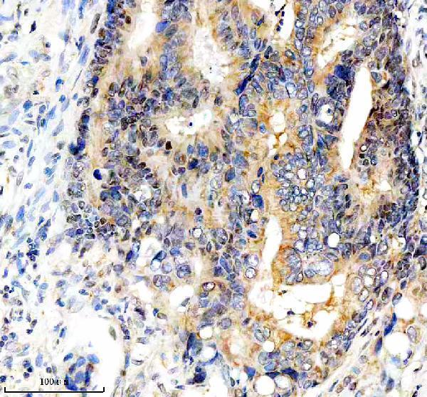
| Sample | Paraffin-embedded human colon cancer tissue; fixative not specified (datasheet A04086-2) |
| Fixation | Image fixative and duration unreported (datasheet A04086-2); verify before use. |
| Sectioning | 4–5 µm sections on charged slides (standard) |
| Deparaffinisation | Xylene, graded ethanol series to water (standard) |
| Antigen retrieval | Heat retrieval: EDTA pH 8.0 (datasheet A04086-2); 20 min, 95–100 °C (standard) |
| Peroxidase block | 3% H2O2, 10 min, room temperature (standard) |
| Blocking | 10% goat serum (datasheet A04086-2) |
| Primary antibody | Rabbit anti-FBXO7, 2-5 μg/ml (datasheet A04086-2) |
| Primary incubation | Overnight at 4 °C (datasheet A04086-2) |
| Detection | HRP-conjugated secondary, DAB chromogen (datasheet A04086-2) |
| Counterstain | Hematoxylin, blue, dehydrate and mount (standard) |
| Expected result | FBXO7-positive staining in glandular cells of appendix (HPA tissue IHC: High). HPA tissue profile: Nuclear and cytoplasmic expression in most tissues. No signal in the no-primary control. |
FBXO7 is predominantly cytoplasmic, with a minor nuclear fraction and possible relocation to depolarized mitochondria (UniProt Q9Y3I1). HPA reports nuclear and cytoplasmic staining in most tissues, including high staining in selected glandular, hematopoietic, endothelial, tubular, adipocyte, and macrophage populations (HPA tissue IHC). Interpret these patterns cautiously: HPA rates its tissue IHC profile Uncertain because antibody staining has low consistency with RNA expression (HPA tissue IHC).
| Nuclear and cytoplasmic staining in the expected cells, with a visible cellular outline and limited background. | This fits HPA’s broad tissue IHC profile (HPA tissue IHC). A stronger cytoplasmic component is compatible with predominantly cytoplasmic FBXO7 (UniProt Q9Y3I1). Record the compartments separately; the HPA tissue profile is rated Uncertain, so appearance alone does not establish antibody specificity (HPA tissue IHC). |
| Staining appears confined to an unexpected compartment, such as a sharp membrane rim or extracellular deposit. | FBXO7 has no transmembrane segment (UniProt Q9Y3I1). Check morphology, counterstain, and control sections before assigning such signal to FBXO7. Mitochondrial relocation is reported after depolarization (UniProt Q9Y3I1); it does not establish that every punctum in an untreated section is specific. |
| A cell population outside the expected HPA pattern stains strongly while nearby expected cells do not. | Compare cell identity on the counterstain and review antibody and detection controls. Cross-reactivity or endogenous chromogenic activity can mimic cell-specific signal (general IHC practice). HPA’s Uncertain tissue IHC rating makes an isolated unexpected pattern especially weak evidence for FBXO7 expression (HPA tissue IHC). |
| Brown signal covers stroma, lumina, or broad areas without recognizable nuclear or cytoplasmic boundaries. | Treat this as background until controls support cellular staining (general IHC practice). Diffuse deposits cannot be assigned the nuclear and cytoplasmic pattern reported by HPA (HPA tissue IHC). Check nonspecific binding, wash quality, and chromogen development within the validated workflow (general IHC practice). |
| Little or no signal appears in a section containing a reported high-staining population. | HPA reports high staining in bone marrow hematopoietic cells, kidney tubule cells, and lung macrophages, among others (HPA tissue IHC). First confirm that the intended cells are present, then check the staining run and antibody controls (general IHC practice). An absent signal does not alone prove absent FBXO7. |
| Tissue and cell choice | HPA reports High staining in appendix, duodenum, and gallbladder glandular cells; bone marrow hematopoietic cells; kidney tubule cells; and lung macrophages (HPA tissue IHC). These are comparison patterns, not guaranteed positive controls: HPA rates the tissue IHC profile Uncertain (HPA tissue IHC). |
| Low or undetected comparison cells | HPA reports no detection in parathyroid glandular cells and spleen red pulp cells, and low staining in cerebellar Purkinje cells, smooth muscle cells, and skeletal myocytes (HPA tissue IHC). Preserve cell-level context when comparing sections; a tissue name alone does not describe every cell in it. |
| IHC antibody evidence | HPA lists IHC as Uncertain for HPA032114, HPA057352, and CAB034296 (HPA antibodies). Its overall tissue profile also has low staining-to-RNA consistency (HPA tissue IHC). Treat agreement with that profile as supportive context, then assess the IHC-validated antibody with appropriate run controls (general IHC practice). |
| Isoforms and epitope coverage | UniProt lists 3 FBXO7 isoforms and an F-box domain at residues 329–375 (UniProt Q9Y3I1). The supplied evidence does not map this antibody’s epitope across isoforms, so staining cannot identify an isoform or justify an isoform-specific negative call. |
| Topology and processing | UniProt describes one chain spanning residues 1–522, with no signal peptide, propeptide, or transmembrane segment (UniProt Q9Y3I1). This supports an intracellular interpretation; the record supplies no cleavage or shedding pattern that would explain extracellular staining. |
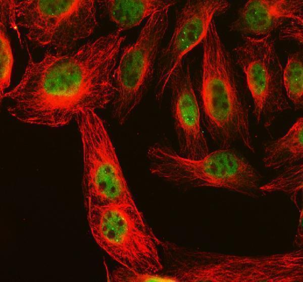
| IF/ICC cross-check? | HPA places FBXO7 in the supported nucleoplasm and cytosol locations in ICC-IF and lists images from A-431 and HEK293 (HPA subcellular). HPA032113 has Supported ICC status (HPA antibodies). This answers the compartment question; ICC-IF evidence does not validate an IHC-P staining run. |
| Situation | Likely cause | Next action |
|---|---|---|
| Expected cells are absent from the section or hard to recognize. | The comparison depends on cell identity: HPA assigns High staining to specific populations, such as kidney tubule cells and lung macrophages, rather than every cell in those tissues (HPA tissue IHC). | Review morphology and counterstain, locate the relevant population, and score it separately from neighboring cells (general IHC practice). Select another section if the intended cells are missing. |
| A reported high-staining population is present but has no visible signal. | A failed IHC run, unsuitable antibody conditions, or a true low result may look alike (general IHC practice); HPA’s High categories are observations with Uncertain overall tissue IHC reliability (HPA tissue IHC). | Check the run control and the IHC-validated antibody’s documented conditions, then repeat if controls failed (general IHC practice). Do not infer absence of FBXO7 from one negative section. |
| Nuclei stain strongly while cytoplasm is consistently blank. | HPA reports both nuclear and cytoplasmic tissue staining (HPA tissue IHC), while UniProt describes predominantly cytoplasmic FBXO7 with a minor nuclear fraction (UniProt Q9Y3I1). An exclusively nuclear result needs verification. | Check nuclear boundaries, chromogen development, and matched control sections (general IHC practice). Report the observed compartment accurately and avoid calling the pattern confirmed solely from nuclear signal. |
| A strong rim, lumen, or extracellular deposit dominates the slide. | This distribution does not match HPA’s nuclear and cytoplasmic profile (HPA tissue IHC) or the lack of a transmembrane segment and signal peptide (UniProt Q9Y3I1). Nonspecific staining is possible (general IHC practice). | Inspect the counterstain and negative detection control; review blocking and wash steps within the established IHC workflow (general IHC practice). Score only signal clearly assigned to intact cells. |
| Macrophages or other isolated cells stain intensely amid weak background. | Lung macrophages are reported High by HPA (HPA tissue IHC), but isolated chromogenic signal can also reflect endogenous detection activity or nonspecific binding (general IHC practice). Cell identity and controls remain decisive. | Confirm morphology, compare the expected compartment, and inspect an appropriate detection control (general IHC practice). Keep macrophage and neighboring cell scores separate; HPA’s tissue IHC reliability remains Uncertain (HPA tissue IHC). |
| ICC-IF suggests cytosolic signal, but the paraffin IHC section gives a different pattern. | HPA supports nucleoplasm and cytosol in ICC-IF, while its tissue IHC profile is Uncertain (HPA subcellular; HPA tissue IHC). Different assay formats and antibodies can yield patterns that need independent evaluation (general IHC practice). | Check each assay against its own controls and document the antibody used (general IHC practice). Use ICC-IF as localization context, not proof that the IHC section is specific. |


Comprehensive Human Protein Atlas IHC scoring per tissue (reliability: Uncertain — Low consistency between antibody staining and RNA expression data.). Rows are taken directly from the HPA tissue chart — click any row's HPA link to view the source.
| Tissue | Cell type | Level | Evidence | Source |
|---|---|---|---|---|
| Appendix | Glandular cells | High | Protein (IHC) | HPA → |
| Bone marrow | Hematopoietic cells | High | Protein (IHC) | HPA → |
| Breast | Adipocytes | High | Protein (IHC) | HPA → |
| Colon | Endothelial cells | High | Protein (IHC) | HPA → |
| Duodenum | Glandular cells | High | Protein (IHC) | HPA → |
Troubleshoot FBXO7 chromogenic IHC in paraffin sections by checking retrieval, compartment-specific staining, controls, and cell-level scoring.
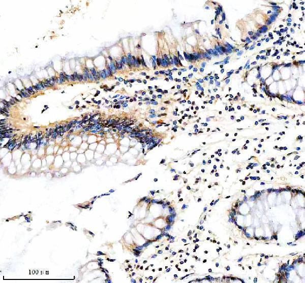
A04086-2 has IHC images from human paraffin sections of colon, colon cancer, and endometrial cancer, plus IF data from U2OS cells (catalog image captions); human and rat reactivity are listed (catalog).
A04086-2 has IHC images from human paraffin sections of colon, colon cancer, and endometrial cancer (A04086-2 IHC captions). The same SKU has an IF image from U2OS cells (A04086-2 IF caption); its application list includes IHC and ICC/IF (catalog).
Which to pick: Choose A04086-2 for human paraffin-section IHC: its own caption documents heat retrieval in EDTA at pH 8.0 and primary antibody at 2 μg/ml (A04086-2 IHC caption); the fixative is unreported (A04086-2 IHC caption). Choose the same SKU for IF/ICC because its IF caption shows staining in U2OS cells at 5 μg/ml (A04086-2 IF caption). For cross-species work, A04086-2 lists human and rat reactivity, but the supplied IHC and IF images show human samples only (catalog reactivity; A04086-2 image captions); clonality is unreported (catalog).