FECH / Ferrochelatase, mitochondrial · IHC design guide

Design Immunohistochemistry for FECH

Plan FECH staining in paraffin sections using the cytoplasmic pattern seen in most tissues and the distinct renal tubular staining as references (HPA tissue IHC). This guide covers antibody dilution, detection and scoring while distinguishing observed tissue staining from FECH’s mitochondrial inner membrane location (UniProt).

Evidence assembled Oct 2026 · For research use; verify linked source records and product datasheet before use
Immunohistochemistry protocol sheet for FECH (IHC for FECH): expected localisation Cytoplasmic tissue staining (HPA tissue IHC); mitochondrial inner membrane at the molecular level (UniProt), antibody A05156-2, validated IHC image, and IHC protocol steps
Printable FECH IHC protocol sheet — expected localisation Cytoplasmic tissue staining (HPA tissue IHC); mitochondrial inner membrane at the molecular level (UniProt), antibody A05156-2, controls and protocol steps. Open the full FECH IHC guide →

FECH Immunohistochemistry Experimental Design Guide

Expected localisation, validated protocols, controls and antibodies — the at-a-glance facts below, then the full design guide.

Must know before staining
Expected localisation Cytoplasmic tissue staining (HPA tissue IHC); mitochondrial inner membrane at the molecular level (UniProt)
Staining pattern Cytoplasmic in most tissues; distinct renal tubular cells (HPA tissue IHC)
Antigen retrieval EDTA pH 8.0 HIER, heat-mediated (datasheet A05156-2)
Positive control ⓘ Kidney+4 more · see all
Negative control ⓘ Adipose tissue+2 more · see all
Important caveats
Reasons your staining may differ from the expected pattern.
Fixation Matched tissue-IHC evidence does not establish the fixation claim. Validate the specimen-specific method before use. (selected-SKU IHC image A05156-2)
Caveat Antibody staining and RNA expression show low consistency (HPA tissue IHC)
Regulation Bone marrow RNA enhanced; IHC impact unclear (HPA tissue RNA)
Isoform / epitope 2 isoforms; mature chain 55–423; epitope coverage unknown (UniProt)
Section 1

Recommended FECH IHC & IF Protocols

The catalog antibody’s IHC-P protocol (datasheet A05156-2) is followed by published FECH IHC methods for meningioma, tissue sections, and glioma (PMC9818642; PMC3048207; PMC9157484).

Recommended immunohistochemistry (IHC-P) protocol parameters
SampleParaffin-embedded human benign Prostate Hyperplasia tissue; fixative not specified (datasheet A05156-2)
FixationImage fixative and duration unreported (datasheet A05156-2); verify before use.
Sectioning4–5 µm sections on charged slides (standard)
DeparaffinisationXylene, graded ethanol series to water (standard)
Antigen retrievalHeat retrieval: EDTA pH 8.0 (datasheet A05156-2); 20 min, 95–100 °C (standard)
Peroxidase block3% H2O2, 10 min, room temperature (standard)
Blocking10% goat serum (datasheet A05156-2)
Primary antibodyRabbit anti-FECH, 1:50 recommended; image 1:100 (datasheet A05156-2)
Primary incubationOvernight at 4 °C (datasheet A05156-2)
DetectionHRP-conjugated secondary, DAB chromogen (datasheet A05156-2)
CounterstainHematoxylin, blue, dehydrate and mount (standard)
Expected resultFECH-positive staining in cells in tubules of kidney (HPA tissue IHC: High). HPA tissue profile: Cytoplasmic expression in most tissues. Distinct expression in a subset of renal tubules. No signal in the no-primary control.
💡Decision noteStart with heat-mediated EDTA pH 8.0 retrieval for the catalog antibody (datasheet A05156-2); published FECH methods also report high-pH retrieval (PMC9157484) or citrate pH 6.0 (PMC9818642).
Section 2

What Is the Expected FECH Staining Pattern?

FECH is associated with the mitochondrial inner membrane and has no annotated transmembrane segment (UniProt P22830 topology). In paraffin section IHC, expect cytoplasmic staining, especially in a subset of renal tubule cells and prostate glandular cells (HPA tissue IHC). HPA rates its tissue IHC profile Approved but reports low consistency between antibody staining and RNA expression, so interpret tissue comparisons cautiously (HPA tissue IHC).

What am I looking at on my slide?
Distinct cytoplasmic staining in a subset of renal tubules; strong staining in prostate glands.This fits HPA's High staining in kidney tubule cells and prostate glandular cells (HPA tissue IHC). A chromogenic section cannot resolve FECH within the mitochondrial inner membrane; the cytoplasmic pattern is the practical IHC readout (UniProt P22830 topology; general IHC practice).
Predominantly nuclear staining, with little cytoplasmic signal.This disagrees with the reported cytoplasmic tissue profile and mitochondrial location (HPA tissue IHC; UniProt P22830). Check the primary antibody control, detection background and scoring before calling the nuclear signal FECH (general IHC practice).
Strong staining in adipocytes, smooth muscle cells or soft tissue fibroblasts.HPA reports FECH as Not detected in these cell types (HPA tissue IHC). Such staining raises concern for cross-reactivity or endogenous detection activity; compare a no-primary control and the expected positive cells on the same run (general IHC practice).
Uniform haze across cells and surrounding section, obscuring cell boundaries.This is difficult to score as FECH because HPA describes cellular cytoplasmic staining, with distinct expression in a subset of renal tubules (HPA tissue IHC). Review blocking, washes and detection background using a no-primary control (general IHC practice).
No staining in renal tubule cells or prostate glandular cells.Both are High staining reference cell types in HPA tissue IHC (HPA tissue IHC). An absent signal there makes a negative call elsewhere uncertain; first review the positive control, reagent performance and chromogenic detection (general IHC practice).
💡Expected FECH appearanceCall a positive result when cytoplasmic stain is distinct in a subset of renal tubule cells or prostate glandular cells, where HPA reports High staining; diffuse haze or predominantly nuclear stain does not match that profile (HPA tissue IHC; UniProt P22830 topology).
How each factor affects the staining
Compartment and optical resolutionFECH is assigned to the mitochondrial inner membrane without an annotated transmembrane segment (UniProt P22830 topology). HPA describes cytoplasmic tissue IHC staining; a chromogenic cytoplasmic signal alone cannot prove inner membrane localization (HPA tissue IHC; general IHC practice).
Cell type selectionKidney tubule cells and prostate glandular cells have High staining; adrenal and appendix glandular cells have Medium staining (HPA tissue IHC). Use the reported cell type, rather than a whole tissue average, when judging whether a section is an informative positive control (general IHC practice).
Expression and validation limitsHPA rates the tissue IHC profile Approved but notes low consistency with RNA expression; its two listed antibodies are IHC Approved, with no ICC status reported (HPA tissue IHC; HPA antibodies). RNA abundance or IHC approval alone should not settle an unexpected cellular pattern.
Mature protein and epitopeUniProt annotates a mature FECH chain spanning residues 55–423 and 2 isoforms (UniProt P22830). The supplied sources do not locate the antibody epitope, so they cannot predict whether processing or isoform choice changes this antibody's staining.
IF/ICC: what pattern is established?UniProt places FECH at the mitochondrial inner membrane, but HPA supplies no ICC-IF images or main subcellular location for this record (UniProt P22830; HPA subcellular). Treat a proposed punctate mitochondrial IF pattern as a hypothesis to validate, not an HPA-observed result.
Why is my staining missing, weak or wrong?
SituationLikely causeNext action
High staining reference cells are blank.A negative run is difficult to interpret when kidney tubule cells or prostate glandular cells lack their reported High signal (HPA tissue IHC).Verify the positive control and primary antibody step, then review retrieval and detection using the antibody's IHC-P instructions; these are general workflow checks, not evidence of FECH-specific retrieval or fixation sensitivity (general IHC practice).
Only scattered renal tubules stain.This can match HPA's distinct expression in a subset of renal tubules (HPA tissue IHC).Score tubule cells individually and compare cellular localization and intensity with the expected cytoplasmic pattern before treating patchiness as a failed run (HPA tissue IHC; general IHC practice).
The entire section has brown haze.Widespread background can hide the cytoplasmic, cell-specific pattern described by HPA (HPA tissue IHC; general IHC practice).Inspect a no-primary control; review blocking, washes and detection conditions before changing the interpretation of tissue staining (general IHC practice).
Nuclei dominate the signal.Predominantly nuclear staining conflicts with mitochondrial FECH localization and HPA's cytoplasmic tissue profile (UniProt P22830; HPA tissue IHC).Compare with a no-primary control and an expected positive cell type; score nuclear staining separately rather than counting it as confirmed FECH (general IHC practice).
Reported negative cell types stain strongly.HPA lists adipocytes, smooth muscle cells and soft tissue fibroblasts as Not detected (HPA tissue IHC).Check no-primary background and cell identification, then compare the same run with kidney tubule or prostate glandular cells before assigning the unexpected signal to FECH (HPA tissue IHC; general IHC practice).

Sample controls for FECH IHC & IF

🧪Run kidney first: tubular cells should stain strongly (HPA: High in cells in tubules). Use adipose tissue as a negative control (HPA: Not detected in adipocytes); on the kidney slide, assess adjacent nontubular cells as internal negatives only if they remain unstained.
Positive control tissue: Kidney (Cells in tubules, HPA High)
Negative control tissue: Adipose tissue (HPA Not detected)
ICC-IF cell lines (HPA subcellular resource): HPA carries no ICC-IF cell line for FECH; derive a cell-line control from the positive tissue's cell type (Cells in tubules) and confirm it by RNA or western blot first.
Technical controls: Include a no-primary, secondary-only control and a concentration-matched rabbit IgG isotype control (selected-SKU caption: rabbit anti-FECH primary). A FECH knockout specimen provides a biological negative where available (standard IHC practice). Quench endogenous peroxidase in kidney sections before HRP/DAB detection (standard chromogenic IHC practice; selected-SKU caption: HRP/DAB).
⚠️Feasibility: No target-specific fixation window or fixation effect is reported, and the selected-SKU paraffin-section caption leaves the fixative unreported. The reported IHC procedure uses heat-mediated EDTA retrieval at pH 8.0; retrieval dependence has not been established (selected-SKU tissue-IHC caption). Frozen sections and IF have no matched procedure here, so their relative ease is unknown; kidney sections warrant an endogenous-peroxidase control for chromogenic detection (standard IHC practice).

HPA tissue IHC evidence for FECH

Comprehensive Human Protein Atlas IHC scoring per tissue (reliability: Approved — Low consistency between antibody staining and RNA expression data.). Rows are taken directly from the HPA tissue chart — click any row's HPA link to view the source.

Positive expression · recommended positive controls

TissueCell typeLevelEvidenceSource
Kidney Cells in tubules High Protein (IHC) HPA →
Prostate Glandular cells High Protein (IHC) HPA →
Adrenal gland Glandular cells Medium Protein (IHC) HPA →
Appendix Glandular cells Medium Protein (IHC) HPA →
Bronchus Respiratory epithelial cells Medium Protein (IHC) HPA →

Undetected expression · recommended negative controls

TissueCell typeLevelEvidenceSource
Adipose tissue Adipocytes Not detected Protein (IHC) HPA →
Smooth muscle Smooth muscle cells Not detected Protein (IHC) HPA →
Soft tissue Fibroblasts Not detected Protein (IHC) HPA →
Section 3

Advanced FECH IHC Tips

Troubleshoot FECH staining by checking retrieval, compartment, and controls before comparing signal across paraffin sections.

Which retrieval condition should I start with for FECH in paraffin sections?
Start with heat-mediated antigen retrieval in EDTA pH 8.0 (datasheet A05156-2). The selected image used this condition before 1:100 primary antibody incubation overnight at 4°C, so keep those variables fixed while assessing retrieval (datasheet A05156-2). Compare retrieval times on adjacent sections, recording tissue integrity and glandular-cell signal (HPA: High in prostate glandular cells; standard IHC practice). If staining remains weak, test another buffer or pH as a documented fallback with matched positive and no-primary controls; those alternatives lack validation in the supplied evidence for this catalog antibody (datasheet A05156-2; standard IHC practice).
How should I troubleshoot FECH staining when fixation history is uncertain?
The selected tissue image is described as paraffin-embedded, but its fixative and fixation duration are unreported; target-specific fixation sensitivity is therefore unknown (datasheet A05156-2). Record the actual fixative, duration, and processing history for each block, then compare adjacent sections under the same EDTA pH 8.0 retrieval and detection conditions (datasheet A05156-2; standard IHC practice). Include a consistently processed positive control and a no-primary control in each run, since apparent losses can arise from processing or detection variation (standard IHC practice). Do not infer a FECH-specific fixation effect from its inner-membrane location, modifications, or HPA staining pattern (UniProt P22830; HPA tissue IHC).
What staining pattern should I expect for FECH in chromogenic IHC?
Expect predominantly cytoplasmic staining at light-microscope resolution, because FECH is assigned to the mitochondrial inner membrane and HPA reports cytoplasmic expression across most tissues (UniProt P22830 subcellular location; HPA tissue IHC). The record reports no transmembrane segment, so a crisp plasma-membrane outline is not the predicted pattern (UniProt P22830 topology). Use glandular cells in prostate or tubular cells in kidney as tissue benchmarks, where HPA reports High staining (HPA: High in prostate glandular cells and kidney tubule cells). Compare these cells with adjacent unstained compartments and a no-primary slide before calling diffuse nuclear or extracellular DAB signal specific (standard IHC practice).
Can this antibody distinguish FECH isoforms or processing states in tissue?
Do not assign an IHC stain to one FECH isoform without epitope validation: the record lists 2 isoforms, while the selected caption gives no epitope sequence (UniProt P22830 isoforms; datasheet A05156-2). The annotated mature chain spans residues 55–423, and modifications include residue 57, 138, and alternate modifications at 415 (UniProt P22830 processing and modified residues). Ask whether the catalog antibody recognizes a sequence shared between isoforms and assess epitope accessibility after tissue processing (standard IHC practice). Until resolved, report staining as FECH immunoreactivity, with the antibody SKU and retrieval conditions, rather than an isoform or modification-specific measurement (datasheet A05156-2; standard IHC practice).
How can IF help resolve ambiguous FECH IHC localisation?
In a separate IF experiment, compare FECH with a mitochondrial marker and a marker of the expected positive cell type, such as prostate glandular or kidney tubule cells (UniProt P22830 subcellular location; HPA: High in those cell populations). Inspect unstained tissue autofluorescence before choosing well-separated fluorophores, placing weaker signal in a channel with less background (standard IF practice). FECH associates with the mitochondrial inner membrane without an annotated transmembrane segment; its epitope side is unspecified, so map the epitope and optimise permeabilisation for access to that side (UniProt P22830 topology; standard IF practice). Include single-label and no-primary controls, and assess mitochondrial morphology (standard IF practice).
How do I separate FECH staining from chromogenic background?
Run a no-primary slide and inspect whether DAB appears on both slides (standard IHC practice). Use a peroxidase block before HRP detection and titrate primary antibody around the caption's 1:100 condition; the caption specifies overnight incubation at 4°C and DAB development (standard IHC practice; datasheet A05156-2). Maintain the reported 10% goat-serum block while assessing one variable at a time, and avoid scoring pigment, precipitate, or staining concentrated at section edges (datasheet A05156-2; standard IHC practice). Compare glandular cells with less reactive neighboring compartments and keep DAB development time consistent across slides (HPA: High in prostate glandular cells; standard IHC practice).
How should I score FECH IHC across samples with different cell composition? ⚠ ANSWER MARKED FOR VERIFICATION
Define the analysis compartment, such as prostate glandular cells or kidney tubule cells, because HPA reports High staining in those populations (HPA tissue IHC). Score the percentage of positive cells and staining intensity with an H-score, or measure DAB-positive area per mm² (standard IHC practice). Normalize counts to eligible cells or measured compartment area, and keep retrieval, antibody incubation, imaging, and threshold settings constant across compared slides (datasheet A05156-2; standard IHC practice). Report excluded edge, necrotic, and artifact regions and use blinded replicate scoring where feasible; HPA's Approved label carries a low RNA–staining consistency note (standard IHC practice; HPA reliability).
When should I doubt a positive FECH IHC signal?
Favor a cellular cytoplasmic pattern in the expected population: FECH is mitochondrial inner-membrane associated, and HPA reports High staining in kidney tubules and prostate glandular cells (UniProt P22830 subcellular location; HPA tissue IHC). Treat isolated nuclear, extracellular, edge-concentrated, or necrotic staining as suspect until it survives matched section and no-primary controls (standard IHC practice). DAB signal persisting without primary antibody may reflect endogenous peroxidase or detection background, so review peroxidase blocking and chromogen timing (standard IHC practice). HPA calls its tissue IHC Approved but reports low consistency with RNA expression; interpret a discordant sample using independent evidence rather than the staining score alone (HPA reliability).
Boster reagents

Best FECH / Ferrochelatase, mitochondrial IHC Antibodies

A05156-2 and A05156 have paraffin-section IHC images from human tissues; A05156 also has a rat heart IHC image and lists IF/ICC, with no IF image supplied (catalog IHC captions, applications, IF image alts).

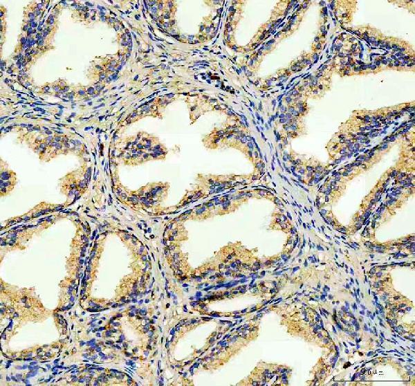
Real IHC data IHC analysis of FECH using anti-FECH antibody (A05156-2). FECH was detected in a paraffin-embedded section of human benign Prostate Hyperplasia tissue. Heat mediated antigen retrieval was performed in EDTA buffer (pH 8.0, epitope retrieval solution). The tissue section was blocked with 10% goat serum. The tissue section was then incubated with 1:100 rabbit anti-FECH Antibody (A05156-2) overnight at 4°C. Peroxidase Conjugated Goat Anti-rabbit IgG was used as secondary antibody and incubated for 30 minutes at 37°C. The tissue section was developed using HRP Conjugated Rabbit IgG Super Vision Assay Kit (Catalog # SV0002) with DAB as the chromogen.
Anti-FECH antibody
Cat # A05156-2
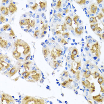
Real IHC data Immunohistochemistry of paraffin-embedded human stomach using FECH antibody at dilution of 1:100 .Perform microwave antigen retrieval with 10 mM PBS buffer pH 7.2 before commencing with IHC staining protocol.
Anti-Ferrochelatase, mitochondrial FECH Antibody
Cat # A05156

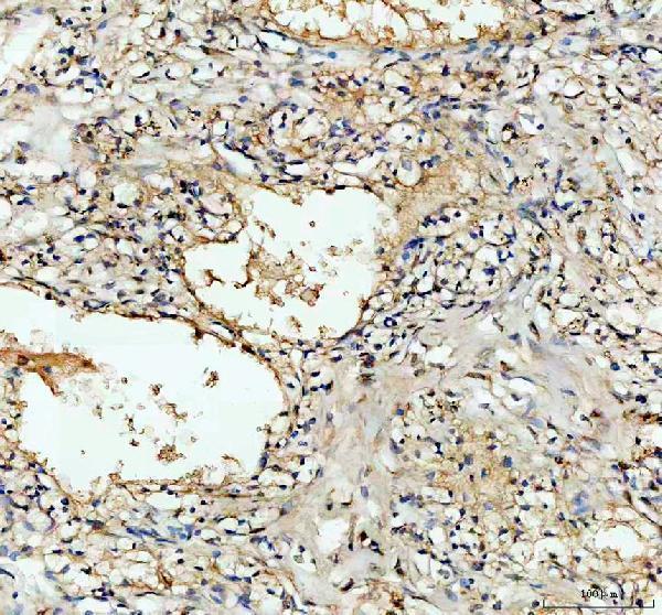
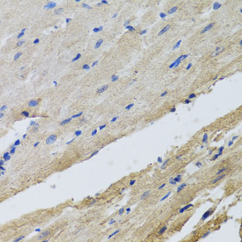
A05156-2 will render with human benign prostate hyperplasia IHC; its catalog also shows human renal cancer IHC (A05156-2 IHC captions). A05156 will render with human stomach IHC; its catalog also shows rat heart IHC and lists IF/ICC (A05156 IHC captions, applications).

Which to pick: For paraffin-section IHC, choose A05156-2 if its documented EDTA pH 8.0 retrieval and DAB workflow suits your assay (A05156-2 IHC captions). For IF/ICC, choose A05156, which lists those applications at 1:50–1:100; no IF image is supplied (A05156 applications, dilution, IF image alts). Both list human, mouse and rat reactivity, while A05156 has IHC images from two species, human and rat; the IHC captions do not report the fixative (catalog reactivity, IHC captions).

Each figure is that product's own IHC / IF validation image from its datasheet.

References

  1. UniProt Consortium. UniProt entry P22830 (HEMH_HUMAN, Ferrochelatase, mitochondrial).
  2. Human Protein Atlas. FECH tissue IHC expression (reliability: Approved).
  3. Human Protein Atlas. FECH subcellular location (ICC-IF): Highest expression in HEL: 29.2 nTPM.
  4. Human Protein Atlas. FECH antibody validation summary (2 antibodies).
  5. Protoporphyrin IX (PpIX) Fluorescence during Meningioma Surgery: Correlations with Histological Findings and Expression of Heme Pathway Molecules. Cancers 2023 — PMC9818642.
  6. Silencing of ferrochelatase enhances 5-aminolevulinic acid-based fluorescence and photodynamic therapy efficacy. British journal of cancer 2011 — PMC3048207.
  7. Heme Biosynthesis Factors and 5-ALA Induced Fluorescence: Analysis of mRNA and Protein Expression in Fluorescing and Non-fluorescing Gliomas. Frontiers in medicine 2022 — PMC9157484.
  8. Ferrochelatase regulates retinal neovascularization. FASEB journal : official publication of the Federation of American Societies for Experimental Biology 2020 — PMC7726024.
  9. PubMed PMID:2260980 — UniProt-cited evidence.
  10. PubMed PMID:14702039 — UniProt-cited evidence.
  11. PubMed PMID:15489334 — UniProt-cited evidence.