FYCO1 / FYVE and coiled-coil domain-containing protein 1 · IHC design guide

Design Immunohistochemistry for FYCO1

Plan FYCO1 paraffin IHC with the catalog antibody at 2–5 μg/ml (datasheet A06691-1) and assess cytoplasmic staining (HPA tissue IHC). Liver hepatocytes and colon glandular cells show high staining, although agreement between staining and RNA is low (HPA tissue IHC).

Evidence assembled Oct 2026 · For research use; verify linked source records and product datasheet before use
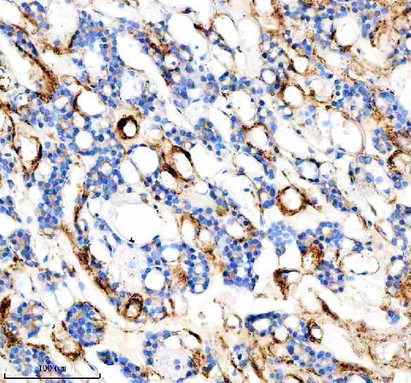
Immunohistochemistry protocol sheet for FYCO1 (IHC for FYCO1): expected localisation Cytoplasmic staining (HPA tissue IHC), antibody A06691-1, validated IHC image, and IHC protocol steps
Printable FYCO1 IHC protocol sheet — expected localisation Cytoplasmic staining (HPA tissue IHC), antibody A06691-1, controls and protocol steps. Open the full FYCO1 IHC guide →

FYCO1 Immunohistochemistry Experimental Design Guide

Expected localisation, validated protocols, controls and antibodies — the at-a-glance facts below, then the full design guide.

Must know before staining
Expected localisation Cytoplasmic staining (HPA tissue IHC)
Staining pattern General cytoplasmic staining across tissues (HPA tissue IHC)
Antigen retrieval EDTA pH 8.0 HIER, heat-mediated (datasheet A06691-1)
Positive control ⓘ Bone marrow+4 more · see all
Negative control ⓘ Caudate+1 more · see all
Important caveats
Reasons your staining may differ from the expected pattern.
Fixation Keep fixation consistent across sections (standard IHC practice; not target-specific)
Caveat Staining and RNA show low consistency (HPA tissue IHC)
Regulation Skeletal-muscle-enhanced RNA (HPA tissue RNA)
Isoform / epitope 4 isoforms; check epitope coverage (UniProt)
Section 1

Recommended FYCO1 IHC & IF Protocols

The catalog antibody has an IHC-P protocol (datasheet: A06691-1). One published FYCO1 IHC protocol stains mouse lens sections (PMC3398491).

Recommended immunohistochemistry (IHC-P) protocol parameters
SampleParaffin-embedded human thyroid cancer tissue; fixative not specified (datasheet A06691-1)
FixationImage fixative and duration unreported (datasheet A06691-1); verify before use.
Sectioning4–5 µm sections on charged slides (standard)
DeparaffinisationXylene, graded ethanol series to water (standard)
Antigen retrievalHeat retrieval: EDTA pH 8.0 (datasheet A06691-1); 20 min, 95–100 °C (standard)
Peroxidase block3% H2O2, 10 min, room temperature (standard)
Blocking10% goat serum (datasheet A06691-1)
Primary antibodyRabbit anti-FYCO1, 2-5 μg/ml (datasheet A06691-1)
Primary incubationOvernight at 4 °C (datasheet A06691-1)
DetectionHRP-conjugated secondary, DAB chromogen (datasheet A06691-1)
CounterstainHematoxylin, blue, dehydrate and mount (standard)
Expected resultFYCO1-positive staining in hematopoietic cells of bone marrow (HPA tissue IHC: High). HPA tissue profile: General cytoplasmic expression. No signal in the no-primary control.
💡Decision noteStart with heat-mediated EDTA pH 8.0 retrieval for the catalog antibody (datasheet: A06691-1); the published mouse lens protocol does not specify retrieval (PMC3398491).
Section 2

What Is the Expected FYCO1 Staining Pattern?

FYCO1 is associated with cytoplasmic vesicles, endosomes and lysosomes; it has no transmembrane segment (UniProt Q9BQS8). In tissue IHC, expect cytoplasmic staining in reported positive cells, including hepatocytes and colon glandular cells (HPA: general cytoplasmic expression; High in both). Treat the pattern as provisional: HPA rates its tissue IHC profile Uncertain because antibody staining and RNA expression have low consistency (HPA: tissue IHC reliability).

What am I looking at on my slide?
Cytoplasmic chromogen in hepatocytes or colon glandular cells, with any discernible vesicle-like puncta confined to cytoplasm.This fits reported High tissue staining and FYCO1's vesicle association (HPA: liver and colon IHC; UniProt Q9BQS8). Puncta are plausible, but routine paraffin IHC may not resolve individual vesicles (general IHC practice).
Strong, predominantly nuclear or sharply plasma-membrane staining, especially when cytoplasm is clear.This conflicts with reported general cytoplasmic expression and vesicle localisation (HPA: tissue IHC and subcellular ICC-IF). Check specificity and detection conditions before assigning this pattern to FYCO1 (general IHC practice).
Prominent staining of glial cells in caudate or hippocampus, particularly alongside weak staining in a positive tissue.HPA reports FYCO1 as Not detected in those glial cells (HPA: caudate and hippocampus IHC). Consider cross-reactivity or endogenous chromogenic activity; the Uncertain tissue IHC rating prevents treating one discordant slide as decisive (HPA: tissue IHC reliability; general IHC practice).
A uniform haze across cells, extracellular spaces and the section edge, without a clear cytoplasmic pattern.This is more consistent with nonspecific background than the reported cell-associated cytoplasmic profile (HPA: tissue IHC profile; general IHC practice). Assess blocking, washes and detection background before scoring expression (general IHC practice).
No detectable cytoplasmic signal in hepatocytes or colon glandular cells on the run.Both are reported High in HPA tissue IHC, so a blank result warrants a run check (HPA: liver and colon IHC). The HPA profile remains Uncertain; absence on one section alone does not establish biological absence (HPA: tissue IHC reliability; general IHC practice).
💡Expected FYCO1 appearanceA credible positive is predominantly cytoplasmic chromogen in reported High cells such as hepatocytes, possibly with puncta; dominant nuclear or extracellular colour is suspect (HPA: liver IHC and cytoplasmic profile; UniProt Q9BQS8; general IHC practice).
How each factor affects the staining
Compartment and resolutionFYCO1 associates with the external surface of autophagosomes and autolysosomes and with endosomes and lysosomes (UniProt Q9BQS8). HPA tissue IHC calls the pattern generally cytoplasmic; punctate detail may be less apparent in chromogenic sections (HPA: tissue IHC profile; general IHC practice).
Tissue and cell choiceHPA reports High staining in hepatocytes, colon glandular cells and several other listed populations, but Not detected in caudate and hippocampal glial cells (HPA: tissue IHC). Interpret within the named cell population rather than scoring an entire organ as uniformly positive or negative (general IHC practice).
Antibody evidenceHPA035526 has Uncertain IHC validation, and the tissue profile has Uncertain reliability (HPA: antibody validation and tissue IHC). Agreement with reported localisation supports interpretation but does not independently establish specificity (general IHC practice).
IF/ICC Q: Should vesicular fluorescence be expected?Yes: HPA reports enhanced vesicle localisation in ICC-IF, with Enhanced ICC validation for HPA035526 and HPA057966 (HPA: subcellular ICC-IF and antibody validation). Those ICC ratings do not upgrade the Uncertain tissue IHC assessment (HPA: antibody validation and tissue IHC).
Why is my staining missing, weak or wrong?
SituationLikely causeNext action
Positive tissue is blank.The run may have lost detectable staining, or that section may differ from HPA's reported High pattern (HPA: liver and colon IHC; general IHC practice).Check a concurrently processed positive section, primary-antibody application, retrieval, detection reagents and counterstain visibility (general IHC practice). Interpret a single negative cautiously because HPA tissue IHC reliability is Uncertain (HPA: tissue IHC reliability).
Chromogen covers the section as a diffuse haze.Insufficient blocking, concentrated detection reagents or inadequate washing can raise background (general IHC practice).Review a primary-omission control, blocking and wash steps; adjust detection conditions using the validated IHC workflow before scoring cytoplasm (general IHC practice).
Nuclei dominate while cytoplasm is faint.The distribution conflicts with FYCO1's reported cytoplasmic and vesicle localisation (HPA: tissue IHC and subcellular ICC-IF; UniProt Q9BQS8).Compare with a primary-omission control and a reported High cell population; review chromogen development and specificity before calling it FYCO1 (HPA: liver or colon IHC; general IHC practice).
Caudate or hippocampal glial cells appear strongly positive.This disagrees with HPA's Not detected calls; nonspecific binding or endogenous detection activity is possible (HPA: caudate and hippocampus IHC; general IHC practice).Inspect the cell identity and a primary-omission control, then compare staining with a reported High tissue on the same run (HPA: tissue IHC; general IHC practice).
Staining appears in many cell types, but cell boundaries are unclear.HPA reports a general cytoplasmic profile across several tissues, so broad staining alone cannot identify the expressing cell (HPA: tissue IHC).Use morphology and the counterstain to score the named cell population; record intensity and subcellular distribution separately (general IHC practice).
A fine punctate pattern is absent despite clear cytoplasmic staining.Chromogenic paraffin IHC may not resolve small vesicles, even though HPA ICC-IF reports vesicle localisation (HPA: subcellular ICC-IF; general IHC practice).Assess whether cytoplasmic staining matches the reported tissue and cell pattern; do not require visible individual vesicles for an IHC call (HPA: tissue IHC; general IHC practice).

Sample controls for FYCO1 IHC & IF

🧪Run colon first: glandular cells should stain (HPA: High in colon glandular cells). Use caudate glial cells as the negative tissue (HPA: Not detected in caudate glial cells); on the colon slide, assess cells outside the glands for background without assuming they lack FYCO1.
Positive control tissue: Bone marrow (Hematopoietic cells, HPA High)
Negative control tissue: Caudate (HPA Not detected)
ICC-IF cell lines (HPA subcellular resource): HPA ICC-IF images show FYCO1 in A-431, RT-4, U2OS, U-251MG, HeLa BAC 5295, HeLa BAC 5296, HeLa , with annotated localisation: Vesicles (enhanced) (HPA subcellular).
Technical controls: Include no-primary (secondary-only), a concentration-matched rabbit IgG isotype control matched to the primary’s clonality (caption: rabbit anti-FYCO1), and FYCO1 knockout material if available. Quench endogenous peroxidase for chromogenic detection and check the colon gland lumina for background deposits (HPA: colon glandular cells; caption: HRP/DAB detection).
⚠️Feasibility: A target-specific fixation window or fixation effect is unreported, and the selected tissue-IHC caption does not state a fixative (caption: fixative not stated). The caption uses EDTA retrieval at pH 8.0, but does not establish that retrieval is required (caption: heat-mediated EDTA retrieval). Frozen sections are not established as easier; IF/ICC has a separate guide, although vesicular staining is documented in ICC-IF images (HPA: vesicles, enhanced).

HPA tissue IHC evidence for FYCO1

Comprehensive Human Protein Atlas IHC scoring per tissue (reliability: Uncertain — Low consistency between antibody staining and RNA expression data. Pending external verification.). Rows are taken directly from the HPA tissue chart — click any row's HPA link to view the source.

Positive expression · recommended positive controls

TissueCell typeLevelEvidenceSource
Bone marrow Hematopoietic cells High Protein (IHC) HPA →
Bronchus Respiratory epithelial cells High Protein (IHC) HPA →
Colon Glandular cells High Protein (IHC) HPA →
Duodenum Glandular cells High Protein (IHC) HPA →
Epididymis Glandular cells High Protein (IHC) HPA →

Undetected expression · recommended negative controls

TissueCell typeLevelEvidenceSource
Caudate Glial cells Not detected Protein (IHC) HPA →
Hippocampus Glial cells Not detected Protein (IHC) HPA →
Section 3

Advanced FYCO1 IHC Tips

Troubleshoot FYCO1 chromogenic IHC in paraffin sections using the catalog antibody’s tissue image and the reported cellular localisation as starting points.

Which retrieval condition should I start with for FYCO1 paraffin-section IHC?
Start with heat-mediated EDTA retrieval at pH 8.0 for FYCO1 paraffin-section IHC (datasheet A06691-1). The selected tissue image used that condition before overnight incubation with 2 μg/ml primary antibody at 4°C (datasheet A06691-1). If staining is weak, first check that section processing, heating, and antibody incubation were consistent across slides (standard IHC practice). Then compare a small retrieval-time series while holding detection and exposure to DAB constant (standard IHC practice). Judge improvement by clearer cytoplasmic staining and preserved tissue morphology, rather than stronger staining alone (HPA: general cytoplasmic expression; standard IHC practice).
Could fixation explain weak FYCO1 staining in paraffin sections?
The selected paraffin-section caption does not state a fixative, so FYCO1-specific fixation sensitivity is unknown (datasheet A06691-1). Do not attribute weak staining to a particular fixative without comparing sections with documented processing histories (standard IHC practice). Inspect adjacent sections for tissue preservation, and run the same retrieval and antibody conditions on sections processed together (standard IHC practice). Keep section thickness, drying, and DAB development comparable when assessing whether a processing change helped (standard IHC practice). The reported cytoplasmic vesicle localisation does not establish how this antibody’s epitope responds to fixation (UniProt Q9BQS8: subcellular location; datasheet A06691-1: epitope not specified).
What FYCO1 staining pattern should I expect in chromogenic IHC?
Expect a predominantly cytoplasmic pattern in tissue sections, with possible punctate emphasis where vesicles can be resolved (HPA: general cytoplasmic expression; HPA subcellular: vesicles). FYCO1 is associated with autophagosomes, endosomes, and lysosomes, and remains on the outer surface of autolysosomes after fusion (UniProt Q9BQS8: subcellular location). That location supports cytoplasmic vesicular staining, but a single DAB section cannot identify the vesicle type (UniProt Q9BQS8: subcellular location; standard IHC practice). Compare staining in the cell population of interest with nearby morphology and counterstain before assigning a compartment (standard IHC practice). Treat isolated nuclear-only staining cautiously because it does not match the reported location (UniProt Q9BQS8: subcellular location).
How should isoforms and an unknown epitope affect FYCO1 IHC interpretation?
FYCO1 has 4 reported isoforms, but the selected tissue caption does not map this antibody’s epitope (UniProt Q9BQS8: isoforms; datasheet A06691-1). Consequently, staining cannot be assigned to one isoform from this IHC result alone (UniProt Q9BQS8: isoforms; datasheet A06691-1: epitope not specified). The protein has a RUN domain at residues 36–169 and a GOLD domain at 1337–1466, which makes epitope position relevant to interpreting variants (UniProt Q9BQS8: domains). Document the antibody identity and compare its disclosed immunogen, if available, with the isoforms under study (standard IHC practice). Avoid treating altered staining as isoform loss until epitope coverage is established (standard IHC practice).
How can IF help check FYCO1 localisation seen by chromogenic IHC?
Use IF as a localisation check alongside paraffin-section IHC, selecting a marker for the expected cell population; an epithelial marker can help assess the selected thyroid cancer tissue (datasheet A06691-1: tissue; standard IF practice). Choose spectrally separated fluorophores and consider a far-red FYCO1 channel if tissue autofluorescence obscures shorter wavelengths (standard IF practice). Because FYCO1 is associated with the external surface of autophagosomes and lacks a transmembrane segment, permeabilisation should allow antibody access to cytosol-facing structures (UniProt Q9BQS8: topology and subcellular location; standard IF practice). Optimise permeabilisation against morphology and background because this antibody’s epitope position is unreported (datasheet A06691-1; standard IF practice).
How do I reduce diffuse brown background without losing FYCO1 signal?
Compare a no-primary control with the stained section to identify signal from secondary detection or endogenous peroxidase (standard IHC practice). Include a peroxidase block before HRP detection, and control DAB development time across comparison slides (standard IHC practice). The selected image used 10% goat serum blocking, 2 μg/ml primary antibody, and an HRP/DAB readout (datasheet A06691-1). If background persists, titrate the primary antibody around that reported concentration while keeping retrieval and development fixed (datasheet A06691-1; standard IHC practice). Preserve plausible cytoplasmic or vesicular signal when judging improvement; uniform staining across unrelated compartments warrants scrutiny (HPA: general cytoplasmic expression; HPA subcellular: vesicles; standard IHC practice).
How should I score FYCO1 staining across paraffin sections? ⚠ ANSWER MARKED FOR VERIFICATION
Define the cell population and scoring region before reading slides, then exclude folds, edges, and necrotic areas consistently (standard IHC practice). Record the percentage of positive cells and staining intensity; an H-score can summarize intensity-weighted positivity on a 0–300 scale (standard IHC practice). For punctate staining that remains separable, report puncta density per mm² of viable tissue or per counted cell (standard IHC practice). Normalise comparisons to the same cell population, viable area, section processing, and DAB development conditions (standard IHC practice). Report the antibody and retrieval condition because the selected image used EDTA at pH 8.0 and 2 μg/ml antibody (datasheet A06691-1).
When is apparent FYCO1 positivity more likely to be artefact?
Give more weight to staining that follows viable cell morphology and a cytoplasmic, potentially vesicular distribution (HPA: general cytoplasmic expression; HPA subcellular: vesicles; standard IHC practice). Question nuclear-only deposits, strong staining at section edges, and brown signal over necrosis or damaged tissue (UniProt Q9BQS8: subcellular location; standard IHC practice). Compare no-primary and peroxidase-block controls to assess detection background and endogenous enzyme activity (standard IHC practice). Check the stained cell population rather than assuming every positive cell represents the intended compartment (standard IHC practice). Interpret tissue patterns cautiously: HPA rates its FYCO1 tissue IHC reliability uncertain because staining and RNA expression have low consistency (HPA: tissue IHC reliability).
Boster reagents

Best FYCO1 / FYVE and coiled-coil domain-containing protein 1 IHC Antibodies

A06691-1 has real IHC data from a paraffin-embedded human thyroid cancer section (catalog image caption). Listed reactivity covers human, mouse and rat (catalog reactivity).

Real IHC data IHC analysis of FYCO1 using anti-FYCO1 antibody (A06691-1). FYCO1 was detected in a paraffin-embedded section of human thyroid cancer tissue. Heat mediated antigen retrieval was performed in EDTA buffer (pH 8.0, epitope retrieval solution). The tissue section was blocked with 10% goat serum. The tissue section was then incubated with 2 μg/ml rabbit anti-FYCO1 Antibody (A06691-1) overnight at 4°C. Peroxidase Conjugated Goat Anti-rabbit IgG was used as secondary antibody and incubated for 30 minutes at 37°C. The tissue section was developed using HRP Conjugated Rabbit IgG Super Vision Assay Kit (Catalog # SV0002) with DAB as the chromogen.
Anti-FYCO1 Antibody ®
Cat # A06691-1

A06691-1 is the only SKU shown; it lists IHC and human, mouse and rat reactivity (catalog applications and reactivity). Its IHC figure shows a paraffin-embedded human thyroid cancer section, with no fixative reported (catalog image caption).

Which to pick: For tissue IHC on paraffin sections, choose A06691-1: its rabbit antibody is listed for IHC at 2–5 μg/ml, and its figure shows EDTA pH 8.0 retrieval on a human thyroid cancer section (catalog datasheet and image caption). There is no supported IF/ICC choice because A06691-1 has neither an IF/ICC application nor an IF figure (catalog applications and image alts). For mouse or rat samples, A06691-1 lists reactivity, but its shown IHC result is human only; the fixative is unreported (catalog reactivity and image caption).

Each figure is that product's own IHC / IF validation image from its datasheet.

References

  1. UniProt Consortium. UniProt entry Q9BQS8 (FYCO1_HUMAN, FYVE and coiled-coil domain-containing protein 1).
  2. Human Protein Atlas. FYCO1 tissue IHC expression (reliability: Uncertain).
  3. Human Protein Atlas. FYCO1 subcellular location (ICC-IF): Localized to vesicles..
  4. Human Protein Atlas. FYCO1 antibody validation summary (2 antibodies).
  5. Spatial expression patterns of autophagy genes in the eye lens and induction of autophagy in lens cells. Molecular vision 2012 — PMC3398491.
  6. Loss of FYCO1 leads to cataract formation. Scientific reports 2021 — PMC8253827.
  7. Nlp promotes autophagy through facilitating the interaction of Rab7 and FYCO1. Signal transduction and targeted therapy 2021 — PMC8050283.
  8. Consensus machine learning identifies cell death gene signature for carotid artery stenosis diagnosis. iScience 2026 — PMC12874109.
  9. PubMed PMID:11896456 — UniProt-cited evidence.
  10. PubMed PMID:17974005 — UniProt-cited evidence.
  11. PubMed PMID:16641997 — UniProt-cited evidence.