GLUD1 / Glutamate dehydrogenase 1, mitochondrial · IHC design guide

Design Immunohistochemistry for GLUD1

Plan chromogenic IHC for GLUD1 in paraffin sections using the catalog antibody’s documented conditions. Compare cytoplasmic staining with the HPA tissue profile, while accounting for its caution that staining may reflect protein from more than one gene (HPA tissue IHC).

Evidence assembled Oct 2026 · For research use; verify linked source records and product datasheet before use
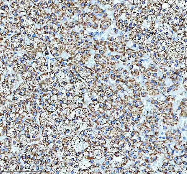
Immunohistochemistry protocol sheet for GLUD1 (IHC for GLUD1): expected localisation Cytoplasmic tissue staining (HPA tissue IHC); mainly mitochondrial, with a minor ER pool (UniProt), antibody A01866-3, validated IHC image, and IHC protocol steps
Printable GLUD1 IHC protocol sheet — expected localisation Cytoplasmic tissue staining (HPA tissue IHC); mainly mitochondrial, with a minor ER pool (UniProt), antibody A01866-3, controls and protocol steps. Open the full GLUD1 IHC guide →

GLUD1 Immunohistochemistry Experimental Design Guide

Expected localisation, validated protocols, controls and antibodies — the at-a-glance facts below, then the full design guide.

Must know before staining
Expected localisation Cytoplasmic tissue staining (HPA tissue IHC); mainly mitochondrial, with a minor ER pool (UniProt)
Staining pattern General cytoplasmic staining (HPA tissue IHC)
Antigen retrieval EDTA pH 8.0 HIER, heat-mediated (datasheet A01866-3)
Positive control ⓘ Adrenal gland+4 more · see all
Negative control ⓘ None in HPA (detected in all 45 tissues); use no-primary + isotype controls
Important caveats
Reasons your staining may differ from the expected pattern.
Fixation Keep fixation consistent across sections (standard IHC practice; not target-specific); Matched tissue-IHC evidence does not establish the fixation claim. Validate the specimen-specific method before use. (selected-SKU IHC image A01866-3)
Caveat Staining may detect protein from more than one gene (HPA tissue IHC)
Regulation Expression regulation is unreported (UniProt)
Isoform / epitope 3 isoforms; epitope effects are unspecified (UniProt)
Section 1

Recommended GLUD1 IHC & IF Protocols

The catalog antibody’s IHC-P protocol is paired with one published GLUD1 staining report from mouse xenograft tumors (PMC13079734).

Recommended immunohistochemistry (IHC-P) protocol parameters
SampleParaffin-embedded human adrenal adenoma tissue; fixative not specified (datasheet A01866-3)
FixationImage fixative and duration unreported (datasheet A01866-3); verify before use.
Sectioning4–5 µm sections on charged slides (standard)
DeparaffinisationXylene, graded ethanol series to water (standard)
Antigen retrievalHeat retrieval: EDTA pH 8.0 (datasheet A01866-3); 20 min, 95–100 °C (standard)
Peroxidase block3% H2O2, 10 min, room temperature (standard)
Blocking10% goat serum (datasheet A01866-3)
Primary antibodyRabbit anti-GLUD1, 2-5 μg/ml (datasheet A01866-3)
Primary incubationOvernight at 4 °C (datasheet A01866-3)
DetectionHRP-conjugated secondary, DAB chromogen (datasheet A01866-3)
CounterstainHematoxylin, blue, dehydrate and mount (standard)
Expected resultGLUD1-positive staining in glandular cells of adrenal gland (HPA tissue IHC: High). HPA tissue profile: General cytoplasmic expression. No signal in the no-primary control.
💡Decision noteStart with heat-mediated EDTA retrieval at pH 8.0 (datasheet A01866-3); the published GLUD1 report does not specify retrieval (PMC13079734).
Section 2

What Is the Expected GLUD1 Staining Pattern?

GLUD1 is predominantly mitochondrial, with a small amount at the endoplasmic reticulum and no transmembrane segment (UniProt P00367: localization and topology). In paraffin section IHC, expect cytoplasmic staining in cells reported as High by HPA, including adrenal glandular cells and bronchial respiratory epithelial cells (HPA: High). HPA rates its tissue staining Approved, with medium agreement between staining and RNA data, and cautions that its antibodies may detect proteins from more than one gene (HPA: reliability).

What am I looking at on my slide?
Cytoplasmic, finely granular staining in adrenal glandular cells or bronchial respiratory epithelial cells.This fits GLUD1's predominantly mitochondrial location and the High staining reported for those cells (UniProt P00367: localization; HPA: High). The tissue pattern supports the interpretation, but HPA's multiple gene antibody caution limits assignment of every stained structure specifically to GLUD1 (HPA: reliability).
Strong, sharply nuclear or exclusively membranous staining, without a convincing cytoplasmic pattern.That distribution conflicts with the predominantly mitochondrial location and lack of a transmembrane segment (UniProt P00367: localization and topology). Check the counterstain and staining controls before treating it as target signal; an unexpected compartment can reflect nonspecific detection or an imaging artefact (standard IHC practice).
Dominant staining in skeletal muscle myocytes, soft tissue fibroblasts, or bone marrow hematopoietic cells.HPA reports Low staining in those cell populations, so a dominant signal there deserves scrutiny rather than automatic GLUD1 attribution (HPA: Low). Compare cell identity and staining in a High reference tissue; antibody cross-reactivity or endogenous chromogen activity can produce misleading signal (HPA: multiple gene caution; standard IHC practice).
Uniform chromogen over cells, empty spaces, and section edges, obscuring cellular detail.That pattern is poor evidence for a mitochondrial cytoplasmic target and suggests background from detection chemistry, incomplete blocking, or excess reagent (UniProt P00367: localization; standard IHC practice). Judge the result against a negative control and repeat with adjusted blocking or detection conditions if background persists (standard IHC practice).
No convincing staining in a well-preserved adrenal glandular or bronchial epithelial reference section.The absence conflicts with HPA's High staining observations in those cells, but does not by itself establish absent GLUD1 (HPA: High). Verify that the reference cells are present, then review antibody and detection performance, retrieval conditions, and the run controls (standard IHC practice).
💡Expected GLUD1 appearanceA plausible positive result is discernible cytoplasmic staining in HPA High cell populations, such as adrenal glandular cells, consistent with predominant mitochondrial localization; isolated nuclear or membranous signal is suspect (HPA: High; UniProt P00367: localization and topology; standard IHC practice).
How each factor affects the staining
Predominant intracellular locationUniProt places most GLUD1 in mitochondria and a small amount at the endoplasmic reticulum, with no transmembrane segment (UniProt P00367: localization and topology). Read a chromogenic cytoplasmic pattern in that context; a paraffin section does not, by itself, resolve individual organelles (standard IHC practice).
Antibody attributionHPA rates tissue IHC Approved but reports medium staining–RNA agreement and cautions that the antibodies target proteins from more than one gene (HPA: reliability). Its IHC status supports use of the observed pattern as a reference, while limiting how confidently staining can be assigned to GLUD1 alone (HPA: reliability).
Cell populationHPA reports High staining in several glandular and epithelial populations, but Low staining in skeletal muscle myocytes, soft tissue fibroblasts, and bone marrow hematopoietic cells (HPA: tissue IHC). Score the named cell population rather than treating the whole tissue as uniformly positive (standard IHC practice).
Processing and isoformsUniProt lists three isoforms and a processed chain spanning residues 54–558 (UniProt P00367: isoforms and processing). Those facts do not identify the antibody epitope or predict which forms a given IHC reagent detects; resolve any epitope question from antibody-specific documentation (standard IHC practice).
Why is my staining missing, weak or wrong?
SituationLikely causeNext action
High reference cells have no visible signal.The result conflicts with HPA High staining but cannot identify a single failed step (HPA: High).Confirm the reference cells and run controls, then check the antibody working conditions, retrieval, and chromogenic detection in the same run (standard IHC practice).
Staining appears mainly nuclear or at cell borders.The location conflicts with predominant mitochondrial localization and absence of a transmembrane segment (UniProt P00367: localization and topology).Inspect the counterstain and negative control; reassess the compartment on a well-preserved section before scoring it as GLUD1 (standard IHC practice).
Low reference populations stain more strongly than High reference populations.This reverses the reported cell-level contrast and may reflect cross-reactivity or technical background (HPA: High and Low; HPA: multiple gene caution).Compare matched sections and cell identities, and check a negative control before assigning the signal to GLUD1 (standard IHC practice).
Brown signal persists in the negative control.Endogenous enzyme activity or nonspecific detection can create chromogen independent of the primary antibody (standard IHC practice).Review the control design, endogenous activity blocking, and detection reagents; interpret the test section only after control background is acceptable (standard IHC practice).
Granular cell staining is buried beneath diffuse background.Excess reagent or insufficient blocking can reduce the visible contrast of cytoplasmic staining (standard IHC practice).Optimize antibody dilution, washing, and blocking with the IHC-validated antibody; retain the same reference tissue while comparing conditions (standard IHC practice).
A cytoplasmic result looks plausible but needs GLUD1-specific attribution.HPA cautions that the relevant antibodies can target proteins from more than one gene, despite Approved tissue IHC status (HPA: reliability).Report the observed cell and compartment pattern with that limitation; seek independent target-specific validation before making a definitive GLUD1 assignment (HPA: reliability; standard IHC practice).

Sample controls for GLUD1 IHC & IF

🧪Run adrenal gland first; glandular cells should stain strongly (HPA: High in adrenal gland glandular cells). HPA detects GLUD1 in all 45 scored tissues, so there is no supported negative tissue; use no-primary and isotype controls, and treat other cells on the positive slide as internal negatives only where they are demonstrably unstained (HPA: no negative tissue rows).
Positive control tissue: Adrenal gland (Glandular cells, HPA High)
Negative control tissue: None in HPA: GLUD1 is detected in all 45 scored tissues. Use a no-primary (secondary-only) and an isotype control instead.
ICC-IF cell lines (HPA subcellular resource): HPA ICC-IF images show GLUD1 in A-431, U-251MG, U2OS, CACO-2, Hep-G2, KOLF2.1J, with annotated localisation: Mitochondria (supported) (HPA subcellular).
Technical controls: Include a no-primary, secondary-only section and a control using nonimmune rabbit IgG matched to the primary antibody’s host and concentration (selected-SKU caption: rabbit anti-GLUD1). A GLUD1 knockout specimen, if available, provides a biological specificity control; quench endogenous peroxidase before HRP/DAB detection, and block endogenous biotin if using biotin-based detection (selected-SKU caption: HRP/DAB detection; standard IHC practice).
⚠️Feasibility: No target-specific fixation window or fixation effect is reported in the supplied evidence, and the selected-SKU paraffin-section caption does not state the fixative (selected-SKU caption: fixative unreported). The reported IHC procedure uses heat retrieval in EDTA at pH 8.0, but its dependency on retrieval has not been established (selected-SKU caption: EDTA heat retrieval). There is no basis here to call frozen sections or IF easier; for adrenal tissue, check that any DAB signal persists after endogenous peroxidase quenching (selected-SKU caption: adrenal adenoma, HRP/DAB; standard IHC practice).

HPA tissue IHC evidence for GLUD1

Comprehensive Human Protein Atlas IHC scoring per tissue (reliability: Approved — Medium consistency between antibody staining and RNA expression data. Caution, targets protein from more than one gene.). Rows are taken directly from the HPA tissue chart — click any row's HPA link to view the source.

Positive expression · recommended positive controls

TissueCell typeLevelEvidenceSource
Adrenal gland Glandular cells High Protein (IHC) HPA →
Appendix Glandular cells High Protein (IHC) HPA →
Breast Glandular cells High Protein (IHC) HPA →
Bronchus Respiratory epithelial cells High Protein (IHC) HPA →
Caudate Glial cells High Protein (IHC) HPA →

Undetected expression · recommended negative controls

TissueCell typeLevelEvidenceSource
None in HPA: GLUD1 is detected in all 45 scored tissues. Use a no-primary (secondary-only) and an isotype control instead.
Section 3

Advanced GLUD1 IHC Tips

Troubleshoot GLUD1 chromogenic IHC in paraffin sections using the catalog antibody’s tissue result, subcellular annotation, and matched controls.

What retrieval should I try first when GLUD1 staining is weak?
Start with heat mediated retrieval in EDTA at pH 8.0 (datasheet A01866-3). That condition was used for the catalog antibody’s paraffin section of human adrenal adenoma, followed by 2 μg/ml primary antibody overnight at 4°C (caption A01866-3). Keep section thickness, heating, cooling, and detection consistent while comparing retrieval runs, so a change in signal has an interpretable cause (standard IHC practice). If staining remains weak, compare a second retrieval condition on adjacent sections as a fallback, alongside a no primary control; neither an alternative buffer nor its pH has target specific support here (standard IHC practice; caption A01866-3).
Could fixation be hiding the GLUD1 epitope?
The selected image identifies a paraffin section but does not report its fixative, so target specific fixation sensitivity is unknown (caption A01866-3). Record the actual fixative, fixation duration, processing history, and section age for each block before attributing weak staining to fixation (standard IHC practice). Compare matched sections with the same EDTA pH 8.0 retrieval and 2 μg/ml primary antibody conditions used in the selected image (caption A01866-3). Include a known staining control in each run, and interpret differences cautiously when tissue processing differs, because retrieval and detection can also change chromogenic signal (standard IHC practice).
Where should convincing GLUD1 staining appear in a tissue section?
Expect predominantly cytoplasmic staining consistent with mitochondria, while a small fraction of GLUD1 may localize to endoplasmic reticulum (UniProt P00367; HPA tissue IHC: general cytoplasmic expression). GLUD1 has no annotated transmembrane segment, so a sharp cell surface rim is inconsistent with its recorded topology (UniProt P00367 topology). At 40× magnification, compare the pattern with cell boundaries and a nuclear counterstain before calling faint granular signal positive (standard IHC practice). Use a no primary control to assess diffuse chromogen, and avoid assigning every cytoplasmic deposit to GLUD1 solely from its position (standard IHC practice; HPA tissue IHC: antibody may target proteins from more than one gene).
Can this stain distinguish GLUD1 isoforms or modified epitopes?
GLUD1 has 3 annotated isoforms and a processed chain spanning residues 54–558 (UniProt P00367). The supplied catalog image demonstrates tissue staining but does not identify an isoform selective or modification selective epitope, so score it as GLUD1 associated immunoreactivity rather than a specific isoform (caption A01866-3; UniProt P00367). Annotated modifications include succinylation at residue 68 and phosphorylation at residue 79, but their effect on this antibody’s staining is unreported (UniProt P00367). If isoform or modification identity matters, establish antibody epitope information and use independent specificity controls before interpreting intensity differences as molecular variants (standard IHC practice).
How can IF help check a puzzling GLUD1 IHC pattern?
On a separate IF/ICC guide workflow, pair GLUD1 with a marker for the expected cell type and assess whether its signal occupies that cell’s cytoplasm (standard IF practice; UniProt P00367 localisation). Choose fluorophores after inspecting tissue autofluorescence and single stain controls, especially if broad background could mask a mitochondrial pattern (standard IF practice; HPA subcellular: mitochondria supported). GLUD1 has no transmembrane segment and is mostly mitochondrial, so permeabilisation must allow antibody access to intracellular epitopes; the antibody’s exact epitope is not supplied (UniProt P00367 topology and localisation). Treat IF agreement as supporting localisation, while retaining IHC controls because the HPA subcellular annotation cautions that its antibodies target proteins from multiple genes (HPA subcellular; standard IHC practice).
How do I reduce diffuse brown staining without losing GLUD1?
Run a no primary control and examine background in the same compartment and tissue region as the test section (standard IHC practice). The selected image used 10% goat serum blocking, 2 μg/ml rabbit primary antibody overnight at 4°C, and peroxidase conjugated secondary antibody for 30 minutes at 37°C (caption A01866-3). In a DAB workflow, control endogenous peroxidase and shorten chromogen development if the no primary section also turns brown (standard IHC practice; caption A01866-3: DAB detection). If background persists, compare primary antibody concentration and washing on adjacent sections while preserving a positive tissue control, since GLUD1 is broadly cytoplasmic by tissue IHC (standard IHC practice; HPA tissue IHC).
What should I score when comparing GLUD1 across specimens? ⚠ ANSWER MARKED FOR VERIFICATION
Define the cell population and compartment before scoring: GLUD1 is mainly mitochondrial, and HPA tissue IHC reports general cytoplasmic expression (UniProt P00367; HPA tissue IHC). For chromogenic sections, record either the percentage of positive target cells and staining intensity as an H-score, or positive cell density per mm² when cell counts are the endpoint (standard IHC practice). Normalize to the number of evaluable target cells or viable tissue area, keeping retrieval, exposure to DAB, and scoring thresholds consistent across sections (standard IHC practice). Report cell type alongside the score because HPA records high signal in several distinct cell populations and cautions about antibody cross recognition (HPA tissue IHC).
How do I separate true GLUD1 signal from artefact?
A plausible result is cytoplasmic staining in an expected cell population, compatible with GLUD1’s mostly mitochondrial localisation; a dominant nuclear or membrane rim pattern warrants scrutiny (UniProt P00367; HPA tissue IHC: general cytoplasmic expression). Compare tissue with the selected adrenal adenoma example and with HPA’s high adrenal glandular cell staining, while remembering those are different specimens (caption A01866-3; HPA tissue IHC). At section edges, necrotic areas, and peroxidase rich regions, inspect a no primary control for nonspecific DAB deposition before calling cells positive (standard IHC practice). Require concordance across morphology, controls, and compartment because HPA reports only medium staining to RNA consistency and cautions that its antibody may recognize proteins from more than one gene (HPA tissue IHC).
Boster reagents

Best GLUD1 / Glutamate dehydrogenase 1, mitochondrial IHC Antibodies

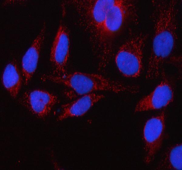
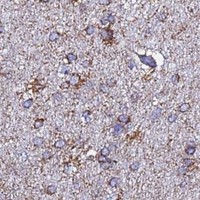
Anti-GLUD1 antibodies have IHC images from human paraffin sections and mouse and rat midbrain sections (catalog IHC captions); A01866-3 also has an IF image from HeLa cells (catalog IF caption).

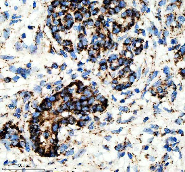
Real IHC data IHC analysis of GLUD1 using anti-GLUD1 antibody (A01866-3). GLUD1 was detected in a paraffin-embedded section of human adrenal adenoma tissue. Heat mediated antigen retrieval was performed in EDTA buffer (pH 8.0, epitope retrieval solution). The tissue section was blocked with 10% goat serum. The tissue section was then incubated with 2 μg/ml rabbit anti-GLUD1 Antibody (A01866-3) overnight at 4°C. Peroxidase Conjugated Goat Anti-rabbit IgG was used as secondary antibody and incubated for 30 minutes at 37°C. The tissue section was developed using HRP Conjugated Rabbit IgG Super Vision Assay Kit (Catalog # SV0002) with DAB as the chromogen.
Anti-GLUD1 Antibody ®
Cat # A01866-3
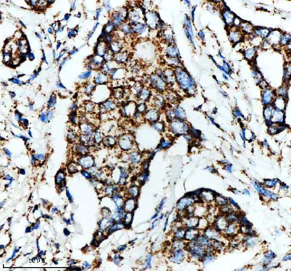
Real IHC data IHC analysis of GLUD1 using anti-GLUD1 antibody (M01866). GLUD1 was detected in a paraffin-embedded section of human breast cancer tissue. Heat mediated antigen retrieval was performed in EDTA buffer (pH 8.0, epitope retrieval solution). The tissue section was blocked with 10% goat serum. The tissue section was then incubated with 1:50 rabbit anti-GLUD1 Antibody (M01866) overnight at 4°C. Peroxidase Conjugated Goat Anti-rabbit IgG was used as secondary antibody and incubated for 30 minutes at 37°C. The tissue section was developed using HRP Conjugated Rabbit IgG Super Vision Assay Kit (Catalog # SV0002) with DAB as the chromogen.
Anti-GLUD1 Rabbit Monoclonal Antibody
Cat # M01866

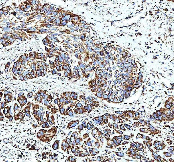
A01866-3 will render with a human adrenal adenoma IHC image; its catalog also shows human esophageal, liver and lung cancer IHC images and a HeLa cell IF image (A01866-3 image captions). M01866 will render with a human breast cancer IHC image; its catalog also shows human pancreas cancer and mouse and rat midbrain IHC images (M01866 image captions).

Which to pick: For tissue IHC, either SKU has paraffin-section images after EDTA pH 8.0 retrieval: A01866-3 at 2 μg/ml and M01866 at 1:50; the fixative is unreported for both (respective IHC image captions). For IF/ICC, choose A01866-3 when an image-based example is useful: it has a HeLa cell IF image, while M01866 lists IF/ICC without an IF image in this payload (catalog applications and IF image captions). For cross-species IHC, M01866 has mouse and rat midbrain images as well as human tissue images; both SKUs list Human, Mouse and Rat reactivity, and M01866 is monoclonal (catalog reactivity, M01866 IHC image captions and clone field).

Each figure is that product's own IHC / IF validation image from its datasheet.

References

  1. UniProt Consortium. UniProt entry P00367 (DHE3_HUMAN, Glutamate dehydrogenase 1, mitochondrial).
  2. Human Protein Atlas. GLUD1 tissue IHC expression (reliability: Approved).
  3. Human Protein Atlas. GLUD1 subcellular location (ICC-IF): Localized to the mitochondria. Caution: Based on antibodies targeting proteins from multiple genes..
  4. Human Protein Atlas. GLUD1 antibody validation summary (3 antibodies).
  5. GLUD1 supports ovarian cancer progression by counteracting anoikis via ARAF/MEK/ERK signaling. NPJ precision oncology 2026 — PMC13079734.
  6. Glutamate Delta-1 Receptor Regulates Inhibitory Neurotransmission in the Nucleus Accumbens Core and Anxiety-Like Behaviors. Molecular neurobiology 2021 — PMC8500932.
  7. Glutamate delta-1 receptor regulates cocaine-induced plasticity in the nucleus accumbens. Translational psychiatry 2018 — PMC6185950.
  8. Dysfunction of Glutamate Delta-1 Receptor-Cerebellin 1 Trans-Synaptic Signaling in the Central Amygdala in Chronic Pain. Cells 2021 — PMC8534524.
  9. PubMed PMID:3426581 — UniProt-cited evidence.
  10. PubMed PMID:3377777 — UniProt-cited evidence.
  11. PubMed PMID:3399399 — UniProt-cited evidence.