This website uses cookies to ensure you get the best experience on our website.


- Table of Contents
Plan chromogenic GNE IHC in paraffin sections using the catalog antibody at 2–5 μg/ml (datasheet A01647-3). Compare cytoplasmic staining with high staining in glandular cells of the colon or duodenum, while accounting for low consistency between staining and RNA expression (HPA tissue IHC).

Expected localisation, validated protocols, controls and antibodies — the at-a-glance facts below, then the full design guide.
| Expected localisation | Cytoplasmic staining in most tissues (HPA tissue IHC) | |
| Staining pattern | Cytoplasmic staining; high in several glandular sites (HPA tissue IHC) | |
| Antigen retrieval | EDTA pH 8.0 HIER, heat-mediated (datasheet A01647-3) | |
| Positive control | Colon+4 more · see all | |
| Negative control | Adipose tissue+4 more · see all |
| Fixation | Keep fixation consistent across paraffin sections (standard IHC practice; not target-specific) | |
| Caveat | Staining has low consistency with RNA expression (HPA tissue IHC) | |
| Regulation | Expression varies by tissue (UniProt) | |
| Isoform / epitope | 5 isoforms; epitope coverage is unspecified (UniProt) |
Start with the catalog antibody’s IHC-P protocol (datasheet A01647-3); the published early gastric tumor protocol provides a second approach (PMC9688572).
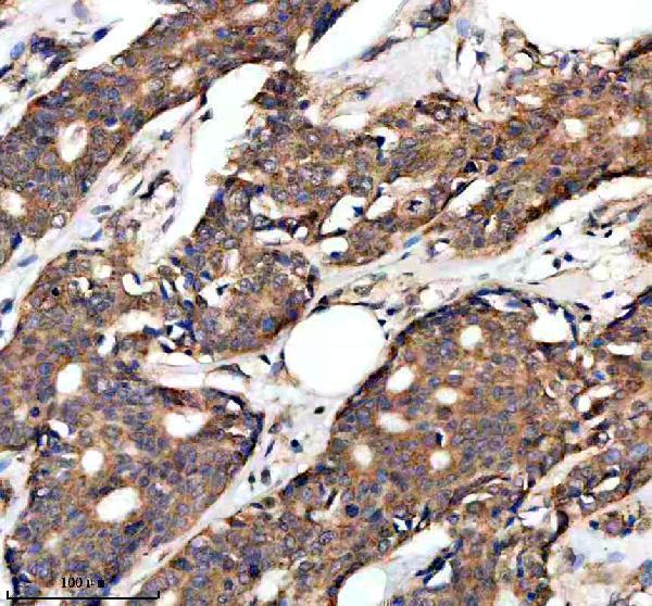
| Sample | Paraffin-embedded human breast cancer tissue; fixative not specified (datasheet A01647-3) |
| Fixation | Image fixative and duration unreported (datasheet A01647-3); verify before use. |
| Sectioning | 4–5 µm sections on charged slides (standard) |
| Deparaffinisation | Xylene, graded ethanol series to water (standard) |
| Antigen retrieval | Heat retrieval: EDTA pH 8.0 (datasheet A01647-3); 20 min, 95–100 °C (standard) |
| Peroxidase block | 3% H2O2, 10 min, room temperature (standard) |
| Blocking | 10% goat serum (datasheet A01647-3) |
| Primary antibody | Rabbit anti-GNE, 2-5 μg/ml (datasheet A01647-3) |
| Primary incubation | Overnight at 4 °C (datasheet A01647-3) |
| Detection | HRP-conjugated secondary, DAB chromogen (datasheet A01647-3) |
| Counterstain | Hematoxylin, blue, dehydrate and mount (standard) |
| Expected result | GNE-positive staining in glandular cells of colon (HPA tissue IHC: High). HPA tissue profile: Cytoplasmic expression in most tissues. No signal in the no-primary control. |
GNE should appear mainly in the cytoplasm, consistent with its cytosolic location and lack of a transmembrane segment (UniProt Q9Y223; HPA: cytoplasmic expression in most tissues). Strong HPA tissue IHC staining occurs in selected glandular cells, lung macrophages and ciliated cells (HPA: High). Interpret these examples with caution: HPA rates its tissue IHC profile Approved but reports low consistency between antibody staining and RNA expression (HPA: reliability description).
| Strong cytoplasmic staining in colon or duodenal glandular cells, with little nuclear signal. | This fits the reported compartment and cell pattern (UniProt Q9Y223: cytosol; HPA: High in colon and duodenal glandular cells). Judge staining in the identified cells, rather than calling an entire section uniformly positive; adjacent cell populations need not share the glandular-cell score (HPA: cell-specific tissue IHC entries). |
| Predominant nuclear staining, or a sharp cell-surface outline without convincing cytoplasmic staining. | That distribution conflicts with the supported cytosolic location (UniProt Q9Y223; HPA ICC-IF: cytosol supported). Treat it as an uncertain IHC result and examine morphology and controls before assigning GNE positivity. Localized ciliary staining is a documented exception to a simple cytoplasm-only reading (HPA: High in fallopian-tube and nasopharyngeal ciliated cells). |
| Strong staining of adipocytes while glandular cells in a known-positive section remain unstained. | Adipocytes are reported as Not detected, whereas several glandular-cell populations are High (HPA: adipose tissue; colon, duodenum, gallbladder, prostate and rectum). The reversal raises concern for nonspecific antibody binding or endogenous chromogen activity; the HPA comparison alone cannot identify which cause applies. |
| Diffuse color across cells, stroma and empty areas, with poorly defined cellular boundaries. | The pattern cannot establish cytosolic GNE localization (UniProt Q9Y223; HPA: cytoplasmic tissue profile). In chromogenic IHC, broad background can arise from nonspecific antibody binding or residual endogenous detection activity (general IHC practice). Evaluate it against matched controls before scoring cells. |
| No convincing staining in colon or duodenal glandular cells. | These are useful positive-reference cell populations because HPA reports High staining in each (HPA: colon and duodenum). A blank result warrants a workflow check, but it does not alone prove that the specimen lacks GNE. HPA also reports low consistency between antibody staining and RNA expression, so RNA abundance is not a substitute for a slide control (HPA: reliability description). |
| Compartment and topology | GNE is cytosolic and has no annotated transmembrane segment (UniProt Q9Y223). Score cellular cytoplasm separately from nuclei and tissue background; membrane-only signal lacks support from these location data. |
| Cell-specific tissue pattern | HPA reports High staining in several glandular-cell populations and lung macrophages, but Not detected in adipocytes and selected other cells (HPA: tissue IHC). Select comparison regions by identified cell type, not by tissue name alone. |
| Ciliary exception | High signal is reported at the cilia axoneme in fallopian-tube and nasopharyngeal ciliated cells; bronchial ciliated-cell tips are Low (HPA: tissue IHC). Assess those structures against their specific HPA pattern before treating all noncytoplasmic staining as artefact. |
| Isoforms and antibody scope | UniProt lists five GNE isoforms with differing reported tissue distributions (UniProt Q9Y223: isoforms 1–5). The supplied HPA antibody statuses do not identify recognized epitopes or establish isoform coverage (HPA: HPA007045 and HPA027258 validation entries). |
| Evidence strength | Both listed antibodies have Approved IHC status, while the overall tissue profile has low staining-to-RNA consistency (HPA: antibody statuses; reliability description). An Approved status supports use of the observed pattern as a reference, without making every tissue intensity definitive. |
| IF/ICC Q: What location should a separate IF/ICC guide expect? | A: Mainly cytosol; HPA marks cytosol as supported and lists ICC-IF images for A-431, U-251MG and U2OS (HPA: subcellular record). Both listed antibodies have Approved ICC status (HPA: antibody statuses). This location evidence supplies no IF/ICC protocol here. |
| Situation | Likely cause | Next action |
|---|---|---|
| A known-positive glandular-cell region has no chromogenic signal. | The slide may have a detection or section-processing problem; the absence conflicts with HPA High staining in colon and duodenal glandular cells (HPA: tissue IHC). | Verify tissue identity and cell morphology, then check the run's positive control and detection reagents (general IHC practice). Review the antibody's documented IHC-P conditions before changing retrieval or dilution; no GNE-specific fixation or retrieval effect is supplied. |
| Signal is mainly nuclear or forms a continuous cell-surface rim. | Those distributions do not match supported cytosolic GNE localization (UniProt Q9Y223; HPA: subcellular record). | Inspect a known-positive cytoplasmic reference and the negative control side by side (general IHC practice). Reassess scoring if the apparent rim comes from cell overlap or section geometry; preserve separately identified ciliary observations (HPA: ciliated-cell entries). |
| Adipocytes or other HPA Not detected cells stain strongly. | The cell-level result conflicts with HPA's adipocyte entry, although a discordant cell pattern alone cannot distinguish cross-reactivity from endogenous detection activity (HPA: adipocytes Not detected). | Confirm cell identity and compare an antibody-omission control for residual detection color (general IHC practice). If color appears only with primary antibody, reassess specificity using another validated reagent where available (HPA: two Approved IHC antibodies). |
| Color spreads across the section and obscures cellular boundaries. | Diffuse background can reflect nonspecific binding or endogenous detection activity in chromogenic IHC (general IHC practice); it is not the described cellular GNE pattern (HPA: cytoplasmic tissue profile). | Examine an antibody-omission control, blocking and washes, then adjust the general IHC workflow according to the detection system (general IHC practice). Score GNE only where cell boundaries and intracellular signal remain interpretable. |
| A low-staining region seems negative despite a positive control. | Several HPA cell entries are Low, including salivary-gland glandular cells and hippocampal neuronal cells (HPA: tissue IHC). A faint result in those cells is less decisive than a failed High reference. | Check the High reference first, then record the low region with its cell type and detection threshold (HPA: tissue IHC; general IHC scoring practice). Avoid converting a Low HPA entry into an expected strong positive. |
| Ciliary staining appears inconsistent with the cytosolic expectation. | HPA reports High axonemal staining in fallopian tube and nasopharynx, alongside a supported cytosolic ICC-IF location (HPA: tissue IHC; HPA: subcellular record). | Identify the ciliated cells and exact subcellular structure before scoring (general IHC practice). Report ciliary and cytoplasmic observations separately, using the tissue-specific HPA entries as context rather than assuming either pattern proves antibody specificity. |


Comprehensive Human Protein Atlas IHC scoring per tissue (reliability: Approved — Low consistency between antibody staining and RNA expression data.). Rows are taken directly from the HPA tissue chart — click any row's HPA link to view the source.
| Tissue | Cell type | Level | Evidence | Source |
|---|---|---|---|---|
| Colon | Glandular cells | High | Protein (IHC) | HPA → |
| Duodenum | Glandular cells | High | Protein (IHC) | HPA → |
| Fallopian tube | Ciliated cells (cilia axoneme) | High | Protein (IHC) | HPA → |
| Gallbladder | Glandular cells | High | Protein (IHC) | HPA → |
| Lung | Macrophages | High | Protein (IHC) | HPA → |
| Tissue | Cell type | Level | Evidence | Source |
|---|---|---|---|---|
| Adipose tissue | Adipocytes | Not detected | Protein (IHC) | HPA → |
| Caudate | Glial cells | Not detected | Protein (IHC) | HPA → |
| Cerebellum | Cells in granular layer | Not detected | Protein (IHC) | HPA → |
| Cervix | Squamous epithelial cells | Not detected | Protein (IHC) | HPA → |
| Ovary | Ovarian stroma cells | Not detected | Protein (IHC) | HPA → |
Troubleshoot GNE staining in paraffin sections by checking retrieval, cytosolic localisation, cell type, controls and scoring before interpreting differences between samples.
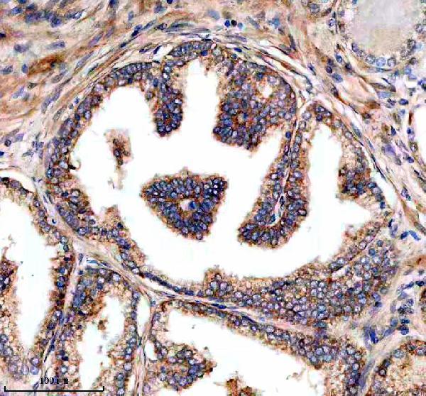
The catalog lists an IHC antibody with paraffin-section images and a separate human IF/ICC antibody without an IF image (A01647-3 IHC captions; M01647 applications and image list).
A01647-3 has IHC images from paraffin-embedded human breast and prostate cancer sections; its catalog reactivity lists human, mouse, and rat (A01647-3 IHC captions; catalog reactivity). M01647 lists human IF/ICC use, but supplies no IF image (M01647 applications, reactivity, and image list).
Which to pick: Choose A01647-3 for chromogenic IHC on paraffin sections: its captions show that use, but do not report the fixative (A01647-3 IHC captions). Choose M01647 for human IF/ICC because it is a rabbit monoclonal listed for those applications; no IF image is supplied (M01647 catalog). For mouse or rat work, A01647-3 lists those species as reactive, but its supplied IHC images show human tissue only (A01647-3 catalog reactivity; IHC captions).