This website uses cookies to ensure you get the best experience on our website.


- Table of Contents
Plan GNG11 IHC on paraffin sections using the reported cytoplasmic and membranous tissue pattern (HPA tissue IHC). This guide covers the 2–5 μg/mL antibody range (datasheet A13707), chromogenic detection (datasheet A13707), and interpretation of uncertain tissue staining (HPA tissue IHC).

Expected localisation, validated protocols, controls and antibodies — the at-a-glance facts below, then the full design guide.
| Expected localisation | Cytoplasmic and membranous tissue staining (HPA tissue IHC) | |
| Staining pattern | Glandular and tubular cells: cytoplasmic, membranous signal (HPA tissue IHC) | |
| Antigen retrieval | EDTA pH 8.0 HIER, heat-mediated (datasheet A13707) | |
| Positive control | Appendix+4 more · see all | |
| Negative control | Adipose tissue+4 more · see all |
| Fixation | Keep fixation consistent across sections (standard IHC practice; not target-specific) | |
| Caveat | Presumed off-target binding; tissue IHC reliability uncertain (HPA tissue IHC) | |
| Regulation | Abundant in tested tissues except brain (UniProt) | |
| Isoform / epitope | No isoforms or TM segment; mature chain 1–70 (UniProt) |
The catalog antibody’s IHC-P protocol (datasheet: A13707) is accompanied by three published GNG11 IHC protocols (PMC12616338; PMC8225293; PMC11382430).
| Sample | Paraffin-embedded human placenta tissue; fixative not specified (datasheet A13707) |
| Fixation | Image fixative and duration unreported (datasheet A13707); verify before use. |
| Sectioning | 4–5 µm sections on charged slides (standard) |
| Deparaffinisation | Xylene, graded ethanol series to water (standard) |
| Antigen retrieval | Heat retrieval: EDTA pH 8.0 (datasheet A13707); 20 min, 95–100 °C (standard) |
| Peroxidase block | 3% H2O2, 10 min, room temperature (standard) |
| Blocking | 10% goat serum (datasheet A13707) |
| Primary antibody | Rabbit anti-GNG11, 2-5μg/ml (datasheet A13707) |
| Primary incubation | Overnight at 4 °C (datasheet A13707) |
| Detection | Streptavidin-biotin complex (SABC), DAB chromogen (datasheet A13707) |
| Counterstain | Hematoxylin, blue, dehydrate and mount (standard) |
| Expected result | GNG11-positive staining in glandular cells of appendix (HPA tissue IHC: Medium). HPA tissue profile: Cytoplasmic and membranous expression in several tissues. No signal in the no-primary control. |
GNG11 is annotated at the cell membrane and has no transmembrane segment (UniProt P61952 topology). In paraffin-section IHC, expect membranous and cytoplasmic staining in selected glandular cells, kidney tubule cells, and smooth muscle cells (HPA: tissue IHC). Treat that pattern as provisional: HPA rates its tissue IHC evidence Uncertain and reports presumed off-target binding (HPA: reliability).
| Membranous staining with some cytoplasmic signal in colon glandular cells or kidney tubule cells. | This fits the reported compartment pattern and the Medium staining observed in those cell groups (HPA: tissue IHC). The membrane component agrees with GNG11's annotated location (UniProt P61952: cell membrane). It supports a plausible result, but HPA's Uncertain reliability prevents treating appearance alone as proof of specificity (HPA: reliability). |
| Predominantly nuclear staining, with little convincing membrane or cytoplasmic signal. | A nuclear-dominant pattern conflicts with the annotated membrane location and reported tissue pattern (UniProt P61952: cell membrane; HPA: tissue IHC). Consider artefact or antibody-dependent off-target binding, especially given the validation caveat (HPA: reliability). Review the negative control and staining distribution before scoring nuclei as GNG11 positive (general IHC practice). |
| Strong staining in adipocytes or bone-marrow hematopoietic cells. | HPA reports GNG11 as Not detected in these respective cell groups (HPA: tissue IHC). Such staining warrants a check for cross-reactivity or endogenous chromogenic activity (general IHC practice). It does not, by itself, establish either cause: the tissue profile is based on an antibody with Uncertain IHC reliability (HPA: antibodies; HPA: reliability). |
| Broad, similar color across cells and extracellular areas, obscuring cell boundaries. | A uniform deposit cannot be assigned confidently to the reported cytoplasmic and membranous cellular pattern (HPA: tissue IHC). Assess no-primary and detection controls, then review blocking, washes, and detection conditions for background (general IHC practice). Do not count diffuse color as a positive cell population while its source remains unresolved (general IHC practice). |
| No staining in colon glandular cells or kidney tubule cells used as a reference. | Those groups showed Medium staining in the supplied HPA observations (HPA: tissue IHC). An absent signal calls for an assay-control check before a biological interpretation: inspect tissue preservation, retrieval and reagent performance using routine IHC controls (general IHC practice). HPA's Uncertain rating means these are observed references, not guaranteed positive controls (HPA: reliability). |
| Subcellular location and topology | GNG11 is annotated at the cell membrane without a transmembrane segment (UniProt P61952 topology). Read staining in the context of the observed cytoplasmic and membranous IHC profile; membrane-only scoring would exclude part of that reported pattern (HPA: tissue IHC). |
| Choice of reference cell group | Colon and rectal glandular cells, kidney tubule cells, and smooth muscle cells have Medium observed staining (HPA: tissue IHC). Adipocytes and bone-marrow hematopoietic cells are reported as Not detected (HPA: tissue IHC). Compare the named cell groups, since a whole-tissue score can conceal which cells carry the signal (general IHC practice). |
| Antibody evidence | The supplied antibody HPA045255 is rated Uncertain for IHC; presumed off-target binding was observed and disregarded in the tissue assessment (HPA: antibodies; HPA: reliability). Interpret an attractive pattern alongside controls and the stated cell-level distribution, without treating HPA staining as independent confirmation of target specificity (general IHC practice). |
| Processing and epitope information | UniProt lists a mature chain at residues 1–70, a 71–73 propeptide, and a modified cysteine at position 70 (UniProt P61952: processing and modified residue). No antibody epitope is supplied, so these annotations cannot establish whether processing or modification affects this IHC result. |
| Fixation and retrieval evidence | No target-specific fixation or retrieval effect is established by the supplied UniProt and HPA records. Apply and document routine paraffin-section retrieval and control procedures as general IHC practice; avoid attributing a weak or absent GNG11 signal to target-specific epitope masking without further evidence. |
| Situation | Likely cause | Next action |
|---|---|---|
| An HPA-observed positive reference has no chromogenic signal. | The assay may have failed, or this sample may differ from the reported observation; the reference pattern itself carries Uncertain reliability (HPA: tissue IHC; HPA: reliability). | Check tissue and detection controls, primary-antibody application, retrieval execution, and counterstain visibility; repeat with a documented control section before assigning a biological negative (general IHC practice). |
| Color is diffuse across the section. | Diffuse deposition is inconsistent with a readable cell-level membranous and cytoplasmic pattern (HPA: tissue IHC). Background from blocking, washing, or detection is possible (general IHC practice). | Compare no-primary and detection controls; review blocking, washes, and chromogen development, then score only signal that resolves to identifiable cells and compartments (general IHC practice). |
| Nuclei appear to be the dominant positive compartment. | That distribution conflicts with the annotated cell-membrane location and the reported cytoplasmic and membranous IHC pattern (UniProt P61952: cell membrane; HPA: tissue IHC). | Check controls and examine whether the deposit tracks nuclei across unrelated cell groups; withhold a GNG11-positive call until the expected cell and compartment pattern is resolved (general IHC practice). |
| Adipocytes or bone-marrow hematopoietic cells stain strongly. | These cell groups were Not detected in the supplied HPA observations; cross-reactivity or endogenous detection activity is possible (HPA: tissue IHC; general IHC practice). | Review the no-primary and detection controls and compare adjacent expected cell groups. Record the discrepancy rather than converting the HPA negative observation into a claim of absolute absence (general IHC practice; HPA: reliability). |
| The expected cell groups stain, but confidence in target identity remains low. | Matching a reported pattern does not resolve the antibody's Uncertain IHC rating or the noted presumed off-target binding (HPA: antibodies; HPA: reliability). | Report the cell group, compartment, intensity, and controls explicitly; qualify the finding as pattern-consistent rather than independently validated GNG11 detection (HPA: tissue IHC; HPA: reliability). |
| Can IF/ICC images settle an ambiguous IHC localization? | The supplied HPA subcellular record has no main location or ICC-IF images, so it provides no image-based localization check (HPA: subcellular). | Keep the paraffin-section IHC interpretation tied to the UniProt membrane annotation and HPA tissue observations (UniProt P61952: cell membrane; HPA: tissue IHC). Consult the separate IF/ICC guide for that application. |


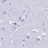
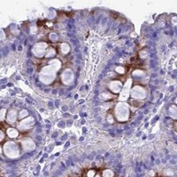


Comprehensive Human Protein Atlas IHC scoring per tissue (reliability: Uncertain — Pending external verification. Presumed off target binding observed and disregarded.). Rows are taken directly from the HPA tissue chart — click any row's HPA link to view the source.
| Tissue | Cell type | Level | Evidence | Source |
|---|---|---|---|---|
| Appendix | Glandular cells | Medium | Protein (IHC) | HPA → |
| Colon | Glandular cells | Medium | Protein (IHC) | HPA → |
| Duodenum | Glandular cells | Medium | Protein (IHC) | HPA → |
| Gallbladder | Glandular cells | Medium | Protein (IHC) | HPA → |
| Kidney | Cells in tubules | Medium | Protein (IHC) | HPA → |
| Tissue | Cell type | Level | Evidence | Source |
|---|---|---|---|---|
| Adipose tissue | Adipocytes | Not detected | Protein (IHC) | HPA → |
| Adrenal gland | Glandular cells | Not detected | Protein (IHC) | HPA → |
| Bone marrow | Hematopoietic cells | Not detected | Protein (IHC) | HPA → |
| Bronchus | Respiratory epithelial cells | Not detected | Protein (IHC) | HPA → |
| Caudate | Glial cells | Not detected | Protein (IHC) | HPA → |
Troubleshoot GNG11 staining in paraffin section chromogenic IHC using the catalog antibody’s tissue example while treating uncertain tissue patterns cautiously.
A13707 has real IHC data from paraffin sections of human placenta, skin cancer, gastric cancer and ovarian cancer (catalog IHC image captions). No IF/ICC data are supplied (catalog applications and image captions).
A13707 is listed for IHC in human samples, with staining shown in paraffin sections of human placenta, skin cancer, gastric cancer and ovarian cancer (catalog IHC image captions and dilution listing). Its broader reactivity lists Human, Mouse and Rat, but the supplied IHC captions show human tissue only (catalog reactivity; catalog IHC image captions).
Which to pick: Choose A13707 for human paraffin section IHC: its captions report EDTA retrieval at pH 8.0 and primary antibody at 2 μg/ml, within the listed IHC range of 2–5 μg/ml (A13707 IHC image captions; catalog IHC dilution). The fixative is unreported, and neither IF/ICC validation nor antibody clonality is provided (A13707 IHC image captions; catalog applications and clone field). A13707 lists Mouse and Rat reactivity, but its IHC dilution is specified for Human and its IHC images show human tissues, so cross species tissue IHC needs separate validation (catalog reactivity, dilution listing and IHC image captions).