This website uses cookies to ensure you get the best experience on our website.


- Table of Contents
Plan HASPIN IHC-P using the nuclear and cytoplasmic tissue pattern (HPA tissue IHC). Testis Leydig cells provide a high-staining reference, but staining has low concordance with RNA expression (HPA tissue IHC).

Expected localisation, validated protocols, controls and antibodies — the at-a-glance facts below, then the full design guide.
| Expected localisation | Nuclear and cytoplasmic tissue staining (HPA tissue IHC) | |
| Staining pattern | Leydig cells stain strongly; several tissues show nuclear and cytoplasmic staining (HPA tissue IHC) | |
| Antigen retrieval | Tris-EDTA pH 9.0 HIER, 95–98 °C, 20 min (rule: nuclear antigen) | |
| Positive control | Testis+4 more · see all | |
| Negative control | Duodenum+4 more · see all |
| Fixation | Use formalin-fixed paraffin sections (selected-SKU IHC image A31862-1); keep fixation consistent. (standard IHC practice; not target-specific) | |
| Caveat | Staining has low concordance with RNA expression (HPA tissue IHC) | |
| Regulation | Detected in proliferating cell lines (UniProt) | |
| Isoform / epitope | 2 isoforms; epitope coverage is unspecified (UniProt) |
The catalog antibody’s IHC-P protocol is accompanied by one published ovarian tissue protocol (PMC7826093).
| Sample | FFPE human cancer tissue (datasheet A31862-1) |
| Fixation | Image formalin-fixed; duration unreported (datasheet A31862-1); verify before use. |
| Sectioning | 4–5 µm sections on charged slides (standard) |
| Deparaffinisation | Xylene, graded ethanol series to water (standard) |
| Antigen retrieval | Heat-induced epitope retrieval in Tris-EDTA buffer, pH 9.0, 20 min at 95–98 °C (standard rule: nuclear antigen) |
| Peroxidase block | 3% H2O2, 10 min, room temperature (standard) |
| Blocking | 10% normal serum of the secondary host, 30 min, room temperature (standard) |
| Primary antibody | Rabbit anti-HASPIN, 1:50-1:100 (datasheet A31862-1) |
| Primary incubation | Overnight at 4 °C (standard) |
| Detection | HRP-polymer secondary, DAB chromogen 5–10 min (standard) |
| Counterstain | Hematoxylin, blue, dehydrate and mount (standard) |
| Expected result | HASPIN-positive staining in leydig cells of testis (HPA tissue IHC: High). HPA tissue profile: Nuclear and cytoplasmic expression in several tissues. No signal in the no-primary control. |
HASPIN is nuclear during interphase and associates with chromosomes and the spindle during mitosis (UniProt Q8TF76); HPA reports nuclear and cytoplasmic tissue staining, with high staining in testis Leydig cells (HPA: tissue IHC). It has no transmembrane segment (UniProt Q8TF76 topology). Treat the pattern as provisional: HPA rates tissue staining Approved but reports low agreement with RNA data and pending external verification (HPA: tissue IHC).
| Distinct nuclear staining, with some cytoplasmic staining, in testis Leydig cells. | This fits HPA's high Leydig-cell staining and its broader nuclear/cytoplasmic tissue profile (HPA: tissue IHC). Nuclear localization also fits interphase HASPIN (UniProt Q8TF76). Score the stained cell type and compartment separately; HPA's Approved rating carries a low RNA-agreement caveat (HPA: tissue IHC). |
| Staining appears concentrated on chromosomes or the spindle in identifiable mitotic cells. | This can fit HASPIN's reported mitotic localization (UniProt Q8TF76). Its role in histone H3 Thr-3 phosphorylation supports a mitotic context, but staining alone does not measure kinase activity or H3T3ph (UniProt Q8TF76; general IHC interpretation). Check the mitotic morphology before assigning this pattern (general IHC practice). |
| Signal outlines cell membranes or fills extracellular spaces without a convincing intracellular pattern. | Treat this as suspect: UniProt places HASPIN in the nucleus, chromosomes and spindle and reports no transmembrane segment (UniProt Q8TF76). Review morphology and controls before calling it HASPIN; deposited chromogen or nonspecific binding can create misleading outlines (general chromogenic IHC practice). |
| Strong staining dominates a cell type that HPA reports as not detected, such as duodenal glandular cells. | That result conflicts with the reported pattern for that specific cell type (HPA: duodenal glandular cells, Not detected). Check staining controls and cell identification for cross-reactivity or endogenous detection activity (general IHC practice). HPA's pending external verification means the discrepancy needs investigation, not an automatic biological ruling (HPA: tissue IHC). |
| Broad, diffuse staining obscures nuclei and tissue boundaries, or testis Leydig cells show no signal. | Diffuse staining is difficult to score by compartment and may reflect background (general IHC practice). Absent Leydig-cell signal conflicts with HPA's high staining report (HPA: testis Leydig cells, High); check section quality, retrieval and detection controls before treating the specimen as HASPIN-negative (general IHC practice). |
| Cell-cycle state | Interphase nuclear localization and mitotic chromosome/spindle association can produce different intracellular appearances (UniProt Q8TF76); compare like cells and recognizable mitotic figures when scoring (general IHC practice). |
| Tissue and cell-type choice | HPA reports high staining in testis Leydig cells, but no detected staining in duodenal glandular cells (HPA: tissue IHC). UniProt reports strong testis expression (UniProt Q8TF76). HPA cautions that antibody staining has low agreement with RNA data (HPA: tissue IHC). |
| Isoforms and antibody recognition | UniProt lists two HASPIN isoforms and a kinase domain at residues 484–798 (UniProt Q8TF76). The supplied sources do not map the IHC antibody's epitope, so isoform recognition cannot be assigned from these records (HPA: antibody validation; UniProt Q8TF76). |
| Fixation and antigen retrieval | Target-specific fixation sensitivity and retrieval conditions are unreported in the supplied UniProt and HPA records. Retrieval can be evaluated as a general paraffin-IHC workflow variable; do not infer a HASPIN-specific effect from tissue staining or topology (general IHC practice; HPA: tissue IHC; UniProt Q8TF76). |
| IF/ICC Q: where should signal appear? | A: HPA supports nucleoplasmic localization in ICC-IF images from A-431 and U-251MG, with no additional location listed (HPA: subcellular). This is IF/ICC evidence; tissue IHC has a broader nuclear/cytoplasmic profile (HPA: tissue IHC). |
| Situation | Likely cause | Next action |
|---|---|---|
| Leydig cells lack visible chromogen in a testis section. | The result conflicts with HPA's high Leydig-cell staining; the supplied sources do not identify a HASPIN-specific fixation cause (HPA: tissue IHC). | Confirm Leydig-cell identification and section quality, then check positive-control staining and the established paraffin-IHC retrieval and detection steps (general IHC practice). |
| Every compartment appears diffusely colored. | Background can hide the nuclear/cytoplasmic pattern HPA reports (HPA: tissue IHC; general IHC practice). | Inspect the no-primary control; review blocking, antibody dilution, washes and chromogen development as general chromogenic-IHC variables (general IHC practice). |
| Signal appears mainly at membranes or outside cells. | This distribution conflicts with HASPIN's intracellular locations and absent transmembrane segment (UniProt Q8TF76). | Compare with a no-primary control, inspect tissue edges and deposits, and score only signal tied to identifiable cells and compartments (general IHC practice). |
| Unexpected cell types stain strongly while the intended pattern is weak. | Cross-reactivity or endogenous detection activity is possible (general IHC practice); HPA also reports low antibody-staining/RNA agreement (HPA: tissue IHC). | Identify the stained cells, compare HPA's cell-type entries, and check no-primary and detection controls before assigning biological expression (HPA: tissue IHC; general IHC practice). |
| Nuclear signal seems absent, but cytoplasmic signal remains. | HPA allows cytoplasmic tissue staining, while UniProt places HASPIN in interphase nuclei; one compartment alone does not settle specificity (HPA: tissue IHC; UniProt Q8TF76). | Inspect nuclear counterstain and cell morphology; compare a documented positive cell type and control sections under the same scoring rules (general IHC practice). |
| A mitotic figure shows little obvious chromosome-associated signal. | UniProt reports chromosome and spindle association during mitosis, but the supplied sources do not establish how often an IHC section should capture a detectable mitotic pattern (UniProt Q8TF76). | Confirm that the figure is mitotic and technically interpretable; use the broader cellular and tissue pattern when assessing the section (general IHC practice; HPA: tissue IHC). |

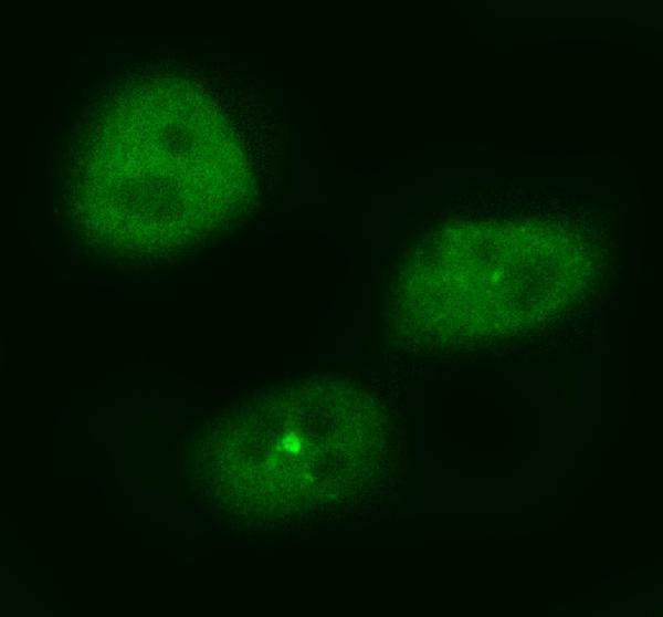
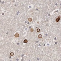


Comprehensive Human Protein Atlas IHC scoring per tissue (reliability: Approved — Low consistency between antibody staining and RNA expression data. Pending external verification.). Rows are taken directly from the HPA tissue chart — click any row's HPA link to view the source.
| Tissue | Cell type | Level | Evidence | Source |
|---|---|---|---|---|
| Testis | Leydig cells | High | Protein (IHC) | HPA → |
| Adipose tissue | Adipocytes | Medium | Protein (IHC) | HPA → |
| Adrenal gland | Glandular cells | Medium | Protein (IHC) | HPA → |
| Breast | Adipocytes | Medium | Protein (IHC) | HPA → |
| Bronchus | Respiratory epithelial cells | Medium | Protein (IHC) | HPA → |
| Tissue | Cell type | Level | Evidence | Source |
|---|---|---|---|---|
| Duodenum | Glandular cells | Not detected | Protein (IHC) | HPA → |
| Liver | Cholangiocytes | Not detected | Protein (IHC) | HPA → |
| Lymph node | Germinal center cells | Not detected | Protein (IHC) | HPA → |
| Pancreas | Exocrine glandular cells | Not detected | Protein (IHC) | HPA → |
| Small intestine | Glandular cells | Not detected | Protein (IHC) | HPA → |
Troubleshoot HASPIN staining in paraffin sections by checking retrieval, compartment, cell type, and controls before interpreting chromogenic signal.
Anti-HASPIN antibodies have IHC data from formalin-fixed, paraffin-embedded human cancer tissue and IF data from MG63 cells (catalog images). Listed reactivity spans human, mouse, and rat (catalog reactivity).
A31862-1 is listed for human IHC-P and has an image of stained breast carcinoma and hepatocarcinoma sections (catalog applications; catalog IHC image). A31862-2 is listed for IF/ICC and has an image of stained MG63 cells (catalog applications; catalog IF image).
Which to pick: Choose A31862-1 for tissue IHC: it is a rabbit polyclonal antibody listed for IHC-P at 1:50–1:100, with an image from formalin-fixed, paraffin-embedded human cancer tissue (catalog host; catalog dilution; catalog IHC image). Choose A31862-2 for IF/ICC: its MG63 cell image documents 5 μg/mL, while its clonality is unreported (catalog IF image; catalog clone). For mouse or rat work, A31862-2 is the only listed reactive option; the supplied IF image shows MG63 cells (catalog reactivity; catalog IF image).