HSD17B7 / 3-keto-steroid reductase/17-beta-hydroxysteroid dehydrogenase 7 · IHC design guide

Design Immunohistochemistry for HSD17B7

Plan paraffin-section chromogenic HSD17B7 IHC using the catalog antibody’s documented assay (datasheet A05850-1). Interpret cytoplasmic tissue staining (HPA tissue IHC) alongside the ER membrane location (UniProt).

Evidence assembled Oct 2026 · For research use; verify linked source records and product datasheet before use
Immunohistochemistry protocol sheet for HSD17B7 (IHC for HSD17B7): expected localisation Cytoplasmic staining (HPA tissue IHC); ER membrane location (UniProt), antibody A05850-1, validated IHC image, and IHC protocol steps
Printable HSD17B7 IHC protocol sheet — expected localisation Cytoplasmic staining (HPA tissue IHC); ER membrane location (UniProt), antibody A05850-1, controls and protocol steps. Open the full HSD17B7 IHC guide →

HSD17B7 Immunohistochemistry Experimental Design Guide

Expected localisation, validated protocols, controls and antibodies — the at-a-glance facts below, then the full design guide.

Must know before staining
Expected localisation Cytoplasmic staining (HPA tissue IHC); ER membrane location (UniProt)
Staining pattern Cytoplasmic staining in most tissues (HPA tissue IHC)
Antigen retrieval EDTA pH 8.0 HIER, heat-mediated (datasheet A05850-1)
Positive control ⓘ Adrenal gland+4 more · see all
Negative control ⓘ Adipose tissue+2 more · see all
Important caveats
Reasons your staining may differ from the expected pattern.
Fixation Keep fixation consistent across sections (standard IHC practice; not target-specific); assess its effect empirically.
Caveat Antibody staining has low consistency with RNA expression (HPA tissue IHC)
Regulation Liver-enhanced RNA expression (HPA tissue RNA)
Isoform / epitope 3 isoforms; check extracellular vs cytoplasmic epitope (UniProt)
Section 1

Recommended HSD17B7 IHC & IF Protocols

Compare the catalog antibody’s IHC-P protocol (datasheet A05850-1) with published HSD17B7 staining of mouse ovarian sections (PMC11121541; PMC5934784) and human prostate tissue (PMC13266810).

Recommended immunohistochemistry (IHC-P) protocol parameters
SampleParaffin-embedded human liver cancer tissue; fixative not specified (datasheet A05850-1)
FixationImage fixative and duration unreported (datasheet A05850-1); verify before use.
Sectioning4–5 µm sections on charged slides (standard)
DeparaffinisationXylene, graded ethanol series to water (standard)
Antigen retrievalHeat retrieval: EDTA pH 8.0 (datasheet A05850-1); 20 min, 95–100 °C (standard)
Peroxidase block3% H2O2, 10 min, room temperature (standard)
Blocking10% goat serum (datasheet A05850-1)
Primary antibodyRabbit anti-HSD17B7, 2-5 μg/ml (datasheet A05850-1)
Primary incubationOvernight at 4 °C (datasheet A05850-1)
DetectionHRP-conjugated secondary, DAB chromogen (datasheet A05850-1)
CounterstainHematoxylin, blue, dehydrate and mount (standard)
Expected resultHSD17B7-positive staining in glandular cells of adrenal gland (HPA tissue IHC: High). HPA tissue profile: Cytoplasmic expression in most tissues. No signal in the no-primary control.
💡Decision noteStart with heat-mediated EDTA at pH 8.0 for the catalog antibody (datasheet A05850-1); published ovarian protocols used citrate at pH 6 (PMC11121541; PMC5934784).
Section 2

What Is the Expected HSD17B7 Staining Pattern?

HSD17B7 is an endoplasmic reticulum membrane protein, so the expected IHC signal is cytoplasmic rather than nuclear (UniProt P56937 topology). HPA reports cytoplasmic expression in most tissues and high staining in selected glandular cells, bronchial ciliated cell bodies, pancreatic exocrine cells and melanocytes (HPA tissue IHC). Interpret these patterns with care: HPA rates the tissue staining Approved but reports low consistency between antibody staining and RNA expression (HPA tissue IHC reliability).

What am I looking at on my slide?
Cytoplasmic staining in adrenal, colonic, duodenal, gallbladder or small-intestinal glandular cells, or pancreatic exocrine cells (HPA tissue IHC).This matches the reported high-staining cell populations (HPA: High in these cells) and is compatible with endoplasmic reticulum membrane localisation (UniProt P56937). Judge signal within the identified cells; a positive section alone does not show that every cell in it should stain.
Predominantly nuclear staining with little cytoplasmic signal in an expected positive cell population.A nuclear-only pattern conflicts with the reported cytoplasmic tissue pattern (HPA tissue IHC) and endoplasmic reticulum membrane assignment (UniProt P56937). Treat it as suspect until the staining controls and tissue morphology have been checked; compartment alone cannot identify the source of an artefact.
Strong staining in adipocytes, parathyroid glandular cells or splenic red-pulp cells (HPA: Not detected in these cells).These cell populations are HPA-reported negatives, so strong signal warrants review for cross-reactivity or endogenous chromogenic detection activity (HPA tissue IHC; general IHC practice). Compare with a no-primary control and a positive tissue on the same run before assigning it to HSD17B7.
Colour spread across stroma, lumens or many cell types without a distinct cytoplasmic pattern.Treat a diffuse field as background when cell boundaries and the expected positive populations cannot be distinguished (HPA tissue IHC; general IHC practice). Examine the no-primary control and review blocking, washing and detection conditions; a dark field by itself cannot establish HSD17B7 localisation.
No cytoplasmic signal in an expected high-staining population, with intact tissue morphology (HPA: High in selected cells).First check whether the run can detect its positive control, then review antibody use and the general IHC workflow. Absence in one section is inconclusive: HPA calls the tissue pattern Approved while noting low staining–RNA consistency, so avoid treating one negative result as proof of absent protein (HPA tissue IHC reliability).
💡Expected HSD17B7 appearanceCall a result positive when identifiable HPA-reported high-staining cells show clear cytoplasmic chromogen (HPA tissue IHC; UniProt P56937); isolated nuclear signal or equally strong colour in HPA-reported negative cells is suspect (HPA tissue IHC; general IHC practice).
How each factor affects the staining
Cell population and tissueHPA reports High staining in adrenal, colonic, duodenal, gallbladder and small-intestinal glandular cells; bronchial ciliated cell bodies, pancreatic exocrine cells and skin melanocytes are also High (HPA tissue IHC). Lung alveolar and prostate glandular cells are Low, while adipocytes are Not detected (HPA tissue IHC). Choose and score the named cells rather than using whole-section colour as the criterion.
Subcellular assignmentUniProt places HSD17B7 at the endoplasmic reticulum membrane with one annotated transmembrane segment at residues 230–250 (UniProt P56937 topology). HPA describes cytoplasmic tissue staining (HPA tissue IHC). These records support a cytoplasmic expectation but do not establish which side of the membrane the catalog antibody recognises; its epitope is unspecified here.
Evidence and antibody scopeHPA lists the supplied antibody, HPA047496, as IHC Approved, and reports low consistency between antibody staining and RNA expression for the tissue profile (HPA antibody validation; HPA tissue IHC reliability). Use the observed cell pattern as a reference with that limitation; do not treat Approved as evidence that every positive cell or signal intensity is independently confirmed.
IF/ICC: what localisation can this section predict?Expect a membrane-associated intracellular pattern from the endoplasmic reticulum assignment (UniProt P56937); HPA's subcellular summary says Membrane but supplies no main-location assignment or ICC-IF image cell lines (HPA subcellular). This supports only a cautious localisation expectation for IF/ICC, not a demonstrated fluorescent pattern or an IF protocol.
Why is my staining missing, weak or wrong?
SituationLikely causeNext action
Expected positive glandular cells are blank while the section looks intact (HPA: High in selected glandular cells).Target-specific fixation effects are not established by the supplied assay evidence. Verify with a matched IHC source before attributing a result to fixation.Check a positive control from an HPA-reported High cell population and the run's detection controls; then review the antibody's IHC instructions and routine processing records before interpreting the blank section.
An expected positive population has only weak colour (HPA: High in selected cells).Weak staining can make a cell-level pattern hard to judge (general IHC practice); HPA's staining–RNA consistency is low, so expression data alone cannot settle the result (HPA tissue IHC reliability).Compare like cell populations in the positive control and test section. Verify routine chromogenic detection and counterstain conditions, then report the observed intensity rather than upgrading it to High from the reference.
Adipocytes or parathyroid glandular cells appear strongly positive (HPA: Not detected in these cells).Cross-reactivity or endogenous detection activity is possible (general IHC practice); the observation conflicts with these HPA tissue results (HPA tissue IHC).Inspect a no-primary control, confirm cell identity from morphology and compare the same run with an HPA-reported High population. Flag persistent unexpected staining as unresolved rather than assigning target expression.
Signal is predominantly nuclear in cells expected to stain cytoplasm.That compartment disagrees with HPA's cytoplasmic tissue profile and UniProt's endoplasmic reticulum membrane assignment (HPA tissue IHC; UniProt P56937).Check morphology, the no-primary control and staining across the expected positive cells. Record the discordance if the nuclear pattern persists; these sources do not establish a nuclear HSD17B7 pattern.
The whole section is hazy or chromogen obscures cell boundaries.Diffuse background or excessive detection can hide the cell-specific cytoplasmic pattern (general IHC practice; HPA tissue IHC).Review the no-primary control, blocking, washing and detection conditions using routine IHC practice. Reassess only when the named cell populations and cytoplasmic signal can be distinguished.
A low-staining lung alveolar or prostate glandular population seems as strong as an expected high-staining control (HPA tissue IHC).The observed relative intensity differs from HPA's Low versus High categories; staining conditions or specimen variation could contribute (HPA tissue IHC; general IHC practice).Score the named cells separately under comparable detection and counterstain conditions, and document the discrepancy. Do not infer a new HSD17B7 tissue ranking from a single section, given HPA's low staining–RNA consistency (HPA tissue IHC reliability).

Sample controls for HSD17B7 IHC & IF

🧪Run adrenal gland first and expect staining in its glandular cells (HPA: High in adrenal gland glandular cells); use parathyroid gland as the negative tissue (HPA: Not detected in parathyroid gland glandular cells). On the adrenal slide, assess stromal cells as an internal background reference; they should lack specific staining, but their HSD17B7 status is not established by the supplied HPA row.
Positive control tissue: Adrenal gland (Glandular cells, HPA High)
Negative control tissue: Adipose tissue (HPA Not detected)
ICC-IF cell lines (HPA subcellular resource): HPA carries no ICC-IF cell line for HSD17B7; derive a cell-line control from the positive tissue's cell type (Glandular cells) and confirm it by RNA or western blot first.
Technical controls: Include no-primary (secondary-only) and concentration-matched rabbit IgG isotype controls (selected A05850-1 IHC caption: rabbit primary antibody), plus an HSD17B7 knockout biological control where available. For chromogenic staining, quench endogenous peroxidase and check blood-rich areas for background signal (standard IHC practice).
⚠️Feasibility: A target-specific fixation window or fixation effect is unreported, and the exact A05850-1 paraffin-section caption does not state the fixative (selected A05850-1 IHC caption: fixative not stated). The caption reports heat-mediated retrieval in EDTA at pH 8.0 for a paraffin section, but does not establish that retrieval is required for every specimen (selected A05850-1 IHC caption: EDTA retrieval). The supplied evidence does not establish whether frozen sections or IF/ICC are easier; assess adrenal tissue background during chromogenic scoring (HPA: High in adrenal gland glandular cells; standard IHC practice).

HPA tissue IHC evidence for HSD17B7

Comprehensive Human Protein Atlas IHC scoring per tissue (reliability: Approved — Low consistency between antibody staining and RNA expression data.). Rows are taken directly from the HPA tissue chart — click any row's HPA link to view the source.

Positive expression · recommended positive controls

TissueCell typeLevelEvidenceSource
Adrenal gland Glandular cells High Protein (IHC) HPA →
Bronchus Ciliated cells (cell body) High Protein (IHC) HPA →
Colon Glandular cells High Protein (IHC) HPA →
Duodenum Glandular cells High Protein (IHC) HPA →
Gallbladder Glandular cells High Protein (IHC) HPA →

Undetected expression · recommended negative controls

TissueCell typeLevelEvidenceSource
Adipose tissue Adipocytes Not detected Protein (IHC) HPA →
Parathyroid gland Glandular cells Not detected Protein (IHC) HPA →
Spleen Cells in red pulp Not detected Protein (IHC) HPA →
Section 3

Advanced HSD17B7 IHC Tips

Troubleshoot HSD17B7 chromogenic IHC in paraffin sections using the tested retrieval and staining conditions, while checking whether signal fits its ER membrane localisation.

Which retrieval conditions should I try first for weak HSD17B7 staining?
Start with heat-mediated retrieval in EDTA at pH 8.0 (datasheet A05850-1). Keep the antibody at the tested 2 μg/ml and overnight 4°C incubation while assessing retrieval, so changes in signal are easier to attribute (datasheet A05850-1; standard IHC practice). Compare retrieved sections with a no-primary control and a similarly processed positive tissue section to distinguish weak antigen exposure from detection background (standard IHC practice). If staining remains weak, adjust heating or cooling within your established retrieval workflow before trialling another buffer as a fallback (standard IHC practice). Judge improvement by clearer cytoplasmic staining in the expected cells without a parallel rise in diffuse background (HPA tissue IHC; standard IHC practice).
Could fixation explain weak or uneven HSD17B7 staining?
Target-specific sensitivity of HSD17B7 to fixation is unknown: the paraffin-section caption does not report a fixative (datasheet A05850-1). Record the fixative, fixation duration, tissue thickness and processing history for each specimen, then compare sections processed together (standard IHC practice). Use the reported EDTA pH 8.0 retrieval and 2 μg/ml primary incubation consistently during that comparison (datasheet A05850-1). Uneven signal across a section warrants inspection for folds, incomplete reagent coverage and tissue damage before attributing it to fixation (standard IHC practice). Include a positive tissue control in each run, since a failed control cannot support a specimen-level conclusion (standard IHC practice).
What staining pattern should HSD17B7 produce in tissue sections?
Assess predominantly cytoplasmic staining in tissue sections (HPA tissue IHC: cytoplasmic expression in most tissues). HSD17B7 is an endoplasmic-reticulum membrane protein with a transmembrane segment at residues 230–250, so a perinuclear or reticular cytoplasmic pattern is biologically plausible (UniProt P56937 topology; standard IHC interpretation). Strong isolated nuclear staining should prompt review of the no-primary control, counterstain and tissue morphology before being scored as HSD17B7 (UniProt P56937 subcellular location; standard IHC practice). Examine the relevant cell population rather than treating every stained cell as equivalent; HPA reports high staining in adrenal glandular cells and pancreatic exocrine glandular cells (HPA tissue IHC). Compare pattern and intensity across matched processing conditions because chromogenic deposits can obscure fine ER detail (standard IHC practice).
How should isoforms and epitope position affect staining interpretation?
HSD17B7 has 3 annotated isoforms, but the supplied antibody caption gives no epitope coordinates or isoform coverage (UniProt P56937 isoforms; datasheet A05850-1). Verify the catalog antibody’s immunogen against each isoform sequence before interpreting absent staining as absent total HSD17B7 (standard IHC practice). The annotated transmembrane segment spans residues 230–250, with residues 1–229 and 251–341 on opposite membrane sides (UniProt P56937 topology). Glycosylation is annotated at residues 37, 178 and 229, but these annotations alone do not establish an effect on this antibody’s staining (UniProt P56937 glycosylation; standard IHC interpretation). If an alternative antibody is available, compare documented epitopes and concordant tissue patterns using the same controls (standard IHC practice).
How can IF help assess a questionable chromogenic HSD17B7 pattern?
Treat IF/ICC as a separate assay: the supplied A05850-1 evidence describes chromogenic staining of a paraffin section, with no IF/ICC validation stated (datasheet A05850-1). For multiplex IF, pair HSD17B7 with a validated marker of the cell population being assessed, such as glandular cells in an HPA-listed glandular tissue (HPA tissue IHC; standard IF practice). Choose fluorophores after measuring the specimen’s autofluorescence and include single-label controls to assess spectral bleed-through (standard IF practice). If the epitope lies within residues 1–229, test permeabilisation sufficient to expose the luminal-facing region; residues 251–341 face the cytoplasm (UniProt P56937 topology; standard IF practice). Confirm epitope location with the antibody’s documentation before using either topology-based condition (standard IF practice).
How can I reduce diffuse or misleading DAB background?
Include a no-primary section to assess nonspecific detection signal and endogenous tissue colour before changing the antibody concentration (standard IHC practice). The reported tissue-section method used 10% goat-serum block, 2 μg/ml primary overnight at 4°C, and a peroxidase-conjugated secondary for 30 minutes at 37°C (datasheet A05850-1). Apply an endogenous-peroxidase block and control DAB development as general chromogenic IHC steps, especially when intrinsic enzyme activity could mimic staining (standard IHC practice). If background persists, check wash coverage, secondary-only signal and the effect of shortening chromogen development one variable at a time (standard IHC practice). Interpret residual signal against expected cytoplasmic staining rather than diffuse deposits across unrelated structures (HPA tissue IHC; standard IHC practice).
How should I quantify HSD17B7 staining across specimens? ⚠ ANSWER MARKED FOR VERIFICATION
Define the anatomical region and cell population before scoring, then use the same definitions for every specimen (standard IHC practice). For cellular staining, report the percentage of positive eligible cells and an H-score based on intensity categories; record unstained eligible cells in the denominator (standard IHC practice). If the question concerns spatial distribution, report positive-cell density per mm² of viable tissue and exclude empty or damaged areas (standard IHC practice). Normalise within comparable cell populations and tissue area, since HPA reports high signal in selected glandular cells but no detection in adipocytes (HPA tissue IHC). Keep retrieval, DAB development, illumination and scoring thresholds consistent, and report controls alongside the measurements (standard IHC practice).
When is an apparent HSD17B7-positive result convincing?
A convincing result shows reproducible cytoplasmic staining in intact cells with appropriate control behaviour (HPA tissue IHC; standard IHC practice). Check whether the stained cell population fits the specimen: HPA reports high staining in adrenal glandular cells and no detection in adipocytes (HPA tissue IHC). Predominantly nuclear signal, section-edge staining or signal confined to necrotic areas warrants review before assigning HSD17B7 positivity (UniProt P56937 subcellular location; standard IHC practice). Use a no-primary section and an endogenous-peroxidase block to distinguish detection artefact from specific DAB staining (standard IHC practice). Interpret intensity cautiously because HPA labels its tissue IHC profile approved while reporting low consistency between antibody staining and RNA expression (HPA tissue IHC).
Boster reagents

Best HSD17B7 / 3-keto-steroid reductase/17-beta-hydroxysteroid dehydrogenase 7 IHC Antibodies

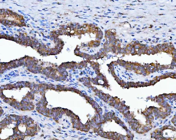
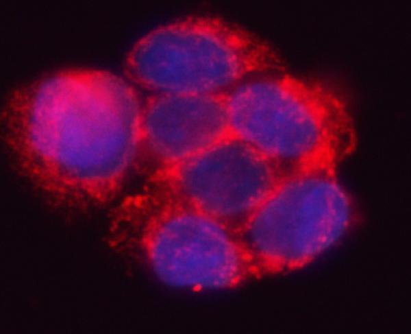
A05850-1 has IHC data from human paraffin sections of liver, prostate, and colorectal cancers (catalog IHC captions), plus IF data from HepG2 cells (catalog IF caption). Rat reactivity is listed (catalog: reactivity).

Real IHC data IHC analysis of HSD17B7 using anti-HSD17B7 antibody (A05850-1). HSD17B7 was detected in a paraffin-embedded section of human liver cancer tissue. Heat mediated antigen retrieval was performed in EDTA buffer (pH 8.0, epitope retrieval solution). The tissue section was blocked with 10% goat serum. The tissue section was then incubated with 2 μg/ml rabbit anti-HSD17B7 Antibody (A05850-1) overnight at 4°C. Peroxidase Conjugated Goat Anti-rabbit IgG was used as secondary antibody and incubated for 30 minutes at 37°C. The tissue section was developed using HRP Conjugated Rabbit IgG Super Vision Assay Kit (Catalog # SV0002) with DAB as the chromogen.
Anti-HSD17B7 Antibody ®
Cat # A05850-1

A05850-1 is shown in IHC on human paraffin sections of liver cancer, prostate cancer, and colorectal adenocarcinoma (catalog IHC captions). It is also shown in IF/ICC on HepG2 cells (catalog IF caption), and its listed reactivity is Human and Rat (catalog: reactivity).

Which to pick: Choose A05850-1 for chromogenic IHC on human paraffin sections: its own IHC captions show EDTA pH 8.0 retrieval and 2 μg/ml primary antibody (catalog IHC captions). The same SKU is listed for IF/ICC at 5 μg/ml and shown on HepG2 cells (catalog: applications, IF dilution, IF caption). A05850-1 lists Rat reactivity, but the supplied IHC and IF images show human samples only (catalog: reactivity, IHC captions, IF caption); the IHC captions do not report the fixative (catalog IHC captions).

Each figure is that product's own IHC / IF validation image from its datasheet.

References

  1. UniProt Consortium. UniProt entry P56937 (DHB7_HUMAN, 3-keto-steroid reductase/17-beta-hydroxysteroid dehydrogenase 7).
  2. Human Protein Atlas. HSD17B7 tissue IHC expression (reliability: Approved).
  3. Human Protein Atlas. HSD17B7 subcellular location (ICC-IF): Membrane.
  4. Human Protein Atlas. HSD17B7 antibody validation summary (1 antibodies).
  5. Normal Ovarian Function in Subfertile Mouse with Amhr2-Cre-Driven Ablation of Insr and Igf1r. Genes 2024 — PMC11121541.
  6. Dysregulation of estrogen signaling and HSD17B7 in the prostate stroma of African American men. Endocrinology 2026 — PMC13266810.
  7. The ovarian estrogen synthesis function was impaired in Y123F mouse and partly restored by exogenous FSH supplement. Reproductive biology and endocrinology : RB&E 2018 — PMC5934784.
  8. HSD17B7 gene in self-renewal and oncogenicity of keratinocytes from Black versus White populations. EMBO molecular medicine 2021 — PMC8261506.
  9. PubMed PMID:10544267 — UniProt-cited evidence.
  10. PubMed PMID:12732193 — UniProt-cited evidence.
  11. PubMed PMID:12975309 — UniProt-cited evidence.