This website uses cookies to ensure you get the best experience on our website.


- Table of Contents
Plan human paraffin IHC for IARS1 using the catalog antibody at 2–5 μg/ml (datasheet A06805-1). Assess cytoplasmic staining in Purkinje cells or lung macrophages (HPA tissue IHC), and interpret intensity with the reported low consistency between antibody staining and RNA expression in mind (HPA tissue IHC).

Expected localisation, validated protocols, controls and antibodies — the at-a-glance facts below, then the full design guide.
| Expected localisation | Cytoplasmic staining in several tissues (HPA tissue IHC) | |
| Staining pattern | Cytoplasmic signal in Purkinje cells and lung macrophages (HPA tissue IHC) | |
| Antigen retrieval | EDTA pH 8.0 HIER, heat-mediated (datasheet A06805-1) | |
| Positive control | Cerebellum+4 more · see all | |
| Negative control | Adipose tissue+4 more · see all |
| Fixation | Keep fixation consistent across paraffin sections (standard IHC practice; not target-specific) | |
| Caveat | Staining has low consistency with RNA expression (HPA tissue IHC) | |
| Regulation | Low tissue specificity at RNA level (HPA tissue RNA) | |
| Isoform / epitope | No annotated isoforms or propeptide; one chain (UniProt) |
The catalog antibody uses EDTA pH 8.0 heat-mediated retrieval (datasheet A06805-1). The published IARS1 protocol below reports cell and tissue preparation and antibody incubation (PMC13584253 methods).
| Sample | Paraffin-embedded human esophageal squamous carcinoma tissue; fixative not specified (datasheet A06805-1) |
| Fixation | Image fixative and duration unreported (datasheet A06805-1); verify before use. |
| Sectioning | 4–5 µm sections on charged slides (standard) |
| Deparaffinisation | Xylene, graded ethanol series to water (standard) |
| Antigen retrieval | Heat retrieval: EDTA pH 8.0 (datasheet A06805-1); 20 min, 95–100 °C (standard) |
| Peroxidase block | 3% H2O2, 10 min, room temperature (standard) |
| Blocking | 10% goat serum (datasheet A06805-1) |
| Primary antibody | Rabbit anti-IARS1, 2-5 μg/ml (datasheet A06805-1) |
| Primary incubation | Overnight at 4 °C (datasheet A06805-1) |
| Detection | HRP-conjugated secondary, DAB chromogen (datasheet A06805-1) |
| Counterstain | Hematoxylin, blue, dehydrate and mount (standard) |
| Expected result | IARS1-positive staining in purkinje cells of cerebellum (HPA tissue IHC: High). HPA tissue profile: Cytoplasmic expression in several tissues. No signal in the no-primary control. |
IARS1 is a cytoplasmic, cytosolic protein with no transmembrane segment (UniProt P41252). In paraffin section IHC, expect cytoplasmic staining in selected cells, including Purkinje cells, cortical and hippocampal neurons, lung macrophages, and glandular cells in the cervix, gallbladder, and salivary gland (HPA: High). Treat the pattern as a guide rather than a definitive specificity test: HPA rates tissue staining Approved but reports low consistency with RNA expression (HPA: tissue IHC reliability).
| Clear cytoplasmic signal in Purkinje cells or lung macrophages, with cell boundaries still readable. | This fits the expected compartment (UniProt P41252: cytosol) and reported high staining in those cells (HPA: High). Compare intensity with nearby cells on the same section; staining alone does not establish antibody specificity (HPA: Approved, low RNA consistency). |
| Signal is predominantly nuclear, with little cytoplasmic staining. | Investigate a compartment mismatch against UniProt's cytosolic assignment (UniProt P41252). HPA reports nucleoplasm in ICC-IF as uncertain, so nuclear signal alone cannot prove an artefact (HPA: subcellular); check the IHC controls before interpreting it. |
| Strong signal appears in a cell population HPA lists as not detected, such as adipocytes. | Consider cross-reactivity or endogenous detection activity and compare a no-primary control (HPA: adipocytes Not detected; general IHC practice). HPA's result is an observed staining level, not proof that every adipocyte lacks IARS1 (HPA: tissue IHC). |
| Diffuse chromogen covers cells and surrounding tissue without a clear cytoplasmic pattern. | Treat this as background until controls separate specific signal from nonspecific binding or endogenous detection activity (general IHC practice). Uniform haze cannot be scored as IARS1 localisation; the supported reference pattern is cytoplasmic (UniProt P41252; HPA: tissue IHC). |
| A proposed positive control, such as cerebellar Purkinje cells, has no detectable signal. | The run is inconclusive: Purkinje cells stain High in HPA IHC, but HPA's Approved rating carries low RNA consistency (HPA: tissue IHC). Check section quality and detection controls, then review retrieval and antibody conditions (general IHC practice). |
| Cell and tissue choice | Use an HPA High population to judge a positive result, such as cortical neurons or salivary glandular cells (HPA: High). Liver hepatocytes are Low in HPA IHC despite UniProt reporting liver protein expression; those statements describe different evidence and need not yield the same slide intensity (HPA: Low; UniProt P41252: tissue specificity). |
| Strength of staining evidence | HPA calls tissue IHC Approved while flagging low consistency with RNA expression; its listed antibody HPA029806 is IHC Approved, with no Enhanced designation supplied (HPA: tissue IHC reliability; HPA: antibody validation). Interpret unexpected staining with controls rather than treating the reference pattern as an absolute map. |
| Topology and processing | UniProt assigns IARS1 to cytoplasm and cytosol, with no transmembrane segment, signal peptide, or propeptide (UniProt P41252). These annotations support a cellular cytoplasmic readout; they do not identify an antibody epitope or predict how retrieval will perform (UniProt P41252 topology; general IHC practice). |
| Q: Does ICC-IF justify a nuclear IHC call? | A: HPA supports cytosol in ICC-IF but labels nucleoplasm uncertain; images are listed for A-431 and U-251MG (HPA: subcellular ICC-IF). Use that observation as context when reviewing nuclear IHC signal, not as a paraffin IHC localisation standard (HPA: tissue IHC profile). |
| Situation | Likely cause | Next action |
|---|---|---|
| No cytoplasmic signal in an HPA High control population. | The IHC run may lack usable antigen exposure or detection; HPA's High result is a reference observation, not a guaranteed result for every run (HPA: tissue IHC; general IHC practice). | Inspect section integrity and detection controls, then optimize antigen retrieval and antibody conditions for the chosen paraffin IHC assay (general IHC practice). |
| Predominantly nuclear staining. | Possible nonspecific signal or a compartment discrepancy; ICC-IF nucleoplasm is uncertain while cytosol is supported (HPA: subcellular; UniProt P41252). | Compare no-primary and positive controls, and check whether cytoplasmic signal remains visible before assigning localisation (general IHC practice). |
| Strong staining in adipocytes or heart cardiomyocytes. | Both are Not detected in HPA tissue IHC; cross-reactivity or endogenous detection activity is possible, but HPA does not establish absolute protein absence (HPA: tissue IHC). | Compare matched no-primary controls and inspect the cell-specific pattern before scoring the unexpected population (general IHC practice). |
| Diffuse staining throughout the section. | Nonspecific binding or endogenous detection activity can obscure the expected cytoplasmic distribution (general IHC practice; UniProt P41252: cytosol). | Check no-primary controls, blocking, washing, and detection conditions; score only distinguishable cellular signal (general IHC practice). |
| Low signal in liver hepatocytes is read as assay failure. | HPA reports hepatocytes as Low in tissue IHC, although UniProt reports liver protein expression (HPA: Low; UniProt P41252: tissue specificity). | Judge run performance against an HPA High cell population and its controls before interpreting weak liver staining (HPA: High; general IHC practice). |
| A not-detected comparison tissue shows faint staining. | HPA's Not detected category describes its observed IHC result; faint signal may reflect background or assay conditions (HPA: tissue IHC; general IHC practice). | Compare the same cell type with a no-primary control and assess whether signal is cytoplasmic and reproducible before calling it positive (UniProt P41252; general IHC practice). |
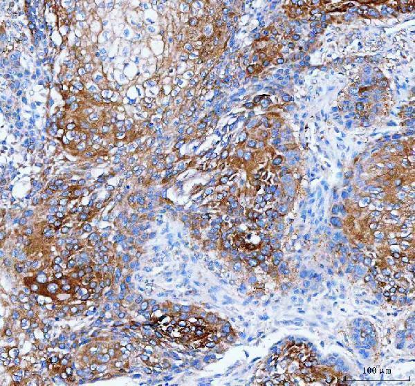
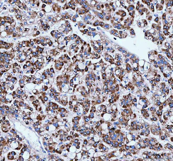


Comprehensive Human Protein Atlas IHC scoring per tissue (reliability: Approved — Low consistency between antibody staining and RNA expression data.). Rows are taken directly from the HPA tissue chart — click any row's HPA link to view the source.
| Tissue | Cell type | Level | Evidence | Source |
|---|---|---|---|---|
| Cerebellum | Purkinje cells | High | Protein (IHC) | HPA → |
| Cerebral cortex | Neuronal cells | High | Protein (IHC) | HPA → |
| Cervix | Glandular cells | High | Protein (IHC) | HPA → |
| Gallbladder | Glandular cells | High | Protein (IHC) | HPA → |
| Hippocampus | Neuronal cells | High | Protein (IHC) | HPA → |
| Tissue | Cell type | Level | Evidence | Source |
|---|---|---|---|---|
| Adipose tissue | Adipocytes | Not detected | Protein (IHC) | HPA → |
| Bone marrow | Hematopoietic cells | Not detected | Protein (IHC) | HPA → |
| Duodenum | Glandular cells | Not detected | Protein (IHC) | HPA → |
| Epididymis | Glandular cells | Not detected | Protein (IHC) | HPA → |
| Esophagus | Squamous epithelial cells | Not detected | Protein (IHC) | HPA → |
Troubleshoot IARS1 staining in paraffin sections by checking retrieval, cytoplasmic localisation, controls and cell-specific scoring (UniProt P41252 localisation; HPA tissue IHC).
A06805-1 has real IHC data from human paraffin sections; no IF/ICC data are supplied (A06805-1 catalog reactivity, applications, and image captions).
A06805-1 is listed for human IHC, with paraffin-section images from human esophageal squamous carcinoma, liver cancer, and lung cancer (A06805-1 catalog and image captions). No IF/ICC validation or figure is listed for A06805-1 (A06805-1 catalog applications and IF image alts).
Which to pick: Choose A06805-1 for human paraffin-section IHC: its image caption documents EDTA pH 8.0 retrieval and 2 μg/ml primary antibody, within the catalog’s 2–5 μg/ml IHC range (A06805-1 image caption; catalog dilution). The fixative is unreported (A06805-1 image caption). No listed SKU supports an IF/ICC or cross-species recommendation: A06805-1 lists human reactivity and IHC, but no IF/ICC application or IF figure (A06805-1 catalog applications, reactivity, and IF image alts).