IDH3B / Isocitrate dehydrogenase [NAD] subunit beta, mitochondrial · IHC design guide

Design Immunohistochemistry for IDH3B

This guide frames IDH3B scoring in paraffin sections around granular cytoplasmic staining (HPA tissue IHC) and mitochondrial localisation (UniProt). A documented human thyroid cancer example uses 2 μg/ml catalog antibody overnight at 4°C (datasheet A09433-3).

Evidence assembled Oct 2026 · For research use; verify linked source records and product datasheet before use
Immunohistochemistry protocol sheet for IDH3B (IHC for IDH3B): expected localisation Granular cytoplasm observed (HPA tissue IHC); mitochondrial protein (UniProt), antibody A09433-3, validated IHC image, and IHC protocol steps
Printable IDH3B IHC protocol sheet — expected localisation Granular cytoplasm observed (HPA tissue IHC); mitochondrial protein (UniProt), antibody A09433-3, controls and protocol steps. Open the full IDH3B IHC guide →

IDH3B Immunohistochemistry Experimental Design Guide

Expected localisation, validated protocols, controls and antibodies — the at-a-glance facts below, then the full design guide.

Must know before staining
Expected localisation Granular cytoplasm observed (HPA tissue IHC); mitochondrial protein (UniProt)
Staining pattern Widespread granular cytoplasmic staining (HPA tissue IHC)
Antigen retrieval EDTA pH 8.0 HIER, heat-mediated (datasheet A09433-3)
Positive control ⓘ Adrenal gland+4 more · see all
Negative control ⓘ None in HPA (detected in all 45 tissues); use no-primary + isotype controls
Important caveats
Reasons your staining may differ from the expected pattern.
Fixation Keep fixation consistent across sections. (standard IHC practice; not target-specific)
Caveat Only medium consistency between staining and RNA (HPA tissue IHC)
Regulation Low tissue specificity (HPA tissue RNA)
Isoform / epitope 3 isoforms (B, A, C); epitope coverage unknown (UniProt)
Section 1

Recommended IDH3B IHC & IF Protocols

The catalog antibody protocol uses EDTA retrieval (datasheet A09433-3); two published IDH3B IHC protocols provide tissue-specific examples (PMC8802454; PMC9963320).

Recommended immunohistochemistry (IHC-P) protocol parameters
SampleParaffin-embedded follicles of human thyroid cancer tissue; fixative not specified (datasheet A09433-3)
FixationImage fixative and duration unreported (datasheet A09433-3); verify before use.
Sectioning4–5 µm sections on charged slides (standard)
DeparaffinisationXylene, graded ethanol series to water (standard)
Antigen retrievalHeat retrieval: EDTA pH 8.0 (datasheet A09433-3); 20 min, 95–100 °C (standard)
Peroxidase block3% H2O2, 10 min, room temperature (standard)
Blocking10% goat serum (datasheet A09433-3)
Primary antibodyRabbit anti-IDH3B, 2-5 μg/ml (datasheet A09433-3)
Primary incubationOvernight at 4 °C (datasheet A09433-3)
DetectionHRP-conjugated secondary, DAB chromogen (datasheet A09433-3)
CounterstainHematoxylin, blue, dehydrate and mount (standard)
Expected resultIDH3B-positive staining in glandular cells of adrenal gland (HPA tissue IHC: High). HPA tissue profile: Ubiquitous cytoplasmic expression in a granular pattern. No signal in the no-primary control.
💡Decision noteStart with heat-mediated EDTA at pH 8.0 (datasheet A09433-3); consider sodium citrate retrieval if adapting the liver tissue protocol (PMC9963320).
Section 2

What Is the Expected IDH3B Staining Pattern?

IDH3B is mitochondrial and has no annotated transmembrane segment (UniProt O43837 topology). In paraffin sections, expect granular cytoplasmic staining across many cell types, including strong staining in bronchial respiratory epithelial cells and several glandular cell populations (HPA tissue IHC). HPA rates the tissue staining evidence Supported, with medium consistency between antibody staining and RNA expression (HPA tissue IHC).

What am I looking at on my slide?
Granular cytoplasmic staining in bronchial respiratory epithelial cells or adrenal glandular cells.This fits the expected compartment and examples of High tissue staining (UniProt O43837; HPA tissue IHC). Judge the pattern within identified cells, since HPA describes expression as ubiquitous rather than restricted to one tissue (HPA tissue IHC).
Predominantly nuclear or sharply plasma membrane staining, with little granular cytoplasmic signal.That compartment conflicts with mitochondrial localisation (UniProt O43837; HPA subcellular ICC-IF). Treat it as a possible artefact or off-target signal and compare it with a known positive section and a no-primary control (general IHC practice).
Strong staining in cells where HPA reports a low level, such as hepatocytes or skeletal myocytes.A stronger result warrants scrutiny; Low is not the same as negative (HPA tissue IHC). Check cell identification and controls before attributing unexpected staining to IDH3B; cross-reactivity or endogenous detection activity are possible explanations (general IHC practice).
A uniform brown haze obscures cell boundaries and the expected granules.An uninterpretable background can arise from detection activity, nonspecific antibody binding, or inadequate washing (general IHC practice). It cannot establish the granular cytoplasmic distribution described for IDH3B (HPA tissue IHC).
No staining in a technically comparable bronchus section's respiratory epithelial cells.HPA reports High staining in those cells, so first assess whether the run worked (HPA tissue IHC; general IHC practice). A single blank section does not by itself establish absence of IDH3B; review the positive control and detection steps (general IHC practice).
💡Expected IDH3B appearanceA convincing positive is granular cytoplasmic staining in identified cells, including strong bronchial respiratory epithelial or glandular cell staining; isolated nuclear staining or uniform brown haze calls for control review (HPA tissue IHC; UniProt O43837; general IHC practice).
How each factor affects the staining
Cellular compartmentUniProt places IDH3B in mitochondria, while HPA describes granular cytoplasmic tissue staining (UniProt O43837; HPA tissue IHC). Read granules in the context of cell morphology; chromogenic IHC alone does not prove organelle colocalisation (general IHC practice).
Tissue and cell distributionHPA reports low tissue specificity and no negative tissue examples in this record (HPA tissue IHC). Its High and Low entries are useful comparisons, but a low entry is not a blank control (HPA tissue IHC).
Antibody evidenceHPA lists IHC as Supported for HPA049387; it lists no IHC status for HPA054180 (HPA antibodies). The overall tissue reliability is Supported with medium staining–RNA consistency, so match the observed pattern to controls rather than treating every positive cell as confirmed (HPA tissue IHC; general IHC practice).
Processing and isoformsUniProt annotates a mature chain at residues 35–385 and isoforms B, A, and C (UniProt O43837). No antibody epitope or isoform-specific staining evidence is supplied, so these annotations cannot predict which isoform a chromogenic signal represents (UniProt O43837; HPA antibodies).
IF/ICC Q: Does its localisation support the IHC interpretation?A: HPA reports mitochondrial ICC-IF localisation with Enhanced evidence, consistent with the granular cytoplasmic IHC profile (HPA subcellular ICC-IF; HPA tissue IHC). It supports a compartment check; the separate IF/ICC guide covers that application.
Why is my staining missing, weak or wrong?
SituationLikely causeNext action
Expected positive cells are blank.A failed detection run or unsuitable assay conditions are possible; HPA reports High bronchial respiratory epithelial and adrenal glandular staining (HPA tissue IHC; general IHC practice).Inspect a known positive section and detection controls, then review the chosen IHC antibody's documented paraffin-section conditions (general IHC practice). Target-specific fixation sensitivity is unreported in the supplied evidence.
Granules are lost in widespread brown background.Nonspecific antibody binding, endogenous detection activity, or inadequate washing may obscure a tissue pattern (general IHC practice). HPA describes a granular cytoplasmic pattern (HPA tissue IHC).Compare no-primary and reagent controls; review blocking, washing, and chromogen development against the established IHC workflow (general IHC practice). Reassess only when cell boundaries and granules can be read.
Most visible signal is nuclear or at cell borders.That distribution disagrees with mitochondrial localisation and raises concern for artefact or off-target binding (UniProt O43837; HPA subcellular ICC-IF; general IHC practice).Check morphology, the no-primary control, and a known positive tissue for the expected granular cytoplasmic signal (HPA tissue IHC; general IHC practice). Do not score an isolated wrong-compartment signal as a positive.
Low-level reference cells appear as strong as high-level reference cells.HPA reports Low hepatocyte and skeletal myocyte staining but High staining in several glandular and epithelial populations (HPA tissue IHC). Excess detection signal or cell-identification error may blur that comparison (general IHC practice).Confirm cell identity and compare sections processed in the same run; inspect controls before interpreting intensity differences (general IHC practice). Keep in mind that HPA's low categories allow genuine staining (HPA tissue IHC).
Strong staining appears in an unexpected cell population.Cross-reactivity or endogenous detection activity is possible, although HPA's ubiquitous profile means an unlisted cell type is not automatically negative (HPA tissue IHC; general IHC practice).Use a no-primary control to assess detection background and compare the staining compartment with the expected granular cytoplasm (general IHC practice; HPA tissue IHC). Describe unresolved signal as uncertain.
Two antibodies give different tissue patterns.HPA supplies a Supported IHC status for HPA049387 but no IHC status for HPA054180; their listed ICC-IF evidence does not establish matching IHC performance (HPA antibodies).Compare each antibody's IHC evidence and its positive and negative controls under the same scoring criteria (HPA antibodies; general IHC practice). Avoid inferring an isoform-specific pattern without epitope evidence (UniProt O43837).

Sample controls for IDH3B IHC & IF

🧪Run adrenal gland first and score its glandular cells for IDH3B staining (HPA: High in adrenal gland glandular cells). HPA detects IDH3B in all 45 scored tissues, so there is no supported negative tissue; use no-primary and isotype controls, and treat any unstained stromal or vascular cells on the positive slide as background comparators, not validated IDH3B-negative cells (HPA: no negative rows; detected in all 45 tissues).
Positive control tissue: Adrenal gland (Glandular cells, HPA High)
Negative control tissue: None in HPA: IDH3B is detected in all 45 scored tissues. Use a no-primary (secondary-only) and an isotype control instead.
ICC-IF cell lines (HPA subcellular resource): HPA ICC-IF images show IDH3B in A-549, U2OS, KOLF2.1J, NIH 3T3, with annotated localisation: Mitochondria (enhanced) (HPA subcellular).
Technical controls: Include a no-primary, secondary-only section; a concentration-matched rabbit IgG isotype control matched to the primary antibody’s clonality if known; and IDH3B-knockout material as a biological negative where available (A09433-3 caption: rabbit anti-IDH3B primary). For adrenal DAB staining, quench endogenous peroxidase and compare brown signal with the no-primary section, including areas with endogenous pigment (A09433-3 caption: HRP/DAB detection; standard IHC practice).
⚠️Feasibility: A target-specific fixation window or fixation effect is unreported, and the selected A09433-3 paraffin-section caption does not state the fixative (A09433-3 caption: fixative unreported). That caption uses heat retrieval in EDTA at pH 8.0 for thyroid cancer tissue; it does not establish a retrieval requirement for adrenal gland (A09433-3 caption: EDTA retrieval). The supplied evidence does not establish that frozen sections or IF are easier than paraffin IHC, although ICC-IF images show mitochondrial localization; adrenal pigment and endogenous peroxidase warrant careful DAB interpretation (HPA: mitochondrial ICC-IF localization; standard IHC practice).

HPA tissue IHC evidence for IDH3B

Comprehensive Human Protein Atlas IHC scoring per tissue (reliability: Supported — Medium consistency between antibody staining and RNA expression data.). Rows are taken directly from the HPA tissue chart — click any row's HPA link to view the source.

Positive expression · recommended positive controls

TissueCell typeLevelEvidenceSource
Adrenal gland Glandular cells High Protein (IHC) HPA →
Appendix Glandular cells High Protein (IHC) HPA →
Breast Glandular cells High Protein (IHC) HPA →
Bronchus Respiratory epithelial cells High Protein (IHC) HPA →
Caudate Neuronal cells High Protein (IHC) HPA →

Undetected expression · recommended negative controls

TissueCell typeLevelEvidenceSource
None in HPA: IDH3B is detected in all 45 scored tissues. Use a no-primary (secondary-only) and an isotype control instead.
Section 3

Advanced IDH3B IHC Tips

Troubleshoot IDH3B staining by checking retrieval, specimen handling, mitochondrial localisation, and controls before comparing signal across sections.

Which retrieval condition should I try first for IDH3B in paraffin sections?
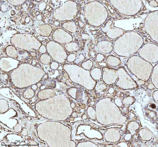
Start with heat-mediated antigen retrieval in EDTA at pH 8.0 for paraffin sections (datasheet A09433-3). This was the retrieval used for the selected IDH3B tissue image; its fixation method was not reported (datasheet A09433-3). Keep heating and cooling conditions consistent across test and control sections, then compare signal in the same cell population (standard IHC practice). The image used 2 μg/ml primary antibody overnight at 4°C, so those conditions provide a reference while assessing retrieval (datasheet A09433-3). If staining remains weak, test another retrieval condition on matched sections and assess background alongside signal (standard IHC practice).
How should I investigate inconsistent staining after fixation?
Target-specific fixation sensitivity for IDH3B is unknown from the supplied evidence (datasheet A09433-3). The selected image identifies a paraffin section but does not report its fixative or fixation duration (datasheet A09433-3). Record fixation and processing histories for each specimen, and compare sections processed alike before changing the antibody concentration (standard IHC practice). Include an established positive section in each run to help distinguish specimen handling from a staining-run problem (standard IHC practice). Do not infer fixation tolerance from the reported mitochondrial localisation or granular tissue pattern; neither observation tests fixation effects (UniProt O43837; HPA tissue IHC).
What staining pattern supports mitochondrial IDH3B localisation in tissue?
Expect a granular cytoplasmic pattern in stained cells, consistent with the tissue profile and mitochondrial localisation reported for IDH3B (HPA tissue IHC; HPA subcellular). IDH3B has no annotated transmembrane segment, so a crisp plasma-membrane outline alone would not fit its recorded topology (UniProt O43837 topology). Assess granules within intact cells at sufficient magnification before calling a section positive (standard IHC practice). Compare the pattern with an appropriate mitochondrial control on adjacent sections when the granules are difficult to resolve (standard IHC practice). Diffuse extracellular deposit or isolated nuclear signal warrants a review of detection background and tissue integrity (standard IHC practice).
Could isoform choice or processing explain uneven IDH3B staining?
IDH3B has three recorded isoforms, B, A, and C, but the supplied antibody caption does not identify the recognized epitope (UniProt O43837 isoforms; datasheet A09433-3). The recorded mature chain spans residues 35–385; that annotation alone does not establish antibody recognition of every isoform (UniProt O43837 processing). An acetylated lysine is annotated at residue 199, but no evidence here connects that modification to staining performance (UniProt O43837 modified residues). Check the antibody’s documented immunogen or epitope before making isoform-specific claims (standard IHC practice). If specificity remains uncertain, assess staining with an independent antibody or a suitable loss-of-target control (standard IHC practice).
How can I check an IDH3B IHC pattern using multiplex IF?
Use IF as a separate localisation check and include a mitochondrial marker to assess whether IDH3B signal follows the expected compartment (UniProt O43837 subcellular; standard IF practice). A marker for the cell population under study can clarify which cells carry signal; HPA reports high staining in several glandular-cell populations (HPA tissue IHC). Choose fluorophores after inspecting tissue autofluorescence, reserving a well-separated channel for the weaker signal (standard IF practice). IDH3B is mitochondrial, but the supplied record does not specify which side of a mitochondrial membrane contains the antibody epitope (UniProt O43837 subcellular; datasheet A09433-3). Titrate permeabilisation against signal and morphology, and establish IF fixation conditions independently because the selected IHC caption reports neither fixation nor IF testing (standard IF practice; datasheet A09433-3).
What should I check when DAB background obscures IDH3B granules?
The selected tissue image used 10% goat serum blocking, a peroxidase-conjugated secondary antibody, and DAB detection (datasheet A09433-3). Check a no-primary control to identify detection-system staining, and include a peroxidase block when using an HRP–DAB workflow (standard IHC practice). Compare the control and test section in the same tissue regions before changing primary antibody concentration (standard IHC practice). If background persists, review blocking, wash stringency, secondary-antibody exposure, and DAB development on matched sections (standard IHC practice). Judge improvement by preservation of granular cytoplasmic signal, the reported IDH3B tissue pattern, rather than by a uniformly darker section (HPA tissue IHC).
How should I score IDH3B staining across paraffin sections? ⚠ ANSWER MARKED FOR VERIFICATION
Define the cell population and viable tissue region before scoring, since IDH3B has a broadly distributed granular cytoplasmic tissue profile (HPA tissue IHC; standard IHC practice). Report the percentage of positive cells and staining intensity, or calculate an H-score from intensity grades 0–3 and their corresponding cell percentages (standard IHC practice). Apply one threshold and imaging setup across comparable sections, with a positive reference and a no-primary control in the run (standard IHC practice). Normalise counts to the number of evaluable cells in the chosen population, or normalise positive-cell density to assessed area in mm² (standard IHC practice). Record compartment-specific staining separately from nonspecific deposits (standard IHC practice).
How do I distinguish true IDH3B staining from tissue artefact?
Call a convincing positive when intact cells show granular cytoplasmic staining compatible with mitochondrial IDH3B (HPA tissue IHC; HPA subcellular). Compare staining among identified cell populations rather than assuming every cell must be equally bright; HPA reports high signal in several glandular and neuronal populations and low signal in hepatocytes, myocytes, and chondrocytes (HPA tissue IHC). Treat isolated nuclear or extracellular colour as suspect because it does not match the reported localisation (UniProt O43837 subcellular; HPA tissue IHC). Check section edges and necrotic areas for local staining artefacts, and review no-primary and peroxidase-block controls for endogenous enzyme signal (standard IHC practice). HPA rates the tissue staining as Supported, with medium agreement between staining and RNA data (HPA tissue IHC).
Boster reagents

Best IDH3B / Isocitrate dehydrogenase [NAD] subunit beta, mitochondrial IHC Antibodies

Both antibodies have real paraffin-section IHC images from human tissue; neither has IF/ICC image evidence (catalog IHC captions; catalog IF image records).

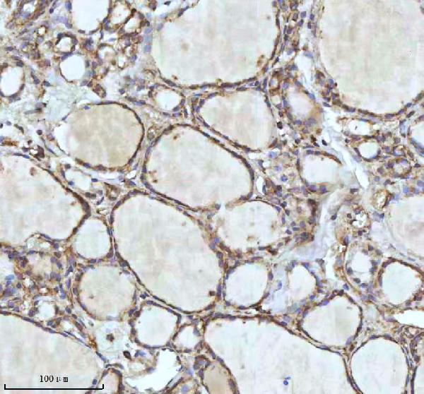
Real IHC data IHC analysis of IDH3B using anti-IDH3B antibody (A09433-3). IDH3B was detected in a paraffin-embedded section of follicles of human thyroid cancer tissue. Heat mediated antigen retrieval was performed in EDTA buffer (pH 8.0, epitope retrieval solution). The tissue section was blocked with 10% goat serum. The tissue section was then incubated with 2 μg/ml rabbit anti-IDH3B Antibody (A09433-3) overnight at 4°C. Peroxidase Conjugated Goat Anti-rabbit IgG was used as secondary antibody and incubated for 30 minutes at 37°C. The tissue section was developed using HRP Conjugated Rabbit IgG Super Vision Assay Kit (Catalog # SV0002) with DAB as the chromogen.
Anti-IDH3B Antibody ®
Cat # A09433-3
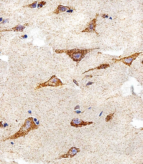
Real IHC data Immunohistochemical analysis of paraffin-embedded H.brain section using IDH3B Antibody (N-term). A09433-2 was diluted at 1:25 dilution. A peroxidase-conjugated goat anti-rabbit IgG at 1:400 dilution was used as the secondary antibody, followed by DAB staining.
Anti-IDH3B Antibody (N-term)
Cat # A09433-2

A09433-3 has an IHC image from paraffin-embedded human thyroid cancer follicles and lists human, mouse, and rat reactivity (A09433-3 image caption; catalog reactivity). A09433-2 has an IHC-P image from a paraffin-embedded human brain section and lists human reactivity (A09433-2 image caption; catalog applications and reactivity).

Which to pick: For human tissue IHC, A09433-3 provides a documented EDTA pH 8.0 retrieval and DAB workflow for its paraffin section; A09433-2 is a polyclonal alternative with a human brain paraffin-section image (A09433-3 image caption; A09433-2 image caption and datasheet). For IF/ICC, neither SKU lists that application or provides an IF image, so there is no validated pick here (catalog applications; catalog IF image records). For mouse or rat work, A09433-3 lists those species, but its IHC image shows human tissue only; the fixative is unreported in both IHC captions (catalog reactivity; A09433-3 image caption; A09433-2 image caption).

Each figure is that product's own IHC / IF validation image from its datasheet.

References

  1. UniProt Consortium. UniProt entry O43837 (IDH3B_HUMAN, Isocitrate dehydrogenase [NAD] subunit beta, mitochondrial).
  2. Human Protein Atlas. IDH3B tissue IHC expression (reliability: Supported).
  3. Human Protein Atlas. IDH3B subcellular location (ICC-IF): Localized to the mitochondria..
  4. Human Protein Atlas. IDH3B antibody validation summary (2 antibodies).
  5. TRPM7 silencing modulates glucose metabolic reprogramming to inhibit the growth of ovarian cancer by enhancing AMPK activation to promote HIF-1α degradation. Journal of experimental & clinical cancer research : CR 2022 — PMC8802454.
  6. A Multi-Omics Analysis of NASH-Related Prognostic Biomarkers Associated with Drug Sensitivity and Immune Infiltration in Hepatocellular Carcinoma. Journal of clinical medicine 2023 — PMC9963320.
  7. A novel mitochondrial metabolism-related gene signature for predicting the prognosis of oesophageal squamous cell carcinoma. Aging 2024 — PMC11210263.
  8. PubMed PMID:10601238 — UniProt-cited evidence.
  9. PubMed PMID:14702039 — UniProt-cited evidence.
  10. PubMed PMID:11780052 — UniProt-cited evidence.