IER3 / Radiation-inducible immediate-early gene IEX-1 · IHC design guide

Design Immunohistochemistry for IER3

Plan chromogenic IHC-P for IER3 using pancreatic exocrine glandular cells as a high-staining reference and cardiac myocytes as a tissue where staining was not detected (HPA tissue IHC). This guide covers observed cytoplasmic staining, membrane topology, fixation consistency, and the catalog antibody's 2–5 μg/mL IHC range (HPA tissue IHC; UniProt; datasheet A04014-1).

Evidence assembled Oct 2026 · For research use; verify linked source records and product datasheet before use
Immunohistochemistry protocol sheet for IER3 (IHC for IER3): expected localisation Cytoplasmic tissue staining (HPA tissue IHC); membrane topology (UniProt), antibody A04014-1, validated IHC image, and IHC protocol steps
Printable IER3 IHC protocol sheet — expected localisation Cytoplasmic tissue staining (HPA tissue IHC); membrane topology (UniProt), antibody A04014-1, controls and protocol steps. Open the full IER3 IHC guide →

IER3 Immunohistochemistry Experimental Design Guide

Expected localisation, validated protocols, controls and antibodies — the at-a-glance facts below, then the full design guide.

Must know before staining
Expected localisation Cytoplasmic tissue staining (HPA tissue IHC); membrane topology (UniProt)
Staining pattern Variable cytoplasmic staining across tissues (HPA tissue IHC)
Antigen retrieval EDTA pH 8.0 HIER, heat-mediated (datasheet A04014-1)
Positive control ⓘ Adrenal gland+4 more · see all
Negative control ⓘ Bone marrow+4 more · see all
Important caveats
Reasons your staining may differ from the expected pattern.
Fixation Keep fixation consistent across sections (standard IHC practice; not target-specific)
Caveat Staining has medium consistency with RNA data (HPA tissue IHC)
Regulation Radiation-inducible IER3 (UniProt)
Isoform / epitope No isoforms annotated; cytoplasmic versus extracellular epitope location matters (UniProt)
Section 1

Recommended IER3 IHC & IF Protocols

The catalog antibody protocol is followed by published IER3 IHC methods for liver and skin sections (PMC8918148; PMC3306129).

Recommended immunohistochemistry (IHC-P) protocol parameters
SampleParaffin-embedded human stomach cancer tissue; fixative not specified (datasheet A04014-1)
FixationImage fixative and duration unreported (datasheet A04014-1); verify before use.
Sectioning4–5 µm sections on charged slides (standard)
DeparaffinisationXylene, graded ethanol series to water (standard)
Antigen retrievalHeat retrieval: EDTA pH 8.0 (datasheet A04014-1); 20 min, 95–100 °C (standard)
Peroxidase block3% H2O2, 10 min, room temperature (standard)
Blocking10% goat serum (datasheet A04014-1)
Primary antibodyRabbit anti-IER3, 2-5 μg/ml (datasheet A04014-1)
Primary incubationOvernight at 4 °C (datasheet A04014-1)
DetectionHRP-conjugated secondary, DAB chromogen (datasheet A04014-1)
CounterstainHematoxylin, blue, dehydrate and mount (standard)
Expected resultIER3-positive staining in glandular cells of adrenal gland (HPA tissue IHC: High). HPA tissue profile: General cytoplasmic expression at variable levels. No signal in the no-primary control.
💡Decision noteStart with EDTA pH 8.0 heat retrieval (datasheet: A04014-1); an HCC study also used EDTA (PMC8918148). Citrate pH 6 is a published alternative (PMC3306129).
Section 2

What Is the Expected IER3 Staining Pattern?

IER3 has a membrane annotation and a single transmembrane segment, with residues 1–82 cytoplasmic and 100–156 extracellular (UniProt P46695 topology). In tissue IHC, expect predominantly cytoplasmic staining at variable levels, including strong staining in selected glandular and epithelial cells (HPA: tissue IHC profile). Treat this as a pattern to check, since HPA rates the tissue IHC evidence Approved with medium consistency between staining and RNA expression (HPA: reliability).

What am I looking at on my slide?
Strong cytoplasmic staining in pancreatic exocrine glandular cells or adrenal glandular cells (HPA: High in both).This fits the reported tissue IHC pattern (HPA: general cytoplasmic expression). Score the relevant cells rather than the whole section; nearby cells may differ in intensity (HPA: variable levels).
Membrane-edge staining accompanies cytoplasmic staining in an otherwise expected cell population (UniProt P46695: membrane topology; HPA: cytoplasmic IHC profile).Membrane association is plausible, but tissue IHC alone does not establish a sharply defined membrane-only pattern. Compare the result with HPA’s reported cytoplasmic staining before calling it typical (HPA: tissue IHC profile).
Strong nuclear-only staining, without the expected cytoplasmic pattern (HPA: tissue IHC profile).This is a compartment mismatch: HPA describes cytoplasmic tissue IHC staining, while UniProt annotates a membrane protein (HPA: tissue IHC profile; UniProt P46695: location). Review controls and antibody performance before interpreting it as IER3.
Strong staining in cardiomyocytes or skeletal myocytes (HPA: Not detected in these cells).This conflicts with the reported tissue pattern and raises concern for cross-reactivity or endogenous chromogenic activity (HPA: tissue IHC; standard IHC practice). Check the detection controls and compare with a known-positive section.
A uniform haze covers cells and surrounding tissue, or a known-positive glandular population is blank (HPA: High in selected glandular cells).A haze obscures cell-specific scoring and suggests background from staining or detection (standard IHC practice). A blank positive-control population makes the run inconclusive; check the IHC workflow before inferring absent IER3.
💡Expected IER3 appearanceCall a positive result when the expected glandular or epithelial cells show distinct, potentially high cytoplasmic staining (HPA: tissue IHC); isolated nuclear signal, uniform haze, or strong staining in reported negative cells is suspect (HPA: tissue IHC; standard IHC practice).
How each factor affects the staining
Compartment and topology (UniProt P46695; HPA: tissue IHC).IER3 has one transmembrane segment at residues 83–99, yet HPA describes cytoplasmic tissue staining. Use the observed IHC profile to judge sections; topology alone does not predict a crisp membrane outline.
Choice of comparison tissue (HPA: tissue IHC).Pancreatic exocrine cells and adrenal glandular cells are reported High; cardiomyocytes and skeletal myocytes are Not detected. HPA also reports low tissue RNA specificity, so a single tissue cannot define universal positivity or absence.
Strength of IHC evidence (HPA: reliability; HPA043847 validation).HPA calls the tissue evidence Approved with medium staining–RNA consistency; HPA043847 is IHC Approved. These ratings support a reference pattern while leaving room for sample and assay differences.
Epitope-dependent interpretation (UniProt P46695: topology and modifications).The antigen includes an extracellular region at residues 100–156, a glycosylation site at 133, and reported phosphorylation sites. Their effect on this antibody’s staining cannot be assigned without its epitope.
IF/ICC cross-check? (HPA: subcellular summary).HPA summarizes IF/ICC location as Membrane but lists no cell-line IF images or main location. This is limited corroboration of membrane association, not a tissue IHC scoring standard (HPA: subcellular record).
Retrieval and detection checks (standard IHC practice).If signal is weak or diffuse, check the documented paraffin IHC retrieval and chromogenic detection steps with suitable controls. No supplied source establishes IER3-specific retrieval conditions or fixation sensitivity.
Why is my staining missing, weak or wrong?
SituationLikely causeNext action
Expected pancreatic exocrine cells show no signal (HPA: High).The assay may have failed, or the sampled cells may be unsuitable for scoring (standard IHC practice).Confirm cell identity, then review retrieval, antibody incubation and chromogen steps against the validated IHC workflow; assess a positive control in the same run (standard IHC practice).
Staining is limited to nuclei (HPA: cytoplasmic IHC profile).The compartment differs from the reported IHC pattern and membrane annotation (HPA: tissue IHC; UniProt P46695).Inspect morphology and controls, then repeat or independently verify the result before assigning it to IER3 (standard IHC practice).
Cardiomyocytes stain strongly (HPA: Not detected).Cross-reactivity or endogenous detection activity is possible (standard IHC practice).Compare with a known-positive tissue and a primary-antibody omission control; check the chromogenic detection block appropriate to the assay (standard IHC practice).
The whole section has a brown haze (standard IHC practice).Nonspecific background or detection activity may obscure the cell-specific pattern (standard IHC practice).Check blocking, washes, primary-antibody conditions and the omission control; score cells only after background permits localisation (standard IHC practice).
A sharp membrane rim appears without cytoplasmic staining (UniProt P46695: membrane; HPA: cytoplasmic IHC).Topology makes membrane association plausible, but the result differs from HPA’s tissue IHC profile (UniProt P46695; HPA: tissue IHC).Check the same cell type in a positive-reference section and review detection controls before calling the rim the expected pattern (standard IHC practice).
An IF/ICC result seems to disagree with the tissue IHC result (HPA: subcellular and tissue records).HPA’s IF/ICC summary says Membrane, while its tissue IHC profile says cytoplasmic; no IF cell-line images are listed (HPA: subcellular; tissue IHC).Interpret each assay against its own controls and readout. Use the tissue IHC profile for paraffin-section scoring (HPA: tissue IHC; standard IHC practice).

Sample controls for IER3 IHC & IF

🧪Run adrenal gland first; its glandular cells should stain (HPA: High in adrenal gland glandular cells). Use heart muscle cardiomyocytes as the negative tissue (HPA: Not detected in cardiomyocytes); on the adrenal slide, treat unstained neighboring cells as a within-slide background reference, without assuming they lack IER3 (HPA: cell-specific glandular-cell result only).
Positive control tissue: Adrenal gland (Glandular cells, HPA High)
Negative control tissue: Bone marrow (HPA Not detected)
ICC-IF cell lines (HPA subcellular resource): HPA carries no ICC-IF cell line for IER3; derive a cell-line control from the positive tissue's cell type (Glandular cells) and confirm it by RNA or western blot first.
Technical controls: Include a no-primary, secondary-only control and a concentration-matched, nonimmune rabbit IgG isotype control (selected-SKU caption: rabbit primary antibody; standard IHC practice); use IER3-knockout material processed in parallel as a biological negative when available (standard IHC practice). Block endogenous peroxidase before HRP/DAB detection and check adrenal pigment against the no-primary slide (selected-SKU caption: HRP/DAB detection; standard IHC practice).
⚠️Feasibility: A target-specific fixation window or fixation effect is unreported, and the exact selected-SKU paraffin-section caption does not state the fixative (selected-SKU caption: fixative not stated). Heat retrieval in EDTA at pH 8.0 was used in that IHC example, but retrieval dependence has not been established (selected-SKU caption: EDTA heat retrieval). The supplied evidence does not show that frozen sections or IF are easier; adrenal pigment can complicate interpretation of brown DAB signal, so compare the positive slide with its no-primary control (HPA: adrenal gland positive tissue; selected-SKU caption: DAB detection; standard IHC practice).

HPA tissue IHC evidence for IER3

Comprehensive Human Protein Atlas IHC scoring per tissue (reliability: Approved — Medium consistency between antibody staining and RNA expression data.). Rows are taken directly from the HPA tissue chart — click any row's HPA link to view the source.

Positive expression · recommended positive controls

TissueCell typeLevelEvidenceSource
Adrenal gland Glandular cells High Protein (IHC) HPA →
Endometrium Glandular cells High Protein (IHC) HPA →
Fallopian tube Glandular cells High Protein (IHC) HPA →
Nasopharynx Respiratory epithelial cells High Protein (IHC) HPA →
Ovary Follicle cells High Protein (IHC) HPA →

Undetected expression · recommended negative controls

TissueCell typeLevelEvidenceSource
Bone marrow Hematopoietic cells Not detected Protein (IHC) HPA →
Heart muscle Cardiomyocytes Not detected Protein (IHC) HPA →
Placenta Decidual cells Not detected Protein (IHC) HPA →
Skeletal muscle Myocytes Not detected Protein (IHC) HPA →
Smooth muscle Smooth muscle cells Not detected Protein (IHC) HPA →
Section 3

Advanced IER3 IHC Tips

Use the catalog antibody’s tissue IHC conditions as a starting point, then assess staining against IER3 localisation and cell specific controls.

How should I adjust retrieval when IER3 staining is weak in paraffin sections?
Start with heat mediated retrieval in EDTA at pH 8.0 for paraffin sections stained with A04014-1 (datasheet A04014-1). Keep heating and cooling conditions consistent across the run, then compare weak sections with a concurrently processed positive control; the documented stomach cancer section was paraffin embedded and stained after this retrieval (datasheet A04014-1). If staining remains weak, vary retrieval duration in small increments while checking tissue preservation, and consider another buffer only as a documented fallback experiment. Hold the primary incubation at 2 μg/ml overnight at 4°C while assessing retrieval, so dilution changes do not obscure its effect (datasheet A04014-1).
Could fixation explain weak or uneven IER3 staining?
Target specific fixation sensitivity is unknown: the selected tissue IHC caption identifies a paraffin section but does not report its fixative (datasheet A04014-1). Record fixative, fixation duration and specimen thickness for each case, and compare sections processed together before attributing a staining difference to IER3 biology. Excessive or uneven fixation can reduce epitope accessibility in IHC generally, so review morphology and a control stain when signal varies between regions. Keep retrieval at EDTA pH 8.0 and primary incubation at 2 μg/ml overnight at 4°C during that comparison (datasheet A04014-1); do not infer an IER3 specific fixation effect from the result alone.
Should I expect membrane or cytoplasmic IER3 staining?
Interpret both patterns cautiously: UniProt annotates IER3 as a membrane protein with a transmembrane segment at residues 83–99 (UniProt P46695 topology), while tissue IHC reports general cytoplasmic expression at variable levels (HPA tissue IHC). Its annotated topology places residues 1–82 on the cytoplasmic side and 100–156 on the extracellular side (UniProt P46695 topology). Because the catalog antibody’s epitope is unspecified in the supplied evidence, a diffuse chromogenic signal cannot by itself establish which side was recognised. Compare membrane accentuation and cytoplasmic staining with morphology, matched controls and the same detection settings before assigning a compartment.
Could epitope position or IER3 modifications change the IHC pattern?
First obtain the antibody’s immunogen or epitope information before explaining a staining change by epitope accessibility; none is supplied here. UniProt annotates 0 isoforms, a transmembrane segment at residues 83–99, and a glycosylation site at residue 133 (UniProt P46695). It also lists modified residues 18, 31, 123 and 126, including MAPK1 linked threonines (UniProt P46695). These annotations identify plausible regions to investigate, but they do not establish that fixation or EDTA retrieval alters this antibody’s binding. Compare serial sections under identical retrieval and primary antibody conditions before attributing a compartment difference to a modification.
How can I check an IER3 signal by multiplex IF?
Treat IF as a separate optimisation: the supplied catalog evidence documents paraffin section chromogenic IHC, and HPA lists no cell lines with ICC/IF images for IER3 (datasheet A04014-1; HPA subcellular). Pair IER3 with a validated marker for the cell population under study; exocrine glandular cells in pancreas are one reported high staining population (HPA tissue IHC). Choose well separated fluorophores, favouring a far red channel when tissue autofluorescence overlaps shorter wavelengths, and include single stain and unstained controls. If the antibody recognises residues 1–82, test permeabilisation for the cytoplasmic side; for residues 100–156, evaluate access without permeabilisation first (UniProt P46695 topology).
What should I check when DAB background obscures IER3 staining?
Review no primary and secondary only controls alongside the stained section to distinguish antibody dependent colour from detection background. The documented procedure used 10% goat serum blocking, a peroxidase conjugated goat anti rabbit secondary for 30 minutes at 37°C, and DAB development (datasheet A04014-1). Apply a peroxidase block and control DAB development as general chromogenic IHC steps, particularly if widespread colour appears outside intact cells. If diffuse staining persists, compare shorter development or lower primary concentration against a concurrently processed positive section; the documented primary condition was 2 μg/ml overnight at 4°C (datasheet A04014-1).
How should I score heterogeneous IER3 staining across sections? ⚠ ANSWER MARKED FOR VERIFICATION
Define the scored cell population and compartment before reading slides; HPA reports variable general cytoplasmic expression in tissue IHC (HPA tissue IHC). For intact target cells, record percent positive and intensity using a fixed threshold, or calculate an H score from intensity categories 0–3 for a possible range of 0–300. If cell numbers vary greatly, report positive cells per mm² and normalise that count to the area of viable, relevant tissue rather than the whole section. Use the same retrieval, DAB development and image settings across cases, and exclude folds, necrotic regions and section edges from scoring.
How do I distinguish convincing IER3 staining from artefact?
Look for staining in intact cells with a consistent compartment and a repeatable pattern across comparable sections; HPA describes variable general cytoplasmic tissue staining, whereas UniProt annotates a membrane protein (HPA tissue IHC; UniProt P46695). Check whether the stained cell population fits the sampled tissue: pancreatic exocrine glandular cells show high staining, while bone marrow hematopoietic cells were not detected in the HPA tissue profile (HPA tissue IHC). Treat edge restricted colour, necrotic debris and signal reproduced in a no primary control as possible artefacts. Investigate widespread DAB colour with a peroxidase control, since the documented chromogenic workflow uses peroxidase detection and DAB (datasheet A04014-1).
Boster reagents

Best IER3 / Radiation-inducible immediate-early gene IEX-1 IHC Antibodies

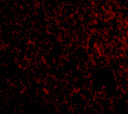
Two rabbit anti-IER3 antibodies have human tissue IHC images (catalog: A04014 and A04014-1); A04014 also has a human brain-cell IF image (catalog: A04014 IF caption) and lists mouse reactivity (catalog: A04014 reactivity).

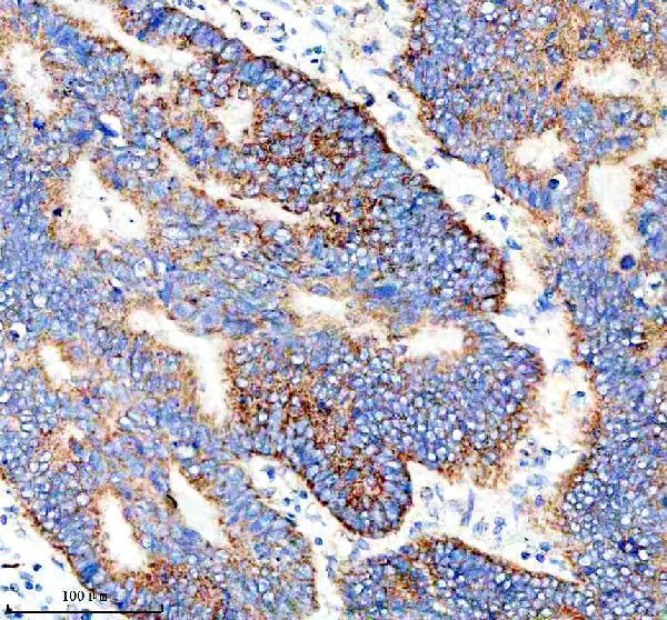
Real IHC data IHC analysis of IER3 using anti-IER3 antibody (A04014-1). IER3 was detected in a paraffin-embedded section of human stomach cancer tissue. Heat mediated antigen retrieval was performed in EDTA buffer (pH 8.0, epitope retrieval solution). The tissue section was blocked with 10% goat serum. The tissue section was then incubated with 2 μg/ml rabbit anti-IER3 Antibody (A04014-1) overnight at 4°C. Peroxidase Conjugated Goat Anti-rabbit IgG was used as secondary antibody and incubated for 30 minutes at 37°C. The tissue section was developed using HRP Conjugated Rabbit IgG Super Vision Assay Kit (Catalog # SV0002) with DAB as the chromogen.
Anti-IER3 Antibody ®
Cat # A04014-1
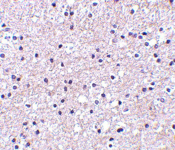
Real IHC data Immunohistochemistry of IEX-1 in human liver tissue with IEX-1 antibody at 2.5 μg/mL.
Anti-IEX-1 IER3 Antibody
Cat # A04014

A04014-1 is shown on a paraffin-embedded human stomach cancer section (A04014-1 IHC caption) and lists human IHC (catalog: A04014-1 applications and reactivity). A04014 is shown by IHC on human liver tissue (A04014 IHC caption) and by IF on human brain cells (A04014 IF caption); it lists human and mouse reactivity (catalog: A04014 reactivity).

Which to pick: For paraffin-section IHC, choose A04014-1 if its documented retrieval and staining conditions are useful: EDTA pH 8.0, 2 μg/ml overnight at 4°C, and HRP/DAB detection (A04014-1 IHC caption); its fixative is unreported (A04014-1 IHC caption). For IF/ICC, choose A04014 as the starting candidate because IF is listed and a human brain-cell IF image is provided (catalog: A04014 applications; A04014 IF caption); ICC-specific validation is unreported (catalog: A04014 applications). For mouse work, A04014 lists mouse reactivity, while its pictured IHC and IF samples are human (catalog: A04014 reactivity; A04014 IHC and IF captions); neither catalog entry specifies a clone (catalog: A04014 and A04014-1 clone fields), and the A04014 liver IHC caption does not report a fixative (A04014 IHC caption).

Each figure is that product's own IHC / IF validation image from its datasheet.

References

  1. UniProt Consortium. UniProt entry P46695 (IEX1_HUMAN, Radiation-inducible immediate-early gene IEX-1).
  2. Human Protein Atlas. IER3 tissue IHC expression (reliability: Approved).
  3. Human Protein Atlas. IER3 subcellular location (ICC-IF): Membrane.
  4. Human Protein Atlas. IER3 antibody validation summary (1 antibodies).
  5. Expression of IER3 in hepatocellular carcinoma: clinicopathology, prognosis, and potential regulatory pathways. PeerJ 2022 — PMC8918148.
  6. Increased expression of immediate early response gene 3 protein promotes aggressive progression and predicts poor prognosis in human bladder cancer. BMC urology 2018 — PMC6154418.
  7. IER3 as an RFX5 transcriptional co-activator promotes hepatocellular carcinoma progression via AKR1B10-mediated p53 transcriptional regulation. Discover oncology 2026 — PMC13230346.
  8. Resistance of Sézary cells to TNF-α-induced apoptosis is mediated in part by a loss of TNFR1 and a high level of the IER3 expression. Experimental dermatology 2012 — PMC3306129.
  9. PubMed PMID:8603392 — UniProt-cited evidence.
  10. PubMed PMID:8653710 — UniProt-cited evidence.
  11. PubMed PMID:9196025 — UniProt-cited evidence.