This website uses cookies to ensure you get the best experience on our website.


- Table of Contents
IKZF1 paraffin IHC shows nuclear staining in immune cells, especially in lymphoid tissue (HPA tissue IHC). This guide covers fixation consistency, the catalog antibody’s 0.5–1 μg/ml IHC dilution (datasheet PB9643), and interpretation across 8 isoforms (UniProt).

Expected localisation, validated protocols, controls and antibodies — the at-a-glance facts below, then the full design guide.
| Expected localisation | Nuclear staining in immune cells (HPA tissue IHC) | |
| Staining pattern | Immune-cell nuclei in most tissues; abundant in lymphoid tissue (HPA tissue IHC) | |
| Antigen retrieval | EDTA pH 8.0 HIER, heat-mediated (datasheet PB9643) | |
| Positive control | Bone marrow+4 more · see all | |
| Negative control | Adipose tissue+4 more · see all |
| Fixation | Keep fixation consistent across sections. (standard IHC practice; not target-specific) | |
| Caveat | Immune-cell nuclei can confound tissue-cell scoring (HPA tissue IHC) | |
| Regulation | Most abundant in lymphoid tissue (HPA tissue IHC) | |
| Isoform / epitope | 8 isoforms; map the antibody epitope across them (UniProt) |
The catalog antibody’s IHC-P protocol is paired with published IKZF1 staining protocols for salivary gland, human tissue, and follicular lymphoma specimens (PMC10545362; PMC10333745; PMC10539171).
| Sample | Paraffin-embedded human tonsil tissue; fixative not specified (datasheet PB9643) |
| Fixation | Image fixative and duration unreported (datasheet PB9643); verify before use. |
| Sectioning | 4–5 µm sections on charged slides (standard) |
| Deparaffinisation | Xylene, graded ethanol series to water (standard) |
| Antigen retrieval | Heat retrieval: EDTA pH 8.0 (datasheet PB9643); 20 min, 95–100 °C (standard) |
| Peroxidase block | 3% H2O2, 10 min, room temperature (standard) |
| Blocking | 10% goat serum (datasheet PB9643) |
| Primary antibody | Rabbit anti-IKZF1, 0.5-1μg/ml (datasheet PB9643) |
| Primary incubation | Overnight at 4 °C (datasheet PB9643) |
| Detection | Streptavidin-biotin complex (SABC), DAB chromogen (datasheet PB9643) |
| Counterstain | Hematoxylin, blue, dehydrate and mount (standard) |
| Expected result | IKZF1-positive staining in hematopoietic cells of bone marrow (HPA tissue IHC: Medium). HPA tissue profile: Nuclear expression of immune cells in most tissues, most abundant in lymphoid tissues. No signal in the no-primary control. |
IKZF1 should appear chiefly in nuclei of immune cells, with the most abundant tissue staining in lymphoid sites (HPA tissue IHC: Enhanced). Resting lymphocytes may show diffuse nuclear distribution; proliferating cells may show pericentromeric heterochromatin localization (UniProt Q13422). IKZF1 has no transmembrane segment (UniProt Q13422 topology). HPA also reports cytoplasmic localization, but its ICC-IF evidence rates cytosol as uncertain (HPA subcellular).
| Immune-cell nuclei stain in lymphoid tissue; germinal center cells in lymph node or tonsil show medium staining (HPA tissue IHC). | This fits the expected compartment and cell distribution (HPA tissue IHC; UniProt Q13422). Score the cells that stain, not the whole section: mixed tissue can contain both positive immune cells and unstained neighboring cells (standard IHC practice). |
| Predominantly cytoplasmic staining appears while nuclei remain unstained. | Treat this as discordant with the main nuclear pattern (HPA tissue IHC; HPA subcellular: supported nucleoplasm). UniProt lists cytoplasm, but HPA rates cytosol uncertain in ICC-IF; cytoplasmic color alone is weak evidence of a specific IHC result (UniProt Q13422; HPA subcellular). |
| Adipocytes or bronchial respiratory epithelial cells stain strongly. | These cell types were not detected in the supplied HPA tissue IHC observations (HPA tissue IHC). Investigate antibody cross-reactivity or endogenous chromogenic detection activity (standard IHC practice). Do not label every nonimmune cell positive as erroneous: placental cytotrophoblasts showed medium staining (HPA tissue IHC). |
| Color spreads across cells, stroma, or the entire section without a clear nuclear pattern. | Diffuse background cannot establish IKZF1 localization (standard IHC interpretation; HPA tissue IHC: nuclear immune-cell profile). Check background with an appropriate detection-only control, then judge whether signal remains restricted to expected cells and nuclei (standard IHC practice). |
| No nuclear signal appears in an otherwise interpretable lymph node or tonsil section. | Germinal center cells showed medium staining in both tissues (HPA tissue IHC), so inspect antibody and detection controls before calling the sample negative (standard IHC practice). A negative field alone cannot distinguish a true absence from a failed run (standard IHC interpretation). |
| Cell population and tissue context (HPA tissue IHC) | HPA describes nuclear immune-cell expression across most tissues and the greatest abundance in lymphoid tissues; bone marrow hematopoietic cells, splenic red-pulp cells, and placental cytotrophoblasts each showed medium staining (HPA tissue IHC). Compare named cell populations rather than treating an entire tissue as uniformly positive or negative (standard IHC interpretation). |
| Nuclear distribution and cell state (UniProt Q13422) | Resting lymphocytes can show diffuse nuclear IKZF1; proliferating cells can show pericentromeric heterochromatin localization, dependent on DNA binding regulated by phosphorylation and dephosphorylation (UniProt Q13422). Different nuclear textures may therefore fit the record; this does not establish a target-specific effect of fixation or retrieval. |
| Isoforms and antibody recognition (UniProt Q13422) | UniProt lists 8 IKZF1 isoforms and says isoform composition modulates transcription regulator activity (UniProt Q13422). The payload gives no antibody epitopes, so it cannot establish which isoforms any IHC-validated antibody detects; avoid assigning a staining difference to one isoform without further evidence. |
| Confidence in the tissue pattern (HPA tissue IHC; HPA antibodies) | The tissue profile is rated Enhanced, with high consistency between antibody staining and RNA expression data (HPA tissue IHC). HPA also lists Enhanced IHC status for HPA035221, HPA035222, and CAB009247 (HPA antibodies). These ratings support the reported pattern, but do not resolve a questionable field on their own. |
| IF/ICC localization Q&A (HPA subcellular) | Where should IKZF1 appear in IF/ICC? Mainly in the nucleoplasm, which HPA marks supported; additional cytosol is marked uncertain (HPA subcellular). The listed ICC-IF image cell lines are HDLM-2, JURKAT, and REH (HPA subcellular). This localization comparison does not supply an IF/ICC protocol. |
| Situation | Likely cause | Next action |
|---|---|---|
| Nuclei are negative in a lymph node or tonsil control (HPA tissue IHC). | A failed staining or detection run is possible when expected medium germinal center signal is absent (HPA tissue IHC; standard IHC practice). | Confirm the control contains germinal center cells, then review antibody dilution, retrieval conditions, and detection reagents against the validated IHC-P procedure (standard IHC practice). The supplied evidence gives no target-specific optimal settings. |
| Signal is mainly cytoplasmic, with little nuclear staining. | The pattern conflicts with HPA's supported nucleoplasmic localization; HPA rates cytosol uncertain in ICC-IF (HPA subcellular). | Compare a known-positive immune-cell control and assess nuclear counterstaining and background before accepting the cytoplasmic signal (standard IHC practice). |
| Adipocytes or bronchial respiratory epithelial cells stain (HPA tissue IHC: not detected). | Cross-reactivity or endogenous detection activity may produce staining outside the reported cell pattern (standard IHC practice). | Use an appropriate detection-only control and compare cell identity with the HPA tissue profile (standard IHC practice; HPA tissue IHC). Account for HPA's placental cytotrophoblast staining before generalizing to all nonimmune cells (HPA tissue IHC). |
| The section has widespread diffuse color that obscures nuclei. | Nonspecific background or endogenous chromogenic activity can complicate interpretation (standard IHC practice). | Check background controls and review blocking, washes, and detection conditions in the IHC workflow; score IKZF1 only where nuclear signal is distinguishable (standard IHC practice; HPA tissue IHC: nuclear profile). |
| Only a few cells stain in an otherwise low-signal field. | The sampled cell mix may differ from a named HPA-positive population; HPA reports low expression in appendix lymphoid tissue but medium staining in several listed populations (HPA tissue IHC). | Identify the stained cells and compare like-for-like tissue compartments; include a separate known-positive section when the sample offers few expected positive cells (standard IHC practice; HPA tissue IHC). |
| IF/ICC shows cytosolic fluorescence without convincing nucleoplasmic signal. | HPA supports nucleoplasm as the main ICC-IF location and marks cytosol uncertain (HPA subcellular). | Treat the result as unresolved, compare localization with the HPA ICC-IF record, and use the separate IF/ICC guide for method choices (HPA subcellular). |


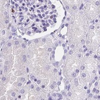


Comprehensive Human Protein Atlas IHC scoring per tissue (reliability: Enhanced — High consistency between antibody staining and RNA expression data.). Rows are taken directly from the HPA tissue chart — click any row's HPA link to view the source.
| Tissue | Cell type | Level | Evidence | Source |
|---|---|---|---|---|
| Bone marrow | Hematopoietic cells | Medium | Protein (IHC) | HPA → |
| Lymph node | Germinal center cells | Medium | Protein (IHC) | HPA → |
| Placenta | Cytotrophoblasts | Medium | Protein (IHC) | HPA → |
| Spleen | Cells in red pulp | Medium | Protein (IHC) | HPA → |
| Tonsil | Germinal center cells | Medium | Protein (IHC) | HPA → |
| Tissue | Cell type | Level | Evidence | Source |
|---|---|---|---|---|
| Adipose tissue | Adipocytes | Not detected | Protein (IHC) | HPA → |
| Adrenal gland | Glandular cells | Not detected | Protein (IHC) | HPA → |
| Breast | Adipocytes | Not detected | Protein (IHC) | HPA → |
| Bronchus | Respiratory epithelial cells | Not detected | Protein (IHC) | HPA → |
| Caudate | Glial cells | Not detected | Protein (IHC) | HPA → |
Troubleshoot IKZF1 staining in paraffin sections by checking retrieval, nuclear localisation, cell identity and background before scoring chromogenic signal.
PB9643 has IHC images from human tonsil and mouse and rat spleen paraffin sections, plus IF in MCF-7 cells (catalog image captions). Both antibodies list human, mouse and rat reactivity (catalog).
M00531-1 lists IHC and human, mouse and rat reactivity, but has no supplied IHC image or specimen (catalog applications, reactivity and image captions). PB9643 has IHC images of human tonsil and mouse and rat spleen paraffin sections, plus an IF image of MCF-7 cells (catalog image captions).
Which to pick: Choose PB9643 for tissue IHC: its own captions document paraffin sections from all three species, EDTA pH 8.0 retrieval and 1 μg/ml primary antibody; the fixative is unreported (PB9643 IHC image captions). Choose PB9643 for IF/ICC because both applications are listed and its IF image shows MCF-7 cells at 5 μg/ml (catalog applications; PB9643 IF image caption). For cross-species IHC, PB9643 has images from human, mouse and rat tissue; M00531-1 offers a rabbit monoclonal, clone AEFA-9, with IHC and all three species listed, but no supplied IHC image (catalog applications, reactivity and image captions).