This website uses cookies to ensure you get the best experience on our website.
- Table of Contents
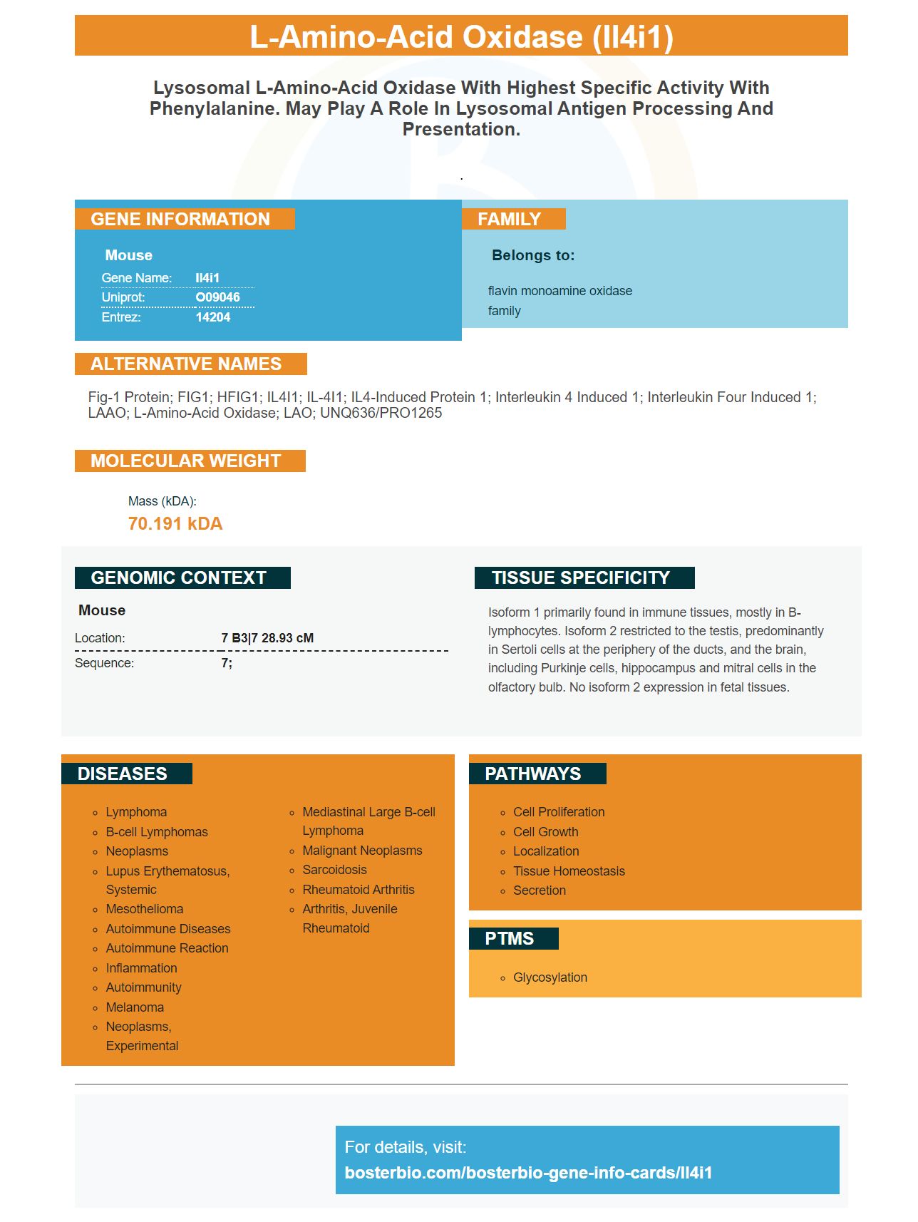
Facts about L-amino-acid oxidase.
.
Mouse | |
---|---|
Gene Name: | Il4i1 |
Uniprot: | O09046 |
Entrez: | 14204 |
Belongs to: |
---|
flavin monoamine oxidase family |
Fig-1 protein; FIG1; hFIG1; IL4I1; IL-4I1; IL4-induced protein 1; interleukin 4 induced 1; interleukin four induced 1; LAAO; L-amino-acid oxidase; LAO; UNQ636/PRO1265
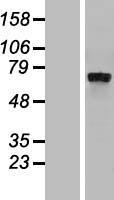
Mass (kDA):
70.191 kDA
Mouse | |
---|---|
Location: | 7 B3|7 28.93 cM |
Sequence: | 7; |
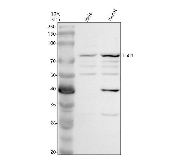
Isoform 1 primarily found in immune tissues, mostly in B-lymphocytes. Isoform 2 restricted to the testis, predominantly in Sertoli cells at the periphery of the ducts, and the brain, including Purkinje cells, hippocampus and mitral cells in the olfactory bulb. No isoform 2 expression in fetal tissues.
Are you interested in learning more about IL4I1? To learn more about its clinical uses, costs, and availability, read this article. After reading this article, you will be able decide if this biomarker is right to use in your research. Continue reading to learn more about this biomarker and how it can help you in research. The results will surprise you!
IL4I1 a glycosylated proteins that is found in bacteria, and mammals, is a member the LAAO family. IL4I1 is responsible in the oxidative degradation of phenylalanine. This enzyme is not able to bind to tryptophan, arginine, or phenylalanine and does not have any inhibitors. Although it may be an anti-inflammatory agent, there is little evidence it plays a role reducing the spread and severity of bacterial infections. IL4I1, on the other side, has been shown to reduce activation of T cells and inhibit proliferative response.
This marker can be used to study tumors that have been resistant chemotherapy. IL4I1 has been found in serum as well as plasma. The IL4I1 marker is difficult to detect due to the presence of matrix background. However, the IL4I1 gene is expressed on B cells in germinal-center B lymphoma. However, the antitumor effects of the IL4I1 gene have not been demonstrated in various types of cancers.
IL4I1 is involved in the immune escape of tumor cells. In mice, IL4I1-producing cells limit the anti-tumor immune response, restricting the proliferation of cytotoxic T cells and Th2 cells, and facilitating the accumulation of Treg cells. In humans, IL4I1 is linked to tumor growth in murine models. Further, melanoma patients are IL4I1-expressing cells that are in close contact with tumor cells.
It has been thoroughly studied and compared to many other markers of inflammation. IL4I1, although less common, is one of most effective markers. Although it's expressed mostly by immune cells, it is not completely immune-specific. Boster Bio: The best uses of the IL4I1 mark
In solid tumors, tumor-associated macrophages produce the IL4I1 marker. This protein is related to CD68 (CD14), CD163 (CD163) and CD163. Strong correlation has been observed with a mean value of 0.63 + 0.15 in CD68 and 0.51+ SD for CD163. There are many potential clinical applications for this marker, from monitoring tumor growth to identifying patients who may be at risk of developing certain cancers.
IL4I1 can be found in human melanoma cells. This molecule is thought to be responsible for tumor growth by modulating the tumor's microenvironment. Moreover, it plays a key role in cancer immune escape. Cancer immunotherapy might be able easily to target IL4I1 transcription. The study results are promising and point to a potential new target for cancer immunotherapy.
A range of inflammatory disorders are linked to the IL4I1 protein. For example, inflammation bowel disease is associated with an inflammatory response mediated via the IL4I1 genome. IL4I1 enhances the activation threshold of T-cells. It promotes priming high-affinity clones, and reduces the response to immunodominant propeptides. This allows for rapid acquisition of effector differentiate.
IL4I1 may be associated with immunoregulation, progression of tumors, and poor prognosis among OC patients. Recent studies show that IL4I1 levels are associated with poor OS among OC patients. In addition, GSEA-based analyses have shown a strong association between high-IL4I1 patients and EMT and G2M checkpoint expression. It is notable that IL4I1 inhibition inhibits the growth of SKOV3- and A2780-cells.
The availability of IL4I1 mRNA performs several important functions. It regulates the activation threshold and limits proliferation of memory T cell cells. It also limits the maturation time of antigen-specific CD8 cells transferred to effector cells and favors the growth of cell types with enhanced memory, similar to endogenous polyclonal cells. These findings offer insight into antigen priming's role in the host.
The IL4I1 mRNA, which is capable of degrading amino acids, has the potential to identify tumor-derived cancer cells. IL4I1 converts amino acids into metabolites and degrades them. IL4I1 is also involved in the formation AHR ligands that act as signaling compounds. Furthermore, IL4I1 promotes the expression of PP, HPP, and kynurenic acid (Kyn).
Availability of the IL4I1 gene is a useful indicator for the presence of AHR. It limits the differentiation of Th2 cells and Th2 cells, alters Th27 effector phenotype, and promotes the polarization of Tregs. It is found in cancer cell lines and is associated with the presence T-cell receptors and AHR. These proteins are related, so cross-talk is possible.
The preferred method detects IL4I1's biological state by altering its expression in cells. The presence of IL4I1 in cells changes its biological state. A change in the expression or activity of the IL4I1 genes indicates AHR-related diseases. To select patients for IL4I1 modulating therapies, the preferred method detects IL4I1.
Scientists have previously looked at the IL4I1 indicator and its relationship with cancer prognosis. This marker has been shown to be associated with poor prognosis of a number cancers, including the endometrial. Transcriptomic data from breast carcinoma biopsies revealed IL4I1 transcripts. Studies on follicularly diagnosed lymphomas showed an inverted relationship between bone marrow and protein expression. In addition, expression of the IL4I1 marker was associated with a poor prognosis in a study of follicular lymphomas.
PMID: 9122225 by Chu C.C., et al. Fig1, an interleukin 4-induced mouse B cell gene isolated by cDNA representational difference analysis.
PMID: 9798653 by Chu C.C., et al. Expressed genes in interleukin-4 treated B cells identified by cDNA representational difference analysis.