IMPG2 / Interphotoreceptor matrix proteoglycan 2 · IHC design guide

Design Immunohistochemistry for IMPG2

Use retinal photoreceptor staining as the IMPG2 IHC-P reference (HPA tissue IHC). Plan consistent fixation and antibody titration, and interpret interphotoreceptor-matrix signal as a molecular expectation (UniProt).

Evidence assembled Oct 2026 · For research use; verify linked source records and product datasheet before use
Immunohistochemistry protocol sheet for IMPG2 (IHC for IMPG2): expected localisation Photoreceptor staining (HPA tissue IHC); interphotoreceptor matrix expected (UniProt), antibody A10288-1, validated IHC image, and IHC protocol steps
Printable IMPG2 IHC protocol sheet — expected localisation Photoreceptor staining (HPA tissue IHC); interphotoreceptor matrix expected (UniProt), antibody A10288-1, controls and protocol steps. Open the full IMPG2 IHC guide →

IMPG2 Immunohistochemistry Experimental Design Guide

Expected localisation, validated protocols, controls and antibodies — the at-a-glance facts below, then the full design guide.

Must know before staining
Expected localisation Photoreceptor staining (HPA tissue IHC); interphotoreceptor matrix expected (UniProt)
Staining pattern Photoreceptor cells; subcellular pattern unreported (HPA tissue IHC)
Antigen retrieval EDTA pH 8.0 HIER, heat-mediated (datasheet A10288-1)
Positive control ⓘ Retina
Negative control ⓘ Adipose tissue+4 more · see all
Important caveats
Reasons your staining may differ from the expected pattern.
Fixation Keep fixation consistent across sections. (standard IHC practice; not target-specific)
Caveat Secreted variants may separate protein staining from tissue RNA (HPA tissue IHC)
Regulation Staining regulation unreported (UniProt)
Isoform / epitope No isoforms annotated; extracellular versus cytoplasmic epitope matters (UniProt)
Section 1

Recommended IMPG2 IHC & IF Protocols

The catalog antibody protocol uses EDTA retrieval (datasheet A10288-1). The published human retinal IHC protocol below documents citrate retrieval (PMC5145218).

Recommended immunohistochemistry (IHC-P) protocol parameters
SampleParaffin-embedded rat retina tissue; fixative not specified (datasheet A10288-1)
FixationImage fixative and duration unreported (datasheet A10288-1); verify before use.
Sectioning4–5 µm sections on charged slides (standard)
DeparaffinisationXylene, graded ethanol series to water (standard)
Antigen retrievalHeat retrieval: EDTA pH 8.0 (datasheet A10288-1); 20 min, 95–100 °C (standard)
Peroxidase block3% H2O2, 10 min, room temperature (standard)
Blocking10% goat serum (datasheet A10288-1)
Primary antibodyRabbit anti-IMPG2, 2-5 μg/ml (datasheet A10288-1)
Primary incubationOvernight at 4 °C (datasheet A10288-1)
DetectionHRP-conjugated secondary, DAB chromogen (datasheet A10288-1)
CounterstainHematoxylin, blue, dehydrate and mount (standard)
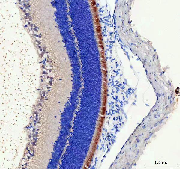
Expected resultIMPG2-positive staining in photoreceptor cells of retina (HPA tissue IHC: High). HPA tissue profile: Expression in photoreceptor cells. No signal in the no-primary control.
💡Decision noteStart with heat-mediated EDTA pH 8.0 for the catalog antibody (datasheet A10288-1). Use citrate pH 6 when reproducing the published protocol (PMC5145218).
Section 2

What Is the Expected IMPG2 Staining Pattern?

IMPG2 should stain retinal photoreceptor cells, where HPA reports High expression with Enhanced tissue IHC reliability (HPA: retina tissue IHC). Membrane-associated signal at photoreceptor inner and outer segments and signal in the surrounding interphotoreceptor matrix are consistent with its annotated locations; its extracellular region spans residues 23–1099 (UniProt Q9BZV3 topology and subcellular location).

What am I looking at on my slide?
Chromogen is concentrated around retinal photoreceptors, including their segment region.This is consistent with HPA’s High photoreceptor-cell staining and UniProt’s membrane and interphotoreceptor-matrix locations (HPA: retina tissue IHC; UniProt Q9BZV3 subcellular location). HPA does not validate each fine subcellular boundary in paraffin sections.
Photoreceptor nuclei show prominent staining, especially without corresponding segment-region signal.Treat nuclear positivity as suspect: HPA explicitly viewed it as off target and disregarded it (HPA: tissue IHC reliability description). Review morphology and compare with a no-primary control before scoring it as IMPG2 (general IHC practice).
Strong staining appears in a nonphotoreceptor cell population or in an HPA-negative tissue.Cross-reactivity or endogenous chromogenic activity is possible (general IHC practice). HPA reports IMPG2 as not detected in the specified negative tissues and High in retinal photoreceptors; those comparisons concern the named cells, not every cell in each tissue (HPA: tissue IHC).
Chromogen is diffuse across the section, including areas without photoreceptors.Widespread background limits interpretation (general IHC practice). Some extracellular signal near photoreceptors is biologically plausible because IMPG2 is annotated in the interphotoreceptor matrix; unstructured staining across unrelated areas is not established as the expected pattern (UniProt Q9BZV3 subcellular location).
No IMPG2 signal is visible in a retinal section containing photoreceptors.A negative result conflicts with the HPA High photoreceptor-cell observation (HPA: retina tissue IHC). Check that photoreceptors are present, then evaluate antibody and detection controls and the retrieval conditions used (general IHC practice). This result alone cannot identify which step failed.
💡Expected IMPG2 appearanceCall a section positive when retinal photoreceptor cells show High segment-associated staining, with plausible adjacent matrix signal (HPA: retina tissue IHC; UniProt Q9BZV3 subcellular location); prominent nuclear staining is a false-positive warning (HPA: tissue IHC reliability description).
How each factor affects the staining
Compartment and epitope positionIMPG2 has one transmembrane segment at 1100–1120, a large extracellular region, and a cytoplasmic tail (UniProt Q9BZV3 topology). Interpret a reagent’s staining against its mapped epitope if that information is available; no epitope position is supplied here.
Extracellular distributionUniProt annotates both photoreceptor-segment membrane and secreted interphotoreceptor-matrix locations (UniProt Q9BZV3 subcellular location). Signal next to a photoreceptor need not be confined to its cell outline; this annotation does not establish a distinct pattern for every paraffin-section preparation.
IHC evidence strengthHPA rates tissue IHC reliability Enhanced; HPA015907 is IHC Enhanced and HPA008779 is IHC Supported (HPA: tissue IHC and antibody validation). These ratings support the reported retinal pattern but do not validate an unlisted reagent or guarantee a particular slide’s result.
Antigen retrieval and fixationNo target-specific retrieval condition or fixation sensitivity is supplied (HPA: tissue IHC; UniProt Q9BZV3). Retrieval should be assessed with appropriate positive and negative controls for the chosen paraffin IHC workflow (general IHC practice); do not infer an IMPG2-specific fixation effect.
Does this establish an IF/ICC pattern?HPA lists “Membrane, Secreted” but provides no ICC-IF images or main location for IMPG2 (HPA: subcellular summary). The IHC observations here therefore do not establish an IF/ICC staining pattern or an IF/ICC protocol.
Why is my staining missing, weak or wrong?
SituationLikely causeNext action
Retinal photoreceptors are unstained.The expected positive population may be absent from the section, or antibody, retrieval, or detection may have failed (HPA: High in retinal photoreceptors; general IHC practice).Confirm photoreceptor morphology and run a documented positive-control section. Review the antibody’s IHC-P instructions and check detection controls before changing one workflow condition at a time (general IHC practice).
Nuclei stain more strongly than photoreceptor segments.HPA disregarded nuclear IMPG2 positivity as off target (HPA: tissue IHC reliability description); nonspecific primary-antibody binding or detection background may contribute (general IHC practice).Do not score nuclei as IMPG2. Compare a no-primary control, inspect segment-region staining, and reassess antibody concentration and blocking within the chosen IHC workflow (general IHC practice).
A broad brown haze obscures retinal layers.General background from antibody concentration, inadequate washing, or the chromogenic detection workflow can obscure localization (general IHC practice); UniProt’s matrix annotation alone does not justify pan-section staining (UniProt Q9BZV3).Use a no-primary control, review wash and blocking steps, and adjust the antibody concentration using the reagent’s IHC-P instructions (general IHC practice). Recheck whether remaining signal follows photoreceptors (HPA: retina tissue IHC).
Staining appears in HPA-negative comparison tissue.The reported cells in several comparison tissues were Not detected (HPA: tissue IHC). Unexpected chromogen may reflect cross-reactivity or endogenous detection activity (general IHC practice).Verify the stained cell type against the HPA entry. Run a no-primary control; if using enzyme detection, check the relevant endogenous-activity block (general IHC practice).
Matrix-adjacent signal is mistaken for nonspecific spread.IMPG2 is annotated as both membrane-associated and secreted into the interphotoreceptor matrix around rods and cones (UniProt Q9BZV3 tissue specificity and subcellular location).Check whether signal tracks the photoreceptor region and compare background elsewhere in the section. Interpret fine boundaries cautiously because HPA reports the cell-level tissue pattern, not a resolved paraffin-section matrix map (HPA: retina tissue IHC).
Two IHC antibodies give different distributions.HPA assigns different antibody-level IHC ratings: HPA015907 Enhanced and HPA008779 Supported (HPA: antibody validation). Different staining alone does not establish which compartment represents IMPG2.Compare each result with photoreceptor enrichment and HPA’s nuclear off-target warning (HPA: tissue IHC). Review each antibody’s IHC-P instructions and matched controls before assigning biological meaning (general IHC practice).

Sample controls for IMPG2 IHC & IF

🧪Run retina first: photoreceptor cells should stain strongly (HPA: High in retinal photoreceptor cells). Use adipose tissue as a negative, with adipocytes at background level (HPA: Not detected in adipocytes); on the retina slide, assess neighboring non-photoreceptor cells as internal background while allowing signal in the interphotoreceptor matrix (UniProt Q9BZV3 localization).
Positive control tissue: Retina (Photoreceptor cells, HPA High)
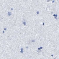
Negative control tissue: Adipose tissue (HPA Not detected)
ICC-IF cell lines (HPA subcellular resource): HPA carries no ICC-IF cell line for IMPG2; derive a cell-line control from the positive tissue's cell type (Photoreceptor cells) and confirm it by RNA or western blot first.
Technical controls: Include a no-primary, secondary-only control and a matched rabbit IgG isotype control for the rabbit catalog antibody (selected-SKU caption: rabbit anti-IMPG2; standard IHC practice); use IMPG2-knockout tissue as a biological specificity control if available (standard IHC practice). Quench endogenous peroxidase for chromogenic IHC and check retinal pigment or tissue autofluorescence in fluorescence images (standard IHC/IF practice).
⚠️Feasibility: A target-specific fixation window and fixation effect are unreported, and the selected-SKU paraffin-section caption does not state a fixative (selected-SKU caption: fixative not stated). The reported IHC used heat retrieval in EDTA at pH 8.0, but a requirement for retrieval has not been established by a comparison in the supplied evidence (selected-SKU caption: EDTA heat retrieval). Whether frozen sections or IF are easier is unreported; retinal pigment and autofluorescence need attention when interpreting fluorescence (standard IHC/IF practice).

HPA tissue IHC evidence for IMPG2

Comprehensive Human Protein Atlas IHC scoring per tissue (reliability: Enhanced — At least one protein variant secreted, tissue location of RNA and protein might differ and correlation is complex. External characterization data supports antibody staining. Nuclear positivity was viewed as off target and disregarded.). Rows are taken directly from the HPA tissue chart — click any row's HPA link to view the source.

Positive expression · recommended positive controls

TissueCell typeLevelEvidenceSource
Retina Photoreceptor cells High Protein (IHC) HPA →

Undetected expression · recommended negative controls

TissueCell typeLevelEvidenceSource
Adipose tissue Adipocytes Not detected Protein (IHC) HPA →
Adrenal gland Glandular cells Not detected Protein (IHC) HPA →
Appendix Glandular cells Not detected Protein (IHC) HPA →
Bone marrow Hematopoietic cells Not detected Protein (IHC) HPA →
Breast Adipocytes Not detected Protein (IHC) HPA →
Section 3

Advanced IMPG2 IHC Tips

Use retinal compartment anatomy and the documented IHC conditions to troubleshoot IMPG2 staining while keeping fixation and IF performance claims within the available evidence.

How should I adjust retrieval when IMPG2 staining is weak in paraffin retina sections?
Start with heat-mediated retrieval in EDTA at pH 8.0 for paraffin retina sections (datasheet A10288-1). The selected image used 2 μg/ml primary antibody overnight at 4°C, so reproduce those conditions before attributing weak staining to retrieval (datasheet A10288-1). Compare retrieved and untreated serial sections while keeping section thickness, detection and imaging conditions consistent (standard IHC practice). If signal remains weak, test retrieval time on adjacent sections and examine whether retinal layers and tissue edges remain intact (standard IHC practice). Score signal in photoreceptor cells and the surrounding interphotoreceptor matrix, rather than treating diffuse retinal staining as recovery of the target (HPA: photoreceptor expression; UniProt Q9BZV3 localisation).
Can fixation explain inconsistent IMPG2 staining across paraffin retina samples?
The selected paraffin retina caption does not report its fixative, so target-specific IMPG2 sensitivity to fixation is unknown (datasheet A10288-1: fixative not stated). Record the fixative, fixation duration, processing schedule and section age for each sample before comparing staining intensity (standard IHC practice). Run matched sections in one staining batch, using the documented EDTA retrieval at pH 8.0 and 2 μg/ml primary concentration as reference conditions (datasheet A10288-1). Inspect morphology and compare the distribution across intact photoreceptor layers, since tissue damage can confound chromogenic scoring (standard IHC practice). Do not infer a preferred fixative or a target-specific fixation effect from the reported retinal expression pattern (HPA: photoreceptor expression; datasheet A10288-1: fixative not stated).
Where should credible IMPG2 staining appear within the retina?
Evaluate staining in photoreceptor cells and the adjacent interphotoreceptor matrix, which occupies the space toward the retinal pigment epithelium (HPA: high in retinal photoreceptors; UniProt Q9BZV3 tissue specificity). IMPG2 is annotated at photoreceptor inner and outer segment membranes and in the extracellular interphotoreceptor matrix (UniProt Q9BZV3 localisation). Its extracellular region spans residues 23–1099, followed by a transmembrane segment at 1100–1120, so epitope position matters when interpreting membrane versus matrix signal (UniProt Q9BZV3 topology). Compare chromogen distribution with preserved retinal layer morphology and an appropriate counterstain on adjacent sections (standard IHC practice). Treat isolated nuclear staining cautiously because the HPA assessment disregarded nuclear positivity as off target (HPA: reliability description).
How could epitope position change the IMPG2 pattern I see?
Check the antibody's documented immunogen or mapped epitope before assigning a particular staining compartment; its position is not supplied in the selected caption (datasheet A10288-1). The annotated extracellular portion covers residues 23–1099, whereas residues 1121–1241 are cytoplasmic (UniProt Q9BZV3 topology). Ten annotated glycosylation sites and two SEA domains lie in the extracellular portion, features to consider when comparing accessibility across preparations (UniProt Q9BZV3 glycosylation and domains; standard IHC practice). The supplied record lists 0 isoforms, so it provides no basis for explaining distinct patterns as annotated isoforms (UniProt Q9BZV3 isoforms). Document whether staining follows photoreceptor membranes, surrounding matrix or unrelated structures before assigning an epitope-based explanation (UniProt Q9BZV3 localisation; standard IHC practice).
How can IF help assess an ambiguous chromogenic IMPG2 pattern?
Use IF as a companion localisation check, while basing this page's chromogenic IHC conditions on the paraffin retina caption (datasheet A10288-1). Multiplex IMPG2 with a validated photoreceptor marker and compare its signal with cell bodies, segment membranes and adjacent matrix (HPA: photoreceptor expression; UniProt Q9BZV3 localisation; standard IF practice). Choose a fluorophore channel after measuring retinal autofluorescence in unstained tissue, and include single-label controls for channel overlap (standard IF practice). If the antibody epitope is cytoplasmic, assess permeabilisation; if extracellular, evaluate signal before adding permeabilisation, since the annotated membrane segment separates residues 23–1099 from 1121–1241 (UniProt Q9BZV3 topology; standard IF practice). The supplied HPA record lists no cell-line ICC/IF images, so IF performance remains unestablished here (HPA: subcellular record).
What controls distinguish retinal background from IMPG2 chromogen signal?
Compare a no-primary section with the stained retina section to reveal signal from detection reagents or endogenous enzyme activity (standard IHC practice). The selected image used peroxidase detection with DAB, so include an appropriate peroxidase block as a general chromogenic workflow step (datasheet A10288-1: detection; standard IHC practice). Its tissue section was blocked with 10% goat serum and received 2 μg/ml primary antibody overnight at 4°C (datasheet A10288-1). If background persists, inspect section edges and damaged areas separately and compare primary concentration and blocking conditions on adjacent sections (standard IHC practice). Retain only staining that aligns with photoreceptors or their surrounding matrix; isolated nuclear positivity warrants skepticism (HPA: photoreceptor expression and nuclear off-target assessment; UniProt Q9BZV3 localisation).
How should I quantify IMPG2 staining without confusing cells and matrix? ⚠ ANSWER MARKED FOR VERIFICATION
Define photoreceptor and interphotoreceptor matrix regions on intact retinal sections before examining the chromogen signal (HPA: photoreceptor expression; UniProt Q9BZV3 localisation; standard IHC practice). For cell-associated staining, report the percentage of positive photoreceptors or an H-score using the same intensity thresholds across samples (standard IHC practice). For matrix staining, measure background-corrected DAB-positive area or optical density and normalise it to the annotated matrix area, such as per mm² (standard IHC practice). Keep section thickness, illumination, counterstain and analysis thresholds consistent, and exclude folds or detached tissue from denominators (standard IHC practice). Report cellular and extracellular measurements separately because IMPG2 is annotated at photoreceptor membranes and in the interphotoreceptor matrix (UniProt Q9BZV3 localisation).
When is a positive IMPG2 IHC result likely to be artefactual?
Judge a positive result by its retinal anatomy: photoreceptor-associated and surrounding matrix staining fits the supplied expression and localisation evidence (HPA: high in retinal photoreceptors; UniProt Q9BZV3 localisation). Predominantly nuclear signal is suspect because the HPA assessment explicitly disregarded nuclear positivity as off target (HPA: reliability description). Compare staining at section edges, damaged or necrotic regions, and areas with lost morphology against intact internal regions before scoring (standard IHC practice). A no-primary control and appropriate peroxidase block help assess reagent background and endogenous enzyme signal in DAB detection (standard IHC practice; datasheet A10288-1: DAB detection). Record a discordant compartment or cell type as unresolved until controls and adjacent sections support its attribution to IMPG2 (standard IHC practice).
Boster reagents

Best IMPG2 / Interphotoreceptor matrix proteoglycan 2 IHC Antibodies

A10288-1 has real IHC data from a paraffin-embedded rat retina section (catalog image caption: A10288-1). Its listed reactivity includes Human, Mouse, and Rat (catalog: reactivity).

Real IHC data IHC analysis of IMPG2 using anti-IMPG2 antibody (A10288-1). IMPG2 was detected in a paraffin-embedded section of rat retina tissue. Heat mediated antigen retrieval was performed in EDTA buffer (pH 8.0, epitope retrieval solution). The tissue section was blocked with 10% goat serum. The tissue section was then incubated with 2 μg/ml rabbit anti-IMPG2 Antibody (A10288-1) overnight at 4°C. Peroxidase Conjugated Goat Anti-rabbit IgG was used as secondary antibody and incubated for 30 minutes at 37°C. The tissue section was developed using HRP Conjugated Rabbit IgG Super Vision Assay Kit (Catalog # SV0002) with DAB as the chromogen.
Anti-IMPG2 Antibody ®
Cat # A10288-1

A10288-1 is shown in IHC on a paraffin-embedded rat retina section (catalog image caption: A10288-1). Human and Mouse reactivity are listed, but the supplied IHC image documents Rat tissue only (catalog: reactivity; catalog image caption: A10288-1).

Which to pick: Choose A10288-1 for paraffin-section IHC: its rat retina caption reports EDTA retrieval at pH 8.0 and 2 μg/ml primary antibody (catalog image caption: A10288-1). For cross-species work, A10288-1 lists Human, Mouse, and Rat reactivity, though its supplied IHC image shows Rat only; the fixative is unreported (catalog: reactivity; catalog image caption: A10288-1). No SKU in this payload has documented IF/ICC validation: A10288-1 has no listed IF application or IF image (catalog: applications; catalog: if_image_alts).

Each figure is that product's own IHC / IF validation image from its datasheet.

References

  1. UniProt Consortium. UniProt entry Q9BZV3 (IMPG2_HUMAN, Interphotoreceptor matrix proteoglycan 2).
  2. Human Protein Atlas. IMPG2 tissue IHC expression (reliability: Enhanced).
  3. Human Protein Atlas. IMPG2 subcellular location (ICC-IF): Membrane, Secreted.
  4. Human Protein Atlas. IMPG2 antibody validation summary (2 antibodies).
  5. Serum Autoantibody Profiling of Patients with Paraneoplastic and Non-Paraneoplastic Autoimmune Retinopathy. PloS one 2016 — PMC5145218.
  6. Identification of the zebrafish homologues of IMPG2, a retinal proteoglycan. Cell and tissue research 2023 — PMC10558372.
  7. Differential neuroglycan C expression during retinal degeneration in Rpe65-/- mice. Molecular vision 2008 — PMC2592529.
  8. PubMed PMID:10542133 — UniProt-cited evidence.
  9. PubMed PMID:10702256 — UniProt-cited evidence.
  10. PubMed PMID:16641997 — UniProt-cited evidence.