ISCU / Iron-sulfur cluster assembly enzyme ISCU · IHC design guide

Design Immunohistochemistry for ISCU

Plan ISCU chromogenic IHC in paraffin sections using its observed cytoplasmic tissue profile (HPA tissue IHC). This guide highlights fixation consistency, staining interpretation, and the 2 annotated isoforms (UniProt).

Evidence assembled Oct 2026 · For research use; verify linked source records and product datasheet before use
Immunohistochemistry protocol sheet for ISCU (IHC for ISCU): expected localisation Cytoplasmic tissue staining (HPA tissue IHC); mitochondrial location (UniProt), antibody A05447-1, validated IHC image, and IHC protocol steps
Printable ISCU IHC protocol sheet — expected localisation Cytoplasmic tissue staining (HPA tissue IHC); mitochondrial location (UniProt), antibody A05447-1, controls and protocol steps. Open the full ISCU IHC guide →

ISCU Immunohistochemistry Experimental Design Guide

Expected localisation, validated protocols, controls and antibodies — the at-a-glance facts below, then the full design guide.

Must know before staining
Expected localisation Cytoplasmic tissue staining (HPA tissue IHC); mitochondrial location (UniProt)
Staining pattern Cytoplasmic staining in most tissues (HPA tissue IHC)
Antigen retrieval EDTA pH 8.0 HIER, heat-mediated (datasheet A05447-1)
Positive control ⓘ Adrenal gland+4 more · see all
Negative control ⓘ Adipose tissue
Important caveats
Reasons your staining may differ from the expected pattern.
Fixation Matched tissue-IHC evidence does not establish the fixation claim. Validate the specimen-specific method before use. (selected-SKU IHC image A05447-1)
Caveat Antibody staining and RNA show medium consistency (HPA tissue IHC)
Regulation Low tissue specificity (HPA tissue RNA)
Isoform / epitope 2 isoforms; epitope coverage is unspecified (UniProt)
Section 1

Recommended ISCU IHC & IF Protocols

The catalog antibody’s IHC-P protocol is accompanied by published ISCU methods for liver cirrhosis and HH, hepatocellular carcinoma, and oropharyngeal carcinoma sections (PMC12446859; PMC4642350; PMC5355133).

Recommended immunohistochemistry (IHC-P) protocol parameters
SampleParaffin-embedded human liver cancer tissue; fixative not specified (datasheet A05447-1)
FixationImage fixative and duration unreported (datasheet A05447-1); verify before use.
Sectioning4–5 µm sections on charged slides (standard)
DeparaffinisationXylene, graded ethanol series to water (standard)
Antigen retrievalHeat retrieval: EDTA pH 8.0 (datasheet A05447-1); 20 min, 95–100 °C (standard)
Peroxidase block3% H2O2, 10 min, room temperature (standard)
Blocking10% goat serum (datasheet A05447-1)
Primary antibodyRabbit anti-ISCU, 1:50 recommended; image 1:100 (datasheet A05447-1)
Primary incubationOvernight at 4 °C (datasheet A05447-1)
DetectionHRP-conjugated secondary, DAB chromogen (datasheet A05447-1)
CounterstainHematoxylin, blue, dehydrate and mount (standard)
Expected resultISCU-positive staining in glandular cells of adrenal gland (HPA tissue IHC: High). HPA tissue profile: Cytoplasmic expression in most tissues. No signal in the no-primary control.
💡Decision noteStart with heat-mediated EDTA retrieval at pH 8.0 (datasheet: A05447-1); pH 9 pressure-cooker retrieval is a published alternative for FFPE sections (PMC5355133: methods).
Section 2

What Is the Expected ISCU Staining Pattern?

ISCU is annotated in mitochondria, cytoplasm and nucleus, with no transmembrane segment (UniProt Q9H1K1). In tissue IHC, expect chiefly cytoplasmic staining in cells that express it, including adrenal and intestinal glandular cells, cerebellar Purkinje cells, and colon endothelial cells (HPA: High in these cell types; cytoplasmic expression in most tissues). Treat intensity comparisons with care: HPA rates the tissue IHC evidence Approved, with medium consistency between antibody staining and RNA expression data (HPA).

What am I looking at on my slide?
Cytoplasmic signal in adrenal glandular cells, with little signal in adipocytes.This fits the observed tissue pattern: adrenal glandular cells stain High, while adipocytes are Not detected (HPA tissue IHC). Judge staining in the named cell types, not from whole-section color alone.
Strong, predominantly nuclear staining with little cytoplasmic signal.This departs from the chiefly cytoplasmic tissue IHC pattern (HPA). Nuclear localization is annotated (UniProt Q9H1K1), so assess controls and antibody specificity before calling every nuclear-positive cell an artefact.
Strong staining in adipocytes or cells outside the expected positive population.Adipocytes were Not detected in the supplied tissue profile (HPA). Unexpected staining can reflect cross-reactivity or endogenous chromogen-generating activity (standard IHC practice); check its distribution against a negative control.
Uniform haze across cells and extracellular spaces.A non-cell-selective deposit is less convincing than HPA's cellular cytoplasmic pattern (HPA tissue IHC). Incomplete blocking, excess primary antibody or detection background are general IHC possibilities (standard IHC practice).
No signal in adrenal glandular cells despite an intact counterstain.These cells are a documented High-staining reference (HPA tissue IHC). First investigate the IHC-P run and its controls; absence of staining alone does not establish that ISCU is absent from the specimen.
💡Expected ISCU appearanceCall a result positive when cytoplasmic staining is clear in an HPA High cell population, such as adrenal glandular cells (HPA tissue IHC); diffuse extracellular color or equally strong adipocyte staining is suspect (HPA: adipocytes Not detected; standard IHC practice).
How each factor affects the staining
Compartment readoutHPA reports cytoplasmic expression in most tissues by IHC and supported mitochondrial and cytosolic localization by ICC-IF (HPA). A chromogenic section may show cytoplasmic color without resolving the mitochondrial component (standard IHC practice).
Cell population and reference tissueHPA records High staining in several glandular populations, cerebellar Purkinje cells and colon endothelial cells; red-pulp spleen cells and marrow hematopoietic cells are Low (HPA tissue IHC). Compare the relevant cells rather than expecting uniform tissue intensity.
Antibody evidenceHPA lists IHC as Approved for HPA038602, HPA057592 and CAB006329 (HPA antibodies). The supplied tissue profile is Approved with medium staining–RNA consistency (HPA); it does not guarantee identical staining with every antibody or specimen.
Processing and epitope positionUniProt annotates the ISCU chain as residues 35–167 and no transmembrane segment (UniProt Q9H1K1). Epitope coordinates are not supplied here, so these annotations cannot predict whether a particular antibody recognizes the processed chain.
IsoformsTwo isoforms are annotated (UniProt Q9H1K1). Their annotation alone does not establish which isoform an IHC antibody detects or explain a tissue-specific staining difference; check antibody-specific epitope information if available.
Antigen retrievalRetrieval is a general variable in paraffin-section IHC (standard IHC practice). Neither the supplied HPA pattern nor the UniProt record establishes an ISCU-specific retrieval requirement or fixation sensitivity; use the IHC-validated antibody's documented conditions.
Why is my staining missing, weak or wrong?
SituationLikely causeNext action
Known-positive glandular cells are blank.An IHC-P run problem is possible because adrenal glandular cells are High in the HPA tissue profile (HPA). The blank result does not identify which step failed.Repeat with a documented positive section and review the catalog antibody's IHC-P conditions, including retrieval, primary dilution and detection (standard IHC practice).
Adipocytes stain as strongly as adjacent positive cells.That conflicts with the supplied adipocyte result, Not detected (HPA tissue IHC). Cross-reactivity or endogenous detection activity is possible (standard IHC practice).Examine a primary-omission control, confirm blocking of endogenous detection activity, and compare staining in the documented positive cell population (standard IHC practice; HPA tissue IHC).
The whole section has brown haze.Background from antibody concentration, blocking or detection can obscure a cellular pattern (standard IHC practice). HPA describes chiefly cytoplasmic tissue staining (HPA).Review wash and blocking steps, then adjust primary dilution within the antibody's documented IHC-P guidance; assess a negative control alongside the section (standard IHC practice).
A strong nuclear pattern dominates.This differs from HPA's usual tissue IHC pattern, although UniProt also annotates nucleus (HPA tissue IHC; UniProt Q9H1K1). The image alone cannot resolve specificity.Compare a documented positive tissue and negative control; if available, compare with an independently validated IHC antibody before assigning a nuclear ISCU pattern (standard IHC practice; HPA antibodies).
Low signal appears in spleen red pulp or marrow hematopoietic cells.Low staining is recorded for those cell populations (HPA tissue IHC); weak color there alone is not evidence of a failed run.Score these cells against a High HPA reference population and the run controls, keeping cell type and section background in view (HPA tissue IHC; standard IHC practice).
Does IF/ICC need to look identical to chromogenic IHC-P?ICC-IF resolves supported mitochondrial and cytosolic localization, whereas the supplied tissue IHC summary calls staining cytoplasmic (HPA subcellular; HPA tissue IHC).Interpret IF/ICC on its own guide page using its supported compartment pattern; use the IHC-P conditions only for the paraffin-section result (HPA subcellular; standard IHC practice).

Sample controls for ISCU IHC & IF

🧪Run adrenal gland first and score its glandular cells, where ISCU staining is high (HPA: High in adrenal gland glandular cells). Use adipose tissue adipocytes as the negative tissue (HPA: Not detected in adipocytes); on the adrenal slide, compare any unstained neighboring cells with the no-primary background, without assuming those cells lack ISCU.
Positive control tissue: Adrenal gland (Glandular cells, HPA High)
Negative control tissue: Adipose tissue (HPA Not detected)
ICC-IF cell lines (HPA subcellular resource): HPA ICC-IF images show ISCU in CACO-2, PC-3, U2OS, Hep-G2, MCF-7, with annotated localisation: Mitochondria (supported), Cytosol (supported) (HPA subcellular).
Technical controls: Include no-primary (secondary-only), a concentration-matched rabbit isotype control (selected-SKU IHC caption: rabbit primary), and ISCU-knockout material or a validated peptide-block control. For DAB detection, quench endogenous peroxidase and check adrenal pigment against the no-primary slide (selected-SKU IHC caption: HRP/DAB detection).
⚠️Feasibility: No target-specific fixation window or fixation effect is reported, and the selected-SKU tissue-IHC caption does not state a fixative; its paraffin section therefore does not establish fixation (selected-SKU tissue-IHC caption: fixative not stated). Heat retrieval in EDTA at pH 8.0 is documented for that paraffin-section example, but retrieval dependence has not been established (selected-SKU tissue-IHC caption: EDTA retrieval). Paraffin IHC has a documented example; the supplied evidence does not establish whether frozen sections or IF would be easier (selected-SKU tissue-IHC caption; HPA: ICC-IF images). The selected A05447-1 tissue-IHC caption documents paraffin sections, but does not specify the fixative (selected-SKU IHC image A05447-1).

HPA tissue IHC evidence for ISCU

Comprehensive Human Protein Atlas IHC scoring per tissue (reliability: Approved — Medium consistency between antibody staining and RNA expression data.). Rows are taken directly from the HPA tissue chart — click any row's HPA link to view the source.

Positive expression · recommended positive controls

TissueCell typeLevelEvidenceSource
Adrenal gland Glandular cells High Protein (IHC) HPA →
Appendix Glandular cells High Protein (IHC) HPA →
Caudate Glial cells High Protein (IHC) HPA →
Cerebellum Purkinje cells High Protein (IHC) HPA →
Colon Endothelial cells High Protein (IHC) HPA →

Undetected expression · recommended negative controls

TissueCell typeLevelEvidenceSource
Adipose tissue Adipocytes Not detected Protein (IHC) HPA →
Section 3

Advanced ISCU IHC Tips

Troubleshoot ISCU staining by checking retrieval, cellular distribution, controls and scoring against the documented tissue IHC result and reference expression patterns.

Which retrieval condition should I try first for ISCU in paraffin sections?
Start with heat-mediated retrieval in EDTA at pH 8.0 for paraffin sections (datasheet A05447-1). The selected image documents ISCU staining in a human liver cancer section after that retrieval, followed by 10% goat serum block and primary antibody at 1:100 overnight at 4°C (caption A05447-1). If staining is weak, check that sections stayed covered during heating and that retrieval and detection runs were consistent before testing an alternative buffer as a fallback (standard IHC practice). Compare any change against a similarly processed positive control, because a stronger DAB signal alone does not establish specific ISCU staining (standard IHC practice).
How should I handle fixation when ISCU staining varies between paraffin blocks?
The selected paraffin-section caption does not report a fixative, so target-specific ISCU sensitivity to fixation is unknown (caption A05447-1). Record each block’s documented fixation and processing history, then compare sections with matched retrieval, antibody incubation and DAB development before attributing a difference to ISCU abundance (standard IHC practice). Use the documented EDTA pH 8.0 heat retrieval and 1:100 primary dilution as reproducible starting conditions for this antibody (caption A05447-1). Include a positive tissue control in each run and review morphology alongside staining, since processing differences can affect both antigen accessibility and interpretation (standard IHC practice).
What ISCU staining pattern is plausible in a chromogenic tissue section?
Expect predominantly cytoplasmic staining in tissue IHC, consistent with the reported pattern across most tissues (HPA tissue IHC: cytoplasmic expression in most tissues). ISCU is a mitochondrial scaffold protein, while its recorded locations also include cytoplasm and nucleus; supported subcellular imaging places it in mitochondria and cytosol (UniProt Q9H1K1 function and location; HPA subcellular). At light-microscope resolution, assess granular or diffuse cytoplasmic DAB against cell morphology rather than claiming that DAB alone proves mitochondrial localisation (standard IHC practice). If nuclear staining dominates, check the positive control, no-primary control and detection background before interpreting that pattern as ISCU (standard IHC practice; UniProt Q9H1K1 location).
Can this antibody distinguish ISCU isoforms or processed protein in tissue?
ISCU has 2 annotated isoforms and a recorded processed chain spanning residues 35–167 (UniProt Q9H1K1 isoforms and processing). The selected tissue caption gives staining conditions but no antibody epitope or isoform specificity, so its DAB signal cannot assign an isoform or distinguish precursor from processed protein (caption A05447-1). Check the antibody’s documented immunogen or epitope mapping before designing an isoform-specific comparison, and keep retrieval and detection identical between sections (standard IHC practice). ISCU has no annotated transmembrane segment; this helps frame cellular localisation but does not establish which molecular form the antibody detects (UniProt Q9H1K1 topology).
How should I adapt the ISCU interpretation for multiplex tissue IF?
For multiplex IF, pair ISCU with a validated marker identifying the cell population being scored, and assess the channels in registered images before assigning signal to cells (standard IF practice). Supported subcellular IF localisation is mitochondrial and cytosolic, so select permeabilisation appropriate for access to an intracellular epitope; the antibody’s precise epitope is unspecified in the selected caption (HPA subcellular; caption A05447-1; standard IF practice). Choose a fluorophore channel with low tissue autofluorescence and include single-label and unstained controls to measure bleed-through and autofluorescence (standard IF practice). The 1:100 overnight condition belongs to the selected chromogenic tissue IHC example and is not an IF validation or prescribed IF dilution (caption A05447-1).
How can I separate genuine ISCU DAB signal from background?
Compare the complete stain with no-primary and detection controls to identify nonspecific secondary signal or endogenous peroxidase activity (standard IHC practice). A peroxidase block before HRP detection and controlled DAB development are general chromogenic workflow steps, while the selected caption specifically reports an HRP-based secondary and DAB readout (standard IHC practice; caption A05447-1). If diffuse staining increases, review blocking, washes and the 1:100 primary dilution used in the selected example without treating that dilution as optimal for every specimen (caption A05447-1; standard IHC practice). Evaluate background beside intact cells, since HPA reports mainly cytoplasmic ISCU staining across tissues (HPA tissue IHC).
How should I score ISCU staining across differently composed tissue sections? ⚠ ANSWER MARKED FOR VERIFICATION
Define the cell compartment and population before scoring, because reported tissue staining is largely cytoplasmic and varies among cell types (HPA tissue IHC). For chromogenic sections, report the percentage of positive target cells and staining intensity, or calculate an H-score from intensity-weighted percentages on a 0–300 scale (standard IHC practice). Normalise each result to the number of evaluable cells in the same defined population, or use positive-cell density per mm² of evaluable tissue when spatial abundance matters (standard IHC practice). Apply the same thresholds and acquisition settings across sections, and exclude folds, necrosis and section edges from evaluable regions (standard IHC practice).
When should an unexpected ISCU-positive area be called an artefact?
First verify that DAB lies within intact cells and is predominantly cytoplasmic, the broad tissue pattern reported for ISCU (HPA tissue IHC). HPA reports high staining in several specified cell populations, including adrenal glandular cells and cerebellar Purkinje cells, but no detection in adipocytes; interpret mixed tissue by cell type rather than by field average (HPA tissue IHC). Treat isolated staining at section edges or in necrotic regions cautiously, and check no-primary and peroxidase controls for detection artefacts (standard IHC practice). Unexpected nuclear-dominant signal warrants control review: UniProt records a nuclear location, whereas supported subcellular imaging identifies mitochondria and cytosol (UniProt Q9H1K1 location; HPA subcellular).
Boster reagents

Best ISCU / Iron-sulfur cluster assembly enzyme ISCU IHC Antibodies

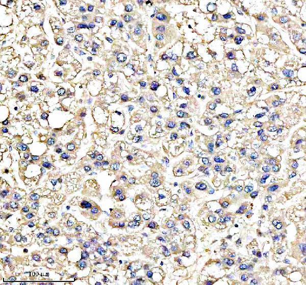
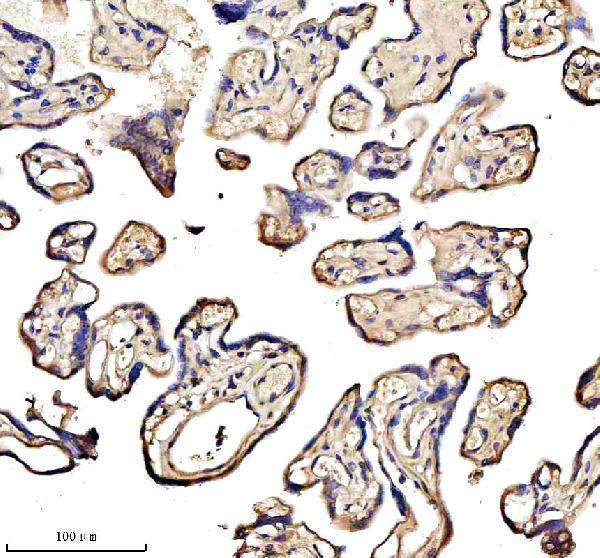
A05447-1 has real IHC images from paraffin-embedded human liver cancer, placenta, breast cancer and rectal cancer tissue (A05447-1 IHC captions); IF/ICC and human, mouse and rat reactivity are listed (catalog applications/reactivity).

Real IHC data IHC analysis of ISCU using anti-ISCU antibody (A05447-1). ISCU was detected in a paraffin-embedded section of human liver cancer tissue. Heat mediated antigen retrieval was performed in EDTA buffer (pH 8.0, epitope retrieval solution). The tissue section was blocked with 10% goat serum. The tissue section was then incubated with 1:100 rabbit anti-ISCU Antibody (A05447-1) overnight at 4°C. Peroxidase Conjugated Goat Anti-rabbit IgG was used as secondary antibody and incubated for 30 minutes at 37°C. The tissue section was developed using HRP Conjugated Rabbit IgG Super Vision Assay Kit (Catalog # SV0002) with DAB as the chromogen.
Anti-ISCU Antibody
Cat # A05447-1

A05447-1 will render with its human liver cancer paraffin-section IHC figure (A05447-1 IHC caption). Its other IHC captions show human placenta, breast cancer and rectal cancer paraffin sections; IF and ICC are listed applications, with no IF image supplied (A05447-1 IHC captions; catalog applications/IF image alts).

Which to pick: For tissue IHC, choose A05447-1: its own caption documents heat retrieval in EDTA at pH 8.0 and staining of a paraffin-embedded human liver cancer section; the fixative is unreported (A05447-1 IHC caption). For IF/ICC, A05447-1 is the listed option, with a 1:50 IF dilution; no IF figure is supplied (catalog applications/IF dilution/IF image alts). For human, mouse or rat samples, A05447-1 is a polyclonal antibody with those species listed as reactive, while its supplied IHC figures show human tissue only (catalog dilution_raw/reactivity; A05447-1 IHC captions).

Each figure is that product's own IHC / IF validation image from its datasheet.

References

  1. UniProt Consortium. UniProt entry Q9H1K1 (ISCU_HUMAN, Iron-sulfur cluster assembly enzyme ISCU).
  2. Human Protein Atlas. ISCU tissue IHC expression (reliability: Approved).
  3. Human Protein Atlas. ISCU subcellular location (ICC-IF): Localized to the cytosol and mitochondria..
  4. Human Protein Atlas. ISCU antibody validation summary (3 antibodies).
  5. miR-210 is a target of hypoxia-inducible factors 1 and 2 in renal cancer, regulates ISCU and correlates with good prognosis. British journal of cancer 2013 — PMC3619073.
  6. Identification and validation of mitochondrial ferroptosis and immune microenvironment-related hub biomarkers in liver cirrhosis by integrated bioinformatics analysis. Science progress 2025 — PMC12446859.
  7. Regulation of iron homeostasis by the p53-ISCU pathway. Scientific reports 2015 — PMC4642350.
  8. Clinically relevant HIF-1α-dependent metabolic reprogramming in oropharyngeal squamous cell carcinomas includes coordinated activation of CAIX and the miR-210/ISCU signaling axis, but not MCT1 and MCT4 upregulation. Oncotarget 2017 — PMC5355133.
  9. PubMed PMID:11060020 — UniProt-cited evidence.
  10. PubMed PMID:16541075 — UniProt-cited evidence.
  11. PubMed PMID:15489334 — UniProt-cited evidence.