This website uses cookies to ensure you get the best experience on our website.


- Table of Contents
Plan KIF11 chromogenic IHC in paraffin sections using the catalog antibody at 2–5 μg/mL (datasheet A01754-2). Compare cytoplasmic staining across the cell subsets highlighted by tissue IHC evidence (HPA tissue IHC).

Expected localisation, validated protocols, controls and antibodies — the at-a-glance facts below, then the full design guide.
| Expected localisation | Cytoplasmic staining in tissue (HPA tissue IHC); spindle-pole localisation is a molecular annotation (UniProt) | |
| Staining pattern | Cytoplasmic in seminiferous, gut and lymphoid cell subsets (HPA tissue IHC) | |
| Antigen retrieval | EDTA pH 8.0 HIER, heat-mediated (datasheet A01754-2) | |
| Positive control | Appendix+4 more · see all | |
| Negative control | Caudate |
| Fixation | Matched tissue-IHC evidence does not establish the fixation claim. Validate the specimen-specific method before use. (selected-SKU IHC image A01754-2) | |
| Caveat | Staining varies between cell subsets (HPA tissue IHC) | |
| Regulation | Expression regulation unannotated (UniProt) | |
| Isoform / epitope | No annotated isoforms; one 1–1056 chain (UniProt) |
The catalog antibody protocol is followed by three published KIF11 IHC protocols (PMC11898751; PMC5696167; PMC5696720).
| Sample | Paraffin-embedded human breast cancer tissue; fixative not specified (datasheet A01754-2) |
| Fixation | Image fixative and duration unreported (datasheet A01754-2); verify before use. |
| Sectioning | 4–5 µm sections on charged slides (standard) |
| Deparaffinisation | Xylene, graded ethanol series to water (standard) |
| Antigen retrieval | Heat retrieval: EDTA pH 8.0 (datasheet A01754-2); 20 min, 95–100 °C (standard) |
| Peroxidase block | 3% H2O2, 10 min, room temperature (standard) |
| Blocking | 10% goat serum (datasheet A01754-2) |
| Primary antibody | Rabbit anti-KIF11, 2-5 μg/ml (datasheet A01754-2) |
| Primary incubation | Overnight at 4 °C (datasheet A01754-2) |
| Detection | HRP-conjugated secondary, DAB chromogen (datasheet A01754-2) |
| Counterstain | Hematoxylin, blue, dehydrate and mount (standard) |
| Expected result | KIF11-positive staining in lymphoid tissue of appendix (HPA tissue IHC: High). HPA tissue profile: Cytoplasmic expression in several tissues. Highest levels in subsets of cells in seminiferous duct, the gastrointestinal tract, in lymphoid tissue and bone marrow. No signal in the no-primary control. |
KIF11 is a cytoplasmic motor associated with the spindle pole and has no transmembrane segment (UniProt P52732). In paraffin section IHC, expect cytoplasmic staining in subsets of cells, including glandular cells of the colon and duodenum, lymph node germinal center cells, and bone marrow hematopoietic cells (HPA: High in each). HPA rates its tissue IHC profile Enhanced, while describing medium consistency between staining and RNA expression (HPA: tissue IHC).
| Cytoplasmic staining is strongest in a subset of glandular, lymphoid, or hematopoietic cells. | This fits the reported compartment and uneven tissue distribution (UniProt P52732: cytoplasm; HPA: cytoplasmic expression in several tissues). Compare cells within the same section: HPA reports High staining in colon glandular cells, lymph node germinal center cells, and bone marrow hematopoietic cells, without implying that every cell must stain (HPA: tissue IHC). |
| Strong staining is predominantly nuclear or confined to a cell membrane, with little cytoplasmic signal. | Treat the compartment mismatch as an unconfirmed result and check controls, morphology, and detection background (UniProt P52732: cytoplasm and spindle pole; no transmembrane segment). A mitotic spindle related pattern may occur in dividing cells, but a nuclear or membrane dominant pattern alone does not match the stated localization (UniProt P52732: function and subcellular location). |
| Cells expected to be weak or negative stain as strongly as the expected positive population. | Check for cross reactivity or endogenous chromogenic detection activity before interpreting this as KIF11 (general IHC practice). For a concrete comparison, HPA reports KIF11 as Not detected in caudate glial cells, while reporting High staining in lymph node germinal center cells (HPA: tissue IHC); these observations are tissue and cell specific. |
| Diffuse color covers extracellular space, several compartments, or most cells without a clear cellular pattern. | This is less convincing than cell associated cytoplasmic staining (UniProt P52732: cytoplasm; HPA: tissue IHC profile). Review the no primary control, blocking, washes, and chromogen development to assess background (general chromogenic IHC practice). Background alone cannot establish KIF11 expression or a target specific fixation effect. |
| A section chosen for an HPA High population has no visible signal in that population. | First confirm that the relevant cells are present and identifiable; HPA levels refer to specified cell populations, not an entire organ (HPA: tissue IHC). If morphology is adequate, review primary antibody application, retrieval conditions, detection reagents, and the positive control (general IHC practice). The supplied sources do not identify a KIF11 specific cause of signal loss. |
| Compartment and cell cycle | KIF11 is annotated in the cytoplasm and at the spindle pole, and its motor function supports bipolar spindle formation during mitosis (UniProt P52732). Assess staining against cellular morphology and avoid requiring a visible spindle in every positive cell; KIF11 also has a reported non mitotic transport function (UniProt P52732). |
| Cell population within the section | HPA describes cytoplasmic expression in several tissues and its highest levels in subsets of cells (HPA: tissue IHC profile). Score the named cell population rather than averaging across the section: colon glandular cells, lymph node germinal center cells, and bone marrow hematopoietic cells are each High, whereas caudate glial cells are Not detected (HPA: tissue IHC). |
| Strength of IHC evidence | HPA calls the tissue profile Enhanced but describes medium consistency between antibody staining and RNA expression (HPA: tissue IHC reliability). Its antibody records differ: HPA010568 is IHC Enhanced, while HPA006916 and CAB017617 are IHC Supported (HPA: antibody validation). Interpret an unexpected compartment or cell population with the validation status of the antibody actually used. |
| Antigen retrieval and detection workflow | Target-specific fixation effects are not established by the supplied assay evidence. Verify with a matched IHC source before attributing a result to fixation. |
| Situation | Likely cause | Next action |
|---|---|---|
| No staining in identifiable colon or duodenal glandular cells, or lymph node germinal center cells. | A failed assay step or unsuitable control section is possible; these populations are reported High, but staining is not specified for every cell (HPA: tissue IHC; general IHC practice). | Verify morphology and run a known positive section alongside a no primary control; then check primary antibody application, retrieval, and detection steps (general IHC practice). Do not assign the failure to KIF11 fixation sensitivity: it is unreported in the supplied sources. |
| Signal appears mainly in nuclei or along plasma membranes. | The dominant compartment conflicts with the annotated cytoplasm and spindle pole and with the absence of a transmembrane segment (UniProt P52732). | Recheck cell boundaries and the no primary control, then compare with a documented cytoplasmic positive population (general IHC practice; HPA: High in colon glandular cells). Treat the unusual localization as unresolved unless independently supported. |
| Caudate glial cells stain strongly. | HPA reports KIF11 as Not detected in caudate glial cells; cross reactivity or background is possible, though a single discrepant section cannot identify the cause (HPA: tissue IHC; general IHC practice). | Confirm glial cell identity and compare the same run with an HPA High cell population and a no primary control (HPA: tissue IHC; general IHC practice). Reassess antibody specificity if strong cell associated staining persists. |
| Chromogen is widespread and obscures cell boundaries. | Excess detection background or endogenous activity can create color unrelated to the expected cytoplasmic distribution (general chromogenic IHC practice; UniProt P52732: cytoplasm). | Inspect no primary and detection controls, then review blocking, washes, and chromogen development (general IHC practice). Score KIF11 only where signal remains clearly cell associated and exceeds background. |
| Only scattered cells are positive in a tissue expected to stain. | Scattered staining may reflect the reported subset pattern rather than assay failure; HPA describes its highest levels in subsets of cells (HPA: tissue IHC profile). | Identify and score the relevant population, then compare its cytoplasmic signal with nearby cells and controls (HPA: tissue IHC; general IHC practice). Do not convert a population specific HPA level into a requirement for uniform staining. |
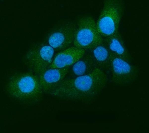
| What pattern should an IF/ICC image show? | HPA reports mainly cytosol and mid piece localization; it also lists mitotic spindle localization as uncertain (HPA: subcellular ICC-IF). These image annotations are distinct from its tissue IHC observations. | For IF/ICC interpretation, look first for the supported cytosol or approved mid piece pattern and treat spindle staining cautiously (HPA: subcellular ICC-IF). Use the separate IF/ICC guide for experimental conditions. |
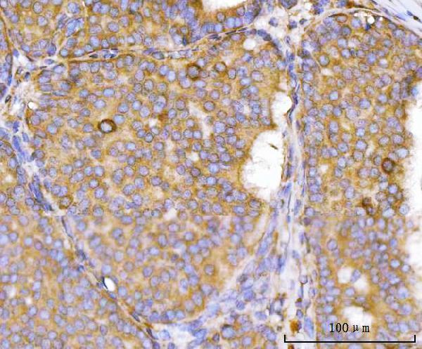
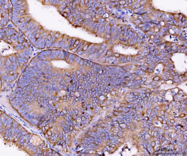
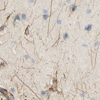


Comprehensive Human Protein Atlas IHC scoring per tissue (reliability: Enhanced — Medium consistency between antibody staining and RNA expression data.). Rows are taken directly from the HPA tissue chart — click any row's HPA link to view the source.
| Tissue | Cell type | Level | Evidence | Source |
|---|---|---|---|---|
| Appendix | Lymphoid tissue | High | Protein (IHC) | HPA → |
| Bone marrow | Hematopoietic cells | High | Protein (IHC) | HPA → |
| Colon | Glandular cells | High | Protein (IHC) | HPA → |
| Duodenum | Glandular cells | High | Protein (IHC) | HPA → |
| Kidney | Cells in tubules | High | Protein (IHC) | HPA → |
| Tissue | Cell type | Level | Evidence | Source |
|---|---|---|---|---|
| Caudate | Glial cells | Not detected | Protein (IHC) | HPA → |
Troubleshoot KIF11 staining in paraffin sections by checking retrieval, cellular localisation, controls, and scoring before interpreting chromogenic signal.
A01754-2 has IHC data from human paraffin sections and IF/ICC data from A431 cells (A01754-2 image captions); its listed reactivity is human (catalog: A01754-2 reactivity).
A01754-2 will render with its human breast cancer paraffin-section IHC figure (A01754-2 IHC caption). The same SKU has an A431 cell IF image (A01754-2 IF caption).
Which to pick: Choose A01754-2 for human paraffin-section IHC: its own caption documents EDTA pH 8 retrieval and chromogenic detection, but does not report the fixative (A01754-2 IHC caption). For IF/ICC, A01754-2 has A431 image data; M01754-3 is a rabbit monoclonal listed for human ICC/IF without a supplied image (A01754-2 IF caption; catalog: M01754-3 host, clone, applications, reactivity, IF image alts). Neither SKU has stated reactivity beyond human, so the payload supports no cross-species recommendation (catalog: both reactivity fields).