KLF5 / Krueppel-like factor 5 · IHC design guide

Design Immunohistochemistry for KLF5

Plan chromogenic KLF5 IHC in paraffin sections using the mainly nuclear tissue pattern as a guide (HPA tissue IHC). Compare cell types within each section and keep fixation consistent when evaluating staining intensity.

Evidence assembled Oct 2026 · For research use; verify linked source records and product datasheet before use
Immunohistochemistry protocol sheet for KLF5 (IHC for KLF5): expected localisation Mainly nuclear in tissue sections (HPA tissue IHC), antibody A00727, validated IHC image, and IHC protocol steps
Printable KLF5 IHC protocol sheet — expected localisation Mainly nuclear in tissue sections (HPA tissue IHC), antibody A00727, controls and protocol steps. Open the full KLF5 IHC guide →

KLF5 Immunohistochemistry Experimental Design Guide

Expected localisation, validated protocols, controls and antibodies — the at-a-glance facts below, then the full design guide.

Must know before staining
Expected localisation Mainly nuclear in tissue sections (HPA tissue IHC)
Staining pattern Mainly nuclear staining in most tissues; cell intensity varies (HPA tissue IHC)
Antigen retrieval Tris-EDTA pH 9.0 HIER, 95–98 °C, 20 min (rule: nuclear antigen)
Positive control ⓘ Adipose tissue+4 more · see all
Negative control ⓘ Hippocampus+1 more · see all
Important caveats
Reasons your staining may differ from the expected pattern.
Fixation Keep formalin fixation consistent across sections. (standard IHC practice; not target-specific)
Caveat Antibody staining has medium consistency with RNA data (HPA tissue IHC)
Regulation RNA is tissue enhanced in esophagus and skin (HPA tissue IHC; RNA)
Isoform / epitope Four isoforms; check whether the epitope is shared (UniProt)
Section 1

Recommended KLF5 IHC & IF Protocols

The catalog antibody protocol is followed by published KLF5 IHC methods for gastric tissue (PMC3553174) and paraffin sections (PMC12044660).

Recommended immunohistochemistry (IHC-P) protocol parameters
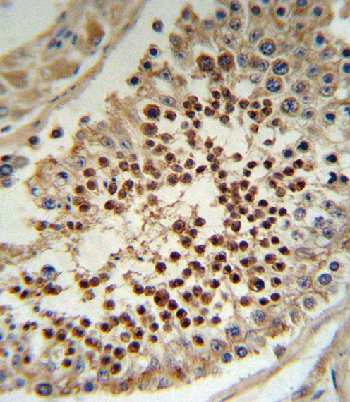
SampleFFPE human testis tissue (datasheet A00727)
FixationImage formalin-fixed; duration unreported (datasheet A00727); verify before use.
Sectioning4–5 µm sections on charged slides (standard)
DeparaffinisationXylene, graded ethanol series to water (standard)
Antigen retrievalHeat-induced epitope retrieval in Tris-EDTA buffer, pH 9.0, 20 min at 95–98 °C (standard rule: nuclear antigen)
Peroxidase block3% H2O2, 10 min, room temperature (standard)
Blocking10% normal serum of the secondary host, 30 min, room temperature (standard)
Primary antibodyRabbit anti-KLF5, 1:10-1:50 (datasheet A00727)
Primary incubationOvernight at 4 °C (standard)
DetectionHRP-polymer secondary, DAB chromogen 5–10 min (standard)
CounterstainHematoxylin, blue, dehydrate and mount (standard)
Expected resultKLF5-positive staining in adipocytes of adipose tissue (HPA tissue IHC: High). HPA tissue profile: Mainly nuclear expression in most tissues. No signal in the no-primary control.
💡Decision noteStart with the page’s Tris-EDTA pH 9.0 retrieval for nuclear KLF5 (UniProt: nucleus); citrate pH 6.0 is a published alternative (PMC12044660).
Section 2

What Is the Expected KLF5 Staining Pattern?

KLF5 is a nuclear transcription factor with no annotated transmembrane segment (UniProt Q13887: subcellular location, function, topology). In paraffin section IHC, expect mainly nuclear staining in the cell populations reported by HPA, including respiratory epithelial cells in bronchus and glandular cells in rectum (HPA: High in both; mainly nuclear expression in most tissues). HPA rates its tissue IHC evidence Approved, with medium consistency between staining and RNA expression (HPA: reliability).

What am I looking at on my slide?
Distinct nuclear staining in bronchial respiratory epithelium or rectal glandular cells, with nuclei identifiable by counterstain.This matches the reported compartment and high staining in those cell populations (HPA: mainly nuclear; High in bronchus and rectum). Compare cells within the section; an apparently dark region alone does not establish which cells are positive (standard IHC interpretation).
Predominantly membranous or widespread cytoplasmic staining, with little convincing nuclear signal.That pattern conflicts with the expected nuclear location (UniProt Q13887: nucleus; HPA: mainly nuclear). Treat it as suspect and review morphology, detection background and antibody specificity before assigning KLF5 positivity (standard IHC interpretation).
Strong staining in a cell population reported as undetected, such as hippocampal glial cells or ovarian stromal cells.HPA reports KLF5 as Not detected in those cells (HPA: hippocampus, glial cells; ovary, ovarian stroma cells). Confirm cell identity and exclude nonspecific antibody binding or endogenous detection activity before interpreting the signal (standard IHC practice).
Diffuse color across nuclei, cytoplasm and surrounding tissue, without clear cellular boundaries.A widespread haze is difficult to reconcile with mainly nuclear KLF5 staining (HPA: tissue IHC profile). Background from detection chemistry or insufficient blocking can obscure a true signal; judge positivity only where cellular localization remains clear (standard IHC practice).
No nuclear staining in bronchial respiratory epithelium or rectal glandular cells.These are reported High populations, so a blank result calls for a technical check (HPA: bronchus and rectum). HPA staining is a reference pattern, not a guarantee for every specimen; first verify tissue identity and a working IHC detection run (standard IHC practice).
💡Expected KLF5 appearanceCall a result positive when recognizable cells show clear nuclear chromogen staining, especially in HPA High populations such as bronchial respiratory epithelium or rectal glandular cells; predominantly membranous staining or diffuse tissue color is suspect (HPA: mainly nuclear, High in bronchus and rectum; UniProt Q13887: nucleus; standard IHC interpretation).
How each factor affects the staining
Choice of reference cellsHigh staining is reported in bronchial respiratory epithelium, rectal glandular cells and cells in testis seminiferous ducts (HPA: tissue IHC). HPA also reports Not detected hippocampal glial cells and ovarian stromal cells; these provide comparison patterns, subject to specimen and assay variation (HPA: tissue IHC; standard IHC practice).
Tissue evidence across sourcesUniProt describes expression only in testis and placenta, while HPA reports High staining in several additional cell populations (UniProt Q13887: tissue specificity; HPA: tissue IHC). For slide interpretation, describe the observed HPA IHC pattern and retain this disagreement rather than treating either source as universal.
Isoform coverage and antibody evidenceFour KLF5 isoforms are listed, but the supplied record gives no epitope or isoform coverage for an IHC antibody (UniProt Q13887: isoforms 1–4). HPA040398 is Approved for IHC and Supported for ICC; those ratings do not establish recognition of every isoform (HPA: antibody validation).
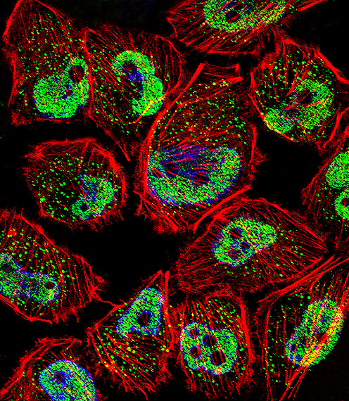
IF/ICC Q: where should fluorescence appear?A: Mainly in the nucleoplasm (HPA: ICC-IF, supported). HPA also notes Golgi apparatus and vesicle localization with uncertain support; interpret those signals cautiously and do not use them to redefine the expected paraffin IHC pattern (HPA: ICC-IF; HPA: tissue IHC profile).
Why is my staining missing, weak or wrong?
SituationLikely causeNext action
Known High cells show no nuclear signal.The section, antibody application or detection run may have failed; HPA High is a reference observation, not a specimen-level guarantee (HPA: bronchus and rectum; standard IHC practice).Confirm the expected cells on the counterstained section, check the assay controls and review the IHC-validated antibody's documented conditions, including retrieval and dilution where provided (standard IHC practice).
Staining is weak in parathyroid gland, endometrial stroma or splenic red pulp.These cell populations are reported Low, so faint staining may reflect their reference pattern (HPA: tissue IHC, Low).Assess the same run using a reported High cell population before calling the assay insensitive; compare nuclear localization and control performance (HPA: tissue IHC, High examples; standard IHC practice).
The slide has diffuse chromogen background.Background may arise from detection chemistry, blocking or an overly concentrated primary antibody (standard IHC practice).Inspect a primary-omission control, review blocking and detection steps, and adjust the antibody concentration within documented IHC conditions; then reassess nuclear contrast (standard IHC practice).
Signal is mainly cytoplasmic or outlines cell membranes.This conflicts with the principal nuclear pattern; location alone cannot identify the technical cause (UniProt Q13887: nucleus; HPA: mainly nuclear).Check cell boundaries and counterstain, inspect control sections, and seek concordant nuclear staining with an independently validated antibody if available (standard IHC practice).
Cells reported as Not detected show strong color.Misidentified cells, nonspecific binding or endogenous detection activity are possible explanations (HPA: hippocampal glial cells and ovarian stromal cells Not detected; standard IHC practice).Recheck morphology and the exact cell population; run a primary-omission control and investigate the detection system before assigning KLF5 positivity (standard IHC practice).
A positive control stains, but the study specimen does not.KLF5 staining varies by tissue and cell population, while HPA reports only medium consistency with RNA expression (HPA: tissue IHC profile and reliability).Record the specimen's cell types and nuclear staining result, compare them with the relevant HPA pattern, and avoid calling the assay failed solely because the study specimen is negative (HPA: tissue IHC; standard IHC interpretation).

Sample controls for KLF5 IHC & IF

🧪Run bronchus first and look for nuclear staining in respiratory epithelial cells (HPA: High in bronchus respiratory epithelial cells; UniProt Q13887: nucleus). Use ovary as the negative tissue and assess its stromal cells (HPA: Not detected in ovarian stroma cells); within the bronchus slide, independently confirmed KLF5-low cells should lack distinct nuclear staining and show only background.
Positive control tissue: Adipose tissue (Adipocytes, HPA High)
Negative control tissue: Hippocampus (HPA Not detected)
ICC-IF cell lines (HPA subcellular resource): HPA ICC-IF images show KLF5 in A-431, U-251MG, U2OS, with annotated localisation: Nucleoplasm (supported) (HPA subcellular).
Technical controls: Include a no-primary, secondary-only control; an isotype control matched to the primary antibody’s host species and clonality; and a KLF5 knockout sample or immunizing-peptide block, if available. For chromogenic bronchus IHC, quench endogenous peroxidase and check inflammatory cells for residual signal before interpreting epithelial staining (HPA: High in bronchus respiratory epithelial cells; standard IHC practice).
⚠️Feasibility: A target-specific fixation window or fixation effect is unreported; the selected A00727 caption explicitly describes formalin-fixed, paraffin-embedded testis stained with peroxidase and DAB (selected A00727 tissue-IHC caption). KLF5-specific antigen-retrieval dependence is unreported, so optimize retrieval empirically for paraffin sections; the supplied evidence does not establish that frozen sections or IF are easier (selected A00727 tissue-IHC caption). In bronchus, residual endogenous peroxidase in inflammatory cells can complicate chromogenic interpretation (standard IHC practice).

HPA tissue IHC evidence for KLF5

Comprehensive Human Protein Atlas IHC scoring per tissue (reliability: Approved — Medium consistency between antibody staining and RNA expression data.). Rows are taken directly from the HPA tissue chart — click any row's HPA link to view the source.

Positive expression · recommended positive controls

TissueCell typeLevelEvidenceSource
Adipose tissue Adipocytes High Protein (IHC) HPA →
Bone marrow Hematopoietic cells High Protein (IHC) HPA →
Bronchus Respiratory epithelial cells High Protein (IHC) HPA →
Cervix Squamous epithelial cells High Protein (IHC) HPA →
Nasopharynx Respiratory epithelial cells High Protein (IHC) HPA →

Undetected expression · recommended negative controls

TissueCell typeLevelEvidenceSource
Hippocampus Glial cells Not detected Protein (IHC) HPA →
Ovary Ovarian stroma cells Not detected Protein (IHC) HPA →
Section 3

Advanced KLF5 IHC Tips

Troubleshoot KLF5 staining in paraffin sections by checking retrieval, nuclear localisation, cell identity and controls before comparing staining scores.

Which retrieval conditions should I try first for KLF5 in paraffin sections?
Use Tris-EDTA pH 9.0 heat-induced retrieval at 95–98 °C for 20 min (page retrieval rule: nuclear antigen). If nuclear staining is weak, compare adjacent sections with a shorter heating time before testing another buffer, while keeping detection and exposure to chromogen constant (standard IHC practice). Allow sections to cool in retrieval buffer, then check that tissue remains attached and nuclear detail is preserved before judging signal strength (standard IHC practice). Score staining in intact nuclei, since KLF5 is a nuclear transcription factor and the supported subcellular location is nucleoplasm (UniProt Q13887: function and location; HPA subcellular: nucleoplasm supported).
How can I troubleshoot weak KLF5 staining after fixation?
Target-specific fixation sensitivity is unknown from the supplied evidence; the selected antibody image shows formalin-fixed, paraffin-embedded human testis, but provides no fixation comparison (A00727 caption). Record fixative, fixation duration, section age and retrieval conditions for each specimen, then compare similarly processed sections in the same staining run (standard IHC practice). If nuclear detail is poor or staining varies across a section, inspect morphology and processing history before increasing antibody concentration or chromogen development (standard IHC practice). Use an appropriate positive-control section to check the run, while recognising that a working control cannot establish how fixation affected each experimental specimen (standard IHC practice).
Where should convincing KLF5 staining appear in IHC sections?
Prioritise nuclear staining because KLF5 is annotated in the nucleus and HPA reports mainly nuclear tissue expression (UniProt Q13887: subcellular location; HPA tissue IHC: profile). In particular, inspect nucleoplasm within morphologically intact cells, since that is the supported subcellular location in HPA imaging (HPA subcellular: nucleoplasm supported). Cytoplasmic or granular signal alone needs caution: Golgi and vesicle locations are listed with uncertain support in IF images, so they do not establish a chromogenic tissue-IHC pattern (HPA subcellular: additional locations uncertain). Compare the suspected signal with negative controls and adjacent cells, using the same counterstain and development conditions to distinguish nuclear reaction product from pigment or debris (standard IHC practice).
How do I assess whether a C-terminal antibody detects every KLF5 isoform?
The record lists 4 KLF5 isoforms, while the selected tissue-IHC reagent is described only as a C-terminal antibody (UniProt Q13887: isoforms; A00727 caption). Those facts do not establish which isoforms retain the recognised epitope, so check the antibody's mapped immunogen or epitope against each isoform sequence before claiming pan-isoform detection (standard antibody-validation practice). KLF5 has no annotated transmembrane segment, signal peptide or glycosylation site in the supplied record; none of these annotations identifies the antibody's binding residues (UniProt Q13887: topology, processing and glycosylation). If isoform specificity matters, confirm the staining interpretation with an independently mapped antibody or an appropriate genetic control (standard antibody-validation practice).
How should I investigate KLF5 localisation by multiplex IF?
For IF as a separate application, pair KLF5 with a marker for the cell population being examined, and score overlap within individual nuclei rather than across neighbouring cells (standard IF practice; UniProt Q13887: nuclear location). Select fluorophores after checking tissue autofluorescence in an unstained section, favouring channels with a clear signal-to-background separation under the available filters (standard IF practice). KLF5 has no transmembrane segment and is chiefly nucleoplasmic, so use fixation-compatible permeabilisation that lets antibody reach the nuclear epitope, then verify that nuclear morphology remains intact (UniProt Q13887: topology; HPA subcellular: nucleoplasm supported; standard IF practice). HPA lists A-431, U-251MG and U2OS IF images, but their localisation does not validate a particular IF fixation or permeabilisation condition for this tissue experiment (HPA subcellular: cell lines with ICC/IF images).
What should I change when KLF5 IHC has diffuse background?
First compare stained sections with a secondary-only control and inspect whether background follows tissue edges, damaged areas or endogenous pigment rather than intact nuclei (standard IHC practice; UniProt Q13887: nuclear location). For peroxidase-based detection, check the endogenous peroxidase block and use the same DAB development time across test and control sections (standard IHC practice; A00727 caption: peroxidase conjugation and DAB staining). If background remains broad, titrate the primary antibody and strengthen washing or protein blocking one variable at a time (standard IHC practice). Retain a positive-control section during optimisation so a cleaner slide is not mistaken for improved specificity when nuclear KLF5 signal has also disappeared (standard IHC practice).
How should I score KLF5 staining across IHC samples? ⚠ ANSWER MARKED FOR VERIFICATION
Define the cell population and counting region before scoring, then record the percentage of cells with convincing nuclear staining alongside intensity categories for an H-score (standard IHC quantification; UniProt Q13887: nuclear location). Use a consistent intensity scale of 0–3 and calculate the H-score as the sum of each intensity score multiplied by its percentage of cells, giving a range of 0–300 (standard IHC quantification). Alternatively, report nuclear-positive cells per mm² when cell density itself is the outcome, with the evaluated tissue area stated (standard IHC quantification). Normalise comparisons to eligible intact cells or measured viable tissue area, and keep retrieval, imaging and scoring thresholds consistent across samples (standard IHC practice).
How can I distinguish true KLF5 staining from artefact?
A plausible positive pattern is nuclear staining in intact cells, consistent with KLF5's nuclear annotation and HPA's mainly nuclear tissue-IHC profile (UniProt Q13887: subcellular location; HPA tissue IHC: profile). Cell identity also matters: HPA reports high staining in cells of seminiferous ducts and no detection in ovarian stromal cells, although its antibody reliability is Approved with medium RNA–staining consistency (HPA tissue IHC: testis, ovary and reliability). Treat isolated edge staining, necrotic debris, diffuse extranuclear colour and signal reproduced by a secondary-only control as potential artefact (standard IHC practice). Investigate suspicious brown signal with morphology and peroxidase controls, because the selected image used peroxidase detection and DAB, while its caption does not evaluate clinical relevance (A00727 caption; standard IHC practice).
Boster reagents

Best KLF5 / Krueppel-like factor 5 IHC Antibodies

Anti-KLF5 antibodies have IHC images from human testis, colon cancer and skin cancer, and mouse and rat small intestine; IF images show human U251 and SiHa cells (catalog image captions).

Real IHC data KLF5 Antibody (C-term) IHC analysis in formalin fixed and paraffin embedded human testis tissue followed by peroxidase conjugation of the secondary antibody and DAB staining. This data demonstrates the use of the KLF5 Antibody (C-term) for immunohistochemistry. Clinical relevance has not been evaluated.
Anti-KLF5 Antibody (C-term)
Cat # A00727

A00727 will render with IHC data from formalin-fixed, paraffin-embedded human testis (A00727 IHC caption). Its IF image shows U251 cells, and its listed reactivity is human (A00727 IF caption; catalog applications and reactivity).

Which to pick: For human tissue IHC, A00727 has documented formalin-fixed, paraffin-embedded testis staining (A00727 IHC caption). For IF/ICC, A00727-1 lists both applications and shows staining in SiHa cells; A00727 also has an IF image from U251 cells (catalog applications; A00727-1 IF caption; A00727 IF caption). For cross-species tissue IHC, choose A00727-1: it is a rabbit polyclonal listed for human, mouse and rat, with paraffin-section images from all three species; the IHC captions do not report a fixative (catalog host, dilution data and reactivity; A00727-1 IHC captions).

Each figure is that product's own IHC / IF validation image from its datasheet.

References

  1. UniProt Consortium. UniProt entry Q13887 (KLF5_HUMAN, Krueppel-like factor 5).
  2. Human Protein Atlas. KLF5 tissue IHC expression (reliability: Approved).
  3. Human Protein Atlas. KLF5 subcellular location (ICC-IF): Mainly localized to the nucleoplasm. In addition localized to the Golgi apparatus and vesicles..
  4. Human Protein Atlas. KLF5 antibody validation summary (1 antibodies).
  5. Helicobacter pylori promotes the expression of Krüppel-like factor 5, a mediator of carcinogenesis, in vitro and in vivo. PloS one 2013 — PMC3553174.
  6. Krüppel-like Factor 5, Increased in Pancreatic Ductal Adenocarcinoma, Promotes Proliferation, Acinar-to-Ductal Metaplasia, Pancreatic Intraepithelial Neoplasia, and Tumor Growth in Mice. Gastroenterology 2018 — PMC5880723.
  7. Krüppel-like factor 5 is important for maintenance of crypt architecture and barrier function in mouse intestine. Gastroenterology 2011 — PMC3186863.
  8. Single-Nucleus RNA Sequencing and Spatial Transcriptomics for Squamous Cell Carcinoma Arising From Ovarian Mature Teratoma. Cancer science 2025 — PMC12044660.
  9. PubMed PMID:10572182 — UniProt-cited evidence.
  10. PubMed PMID:23134681 — UniProt-cited evidence.
  11. PubMed PMID:14702039 — UniProt-cited evidence.