This website uses cookies to ensure you get the best experience on our website.


- Table of Contents
Plan KYNU staining in paraffin sections using hepatocytes and renal tubules as high-expression references (HPA tissue IHC). Assess cytoplasmic staining by cell type and keep fixation consistent across sections (HPA tissue IHC; standard IHC practice).

Expected localisation, validated protocols, controls and antibodies — the at-a-glance facts below, then the full design guide.
| Expected localisation | Cytoplasmic staining in several tissues (HPA tissue IHC) | |
| Staining pattern | Cytoplasmic signal in hepatocytes, renal tubules and lung macrophages (HPA tissue IHC) | |
| Antigen retrieval | Citrate pH 6.0 HIER, 95–98 °C, 20 min (rule: cytoplasmic / membrane antigen) | |
| Positive control | Kidney+4 more · see all | |
| Negative control | Bone marrow+4 more · see all |
| Fixation | Keep fixation conditions consistent across sections. (standard IHC practice; not target-specific) | |
| Caveat | Staining and RNA show medium consistency; verify signal (HPA tissue IHC) | |
| Regulation | Abundance varies by cell type (HPA tissue IHC) | |
| Isoform / epitope | 2 isoforms; epitope coverage is unspecified (UniProt) |
The catalog antibody’s IHC-P protocol is accompanied by published KYNU IHC conditions for nevus, melanoma, lung adenocarcinoma, and other tissue sections (PMC9329910; PMC9139317; PMC7590459).
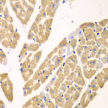
| Sample | Paraffin-embedded mouse heart tissue; fixative not specified (datasheet A06885) |
| Fixation | Image fixative and duration unreported (datasheet A06885); verify before use. |
| Sectioning | 4–5 µm sections on charged slides (standard) |
| Deparaffinisation | Xylene, graded ethanol series to water (standard) |
| Antigen retrieval | Heat-induced epitope retrieval in citrate buffer, pH 6.0, 20 min at 95–98 °C (standard rule: cytoplasmic / membrane antigen) |
| Peroxidase block | 3% H2O2, 10 min, room temperature (standard) |
| Blocking | 10% normal serum of the secondary host, 30 min, room temperature (standard) |
| Primary antibody | Rabbit anti-KYNU, 1:50-1:100 (datasheet A06885) |
| Primary incubation | Overnight at 4 °C (standard) |
| Detection | HRP-polymer secondary, DAB chromogen 5–10 min (standard) |
| Counterstain | Hematoxylin, blue, dehydrate and mount (standard) |
| Expected result | KYNU-positive staining in cells in tubules of kidney (HPA tissue IHC: High). HPA tissue profile: Cytoplasmic expression in several tissues, high expression in hepatocytes, renal tubules and lymphoid tissues. No signal in the no-primary control. |
KYNU is a cytosolic protein with no transmembrane segment (UniProt Q16719). In paraffin-section IHC, expect cytoplasmic staining, particularly in hepatocytes, renal tubular cells, lung macrophages and placental trophoblastic cells (HPA tissue IHC). HPA rates tissue staining reliability Enhanced, while noting medium consistency with RNA data and pending external verification (HPA tissue IHC).
| Strong cytoplasmic staining in hepatocytes or renal tubular cells (HPA tissue IHC). | This matches two HPA High patterns and KYNU’s cytosolic location (HPA tissue IHC; UniProt Q16719). Assess the named cells, since staining across an entire section can obscure whether the expected cells are positive (general IHC practice). |
| Predominantly nuclear staining with little cytoplasmic signal. | This does not match the principal IHC pattern (HPA tissue IHC). HPA reports an uncertain nucleoplasmic location in ICC-IF, so nuclear signal alone cannot establish KYNU localisation (HPA subcellular). Check controls and detection background (general IHC practice). |
| Strong staining in cells reported as undetected, such as bone-marrow hematopoietic cells (HPA tissue IHC). | The cell assignment conflicts with HPA’s observation and warrants a specificity check (HPA tissue IHC). Cross-reactivity or endogenous detection activity are possible explanations, not conclusions from this slide alone (general IHC practice). |
| Weak, diffuse colour across cells and surrounding tissue, without a clear cytoplasmic pattern. | This is difficult to score as KYNU when expected positive cells cannot be distinguished (HPA tissue IHC; general IHC practice). Compare a no-primary control and review blocking, washing and detection conditions (general IHC practice). |
| No staining in hepatocytes or renal tubular cells (HPA tissue IHC). | A blank known-positive cell population makes this IHC run inconclusive (HPA tissue IHC; general IHC practice). Review tissue identity, antibody and detection steps before interpreting an unstained test specimen as KYNU-negative (general IHC practice). |
| Cell type and tissue choice (HPA tissue IHC). | HPA reports High staining in hepatocytes, renal tubular cells, lung macrophages, placental trophoblastic cells and tonsillar non-germinal center cells; other cells in those tissues need separate interpretation (HPA tissue IHC). |
| Evidence strength (HPA tissue IHC; HPA antibodies). | The listed antibody HPA031686 has Enhanced IHC validation (HPA antibodies). HPA also describes medium agreement between tissue staining and RNA data, with external verification pending; an individual section still needs controls (HPA tissue IHC; general IHC practice). |
| Compartment and topology (UniProt Q16719; HPA subcellular). | KYNU is cytosolic and has no transmembrane segment (UniProt Q16719). HPA’s ICC-IF evidence supports cytosol, while its additional nucleoplasmic assignment is uncertain; avoid making nuclear staining the primary IHC criterion (HPA subcellular). |
| Isoforms and antibody coverage (UniProt Q16719). | UniProt lists two KYNU isoforms (UniProt Q16719). The supplied evidence gives no antibody epitope or isoform-specific IHC performance, so an unexpected staining pattern cannot be assigned to either isoform from these records. |
| Situation | Likely cause | Next action |
|---|---|---|
| Known-positive liver or kidney cells are blank (HPA tissue IHC). | An IHC run or specimen problem is possible; HPA’s High pattern does not identify which step failed (HPA tissue IHC; general IHC practice). | Verify tissue and cell identity, then check the antibody, retrieval and detection steps against the chosen IHC-P procedure (general IHC practice). |
| A chromogenic deposit appears broadly, including where cells lack the expected cytoplasmic pattern. | Endogenous detection activity or nonspecific reagent binding may contribute (general IHC practice). | Compare no-primary and reagent controls; review blocking, washing and, if applicable, endogenous enzyme blocking (general IHC practice). |
| Colour is mainly nuclear. | The principal KYNU location is cytosolic; an additional nucleoplasmic ICC-IF location is uncertain (UniProt Q16719; HPA subcellular). | Recheck localisation in a High-staining HPA cell type and inspect controls before accepting a nuclear IHC result (HPA tissue IHC; general IHC practice). |
| Unexpected cells stain strongly in an otherwise plausible section. | HPA levels are cell-specific; for example, bone-marrow hematopoietic cells are reported as Not detected (HPA tissue IHC). | Score the actual cell population, compare an HPA High reference population and use detection controls to investigate unexpected colour (HPA tissue IHC; general IHC practice). |
| A weak sample is being called negative. | HPA reports Low staining in some epithelia, including bronchial respiratory and colonic glandular cells (HPA tissue IHC). | Compare the sample with a High reference cell type in the same run and record intensity by cell type before assigning a negative score (HPA tissue IHC; general IHC practice). |
| Can IF/ICC show nuclear KYNU? | HPA supports cytosol as the main ICC-IF location and marks the additional nucleoplasmic location uncertain (HPA subcellular). | Treat cytosolic signal as the primary expectation, assess nuclear signal with controls and use the separate IF/ICC guide for assay setup (HPA subcellular; general IF practice). |


Comprehensive Human Protein Atlas IHC scoring per tissue (reliability: Enhanced — Medium consistency between antibody staining and RNA expression data. Pending external verification.). Rows are taken directly from the HPA tissue chart — click any row's HPA link to view the source.
| Tissue | Cell type | Level | Evidence | Source |
|---|---|---|---|---|
| Kidney | Cells in tubules | High | Protein (IHC) | HPA → |
| Liver | Hepatocytes | High | Protein (IHC) | HPA → |
| Lung | Macrophages | High | Protein (IHC) | HPA → |
| Placenta | Trophoblastic cells | High | Protein (IHC) | HPA → |
| Tonsil | Non-germinal center cells | High | Protein (IHC) | HPA → |
| Tissue | Cell type | Level | Evidence | Source |
|---|---|---|---|---|
| Bone marrow | Hematopoietic cells | Not detected | Protein (IHC) | HPA → |
| Cerebellum | Cells in granular layer | Not detected | Protein (IHC) | HPA → |
| Epididymis | Glandular cells | Not detected | Protein (IHC) | HPA → |
| Fallopian tube | Glandular cells | Not detected | Protein (IHC) | HPA → |
| Hippocampus | Glial cells | Not detected | Protein (IHC) | HPA → |
Troubleshoot KYNU chromogenic IHC in paraffin sections by checking retrieval, staining location, tissue controls and scoring before interpreting signal.
Anti-KYNU antibodies list IHC and IF/ICC use in human, mouse, and rat; real IHC data show paraffin-embedded mouse heart, while no IF image is provided (catalog applications/reactivity; A06885 image captions).
A06885 is the sole rendered card; its own figure documents IHC on paraffin-embedded mouse heart at 1:100 after microwave antigen retrieval in 10 mM PBS, pH 7.2 (A06885 IHC caption). A06885 also lists IF/ICC and human, mouse, and rat reactivity, but has no IF figure in the payload (A06885 catalog applications/reactivity; A06885 IF image alts).
Which to pick: Choose A06885 for paraffin-section IHC because its own figure shows mouse heart staining; the caption does not report the fixative (A06885 IHC caption). For IF/ICC, both A06885 and A06885-1 list those applications, but neither provides an IF image; both are rabbit antibodies with no clone specified (catalog applications/host/clone; IF image alts). For cross-species planning, either SKU lists human, mouse, and rat reactivity, while only A06885 supplies its own tissue IHC figure (catalog reactivity; IHC image alts).