This website uses cookies to ensure you get the best experience on our website.
- Table of Contents
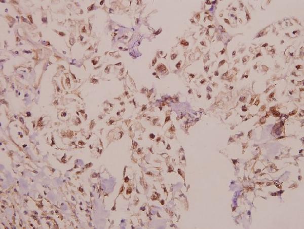
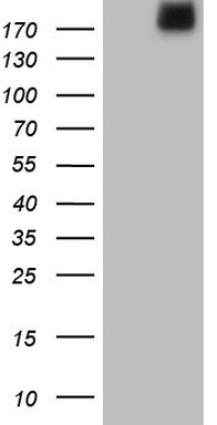
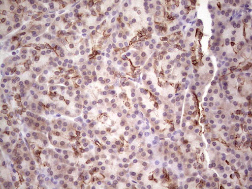
Facts about Laminin subunit alpha-4.
Human | |
---|---|
Gene Name: | LAMA4 |
Uniprot: | Q16363 |
Entrez: | 3910 |
Belongs to: |
---|
No superfamily |
DKFZp686D23145; LAMA3LAMA4*-1; LAMA4; laminin alpha 4 chain; Laminin alpha 4; laminin subunit alpha-4; laminin, alpha 4; Laminin-14 subunit alpha; Laminin-8 subunit alpha; Laminin-9 subunit alpha
Mass (kDA):
202.524 kDA
Human | |
---|---|
Location: | 6q21 |
Sequence: | 6; NC_000006.12 (112107931..112254722, complement) |
In adult, strong expression in heart, lung, ovary small and large intestines, placenta, liver; weak or no expression in skeletal muscle, kidney, pancreas, testis, prostate, brain. High expression in fetal lung and kidney. Expression in fetal and newborn tissues is observed in certain mesenchymal cells in tissues such as smooth muscle and dermis.
Secreted, extracellular space, extracellular matrix, basement membrane. Major component.
You're here because you want to optimize your Boster bio workflow. In this article, I will be discussing the LAMA4 Marker, Anti Laminin Antibody, as well as Luciferase. These three chemicals, which are essential components of boster Bio, can be used in a variety of molecular researches. These products are also available to scientists throughout the world.
Sample preparation is the key step to producing a clear Westernblot. Your choice of method to extract protein can make the difference in creating a beautiful blot. Boster has a wide range of specialized extraction and lysis buffers. The Boster guide will help you to choose the right one. Because of the differences in equipment requirements, speed, and efficiency, transfer methods are critical to Western blot preparation. It is important to choose the right method.
Anti-Laminin Antibody recognizes Laminin, a monoclonal protein that is a major component in the basal layer, which is the protein network that supports most organs and cells of the body. These proteins are important for many biological functions. They can modulate cell function as well as differentiation and adhesion. Anti-Laminin antibody makes a great choice when researching extracellular matrix proteins.
There are several tests that can detect serum antibody against laminin 332, which have been validated and developed by specialized laboratories. ELISA, immunoprecipitation by cultured keratinocytes, IB applying purified or recombinant lamine a3 chain are all methods for detecting serum antibodies against laminin 332. Although none are commercially accessible, the differences between these assays could be attributed to the various extracts used in laboratories. Other variations in the results could be due to detection antibodies against total IgG or IgG4, respectively.
The diagnostic indirect immunofluorescence method (IIF), which is the first test for laminin-based immunotherapy, is the most important. It involves exposing normal skin to 1M NaCl solution and generating laminalucida cleavages of the epidermal cellar membrane. Anti-Laminin-332 autoantibodies are found to be concentrated in the epidermal area of the artificial cut.
ELISA is another method to test for MMP. This technique uses large numbers sera from MMP patients. An immunoblotting test with laminin 332 should confirm a positive ELISA. This technique has many benefits over other immunoassays. The enzyme immunoassays take less time and can be automated, as opposed to immunoblotting. The method also yields numeric results, making them valuable substrates for the quantitative detection of MMP autoantibodies.
The luciferase-assay can provide precise results in a very short time. Luciferase gives the exact volume of a gene product, unlike chromatin antibodyprecipitation. This method does however not measure whether the protein interacts directly with DNA. It is easy to use and simple, making it a popular choice in research.
Luciferases are a group of enzymes that have evolved in many ways over the history of animals. They differ in their expression patterns and substrate specificity. Some organisms emit light indefinitely, while others produce bioluminescent Flashes. Luminescence in dinoflagellates occurs in flashes and is usually very short-lived. The luminescence of jellyfish, however lasts tens or more seconds, making it a viable candidate in a Boster Bio.
Researchers can use the luciferase test to help them understand gene expression. It uses the luciferase genes behind the promoter for transcription research within that region. Luminescence is proportional to the strength of the promoter. The stronger the promoter, the more light is produced. This method can be useful in troubleshooting because it can help determine the protein function.
To prepare crude Luciferase Extracts from Anesthetized Fish, dissected Light Organs of the Thoracic Cavity were first rinsed using 300 ml Deionized Water. Next, they were homogenized by 2.5 ml extraction buffer containing 20mM trisHCl, 50mM sodium chloride, as well as 1:200 Protease Inhibitor Cocktail III (pH8.0). After centrifugation the supernatant is suspended in the same buffer. It is then filtered with a 0.20mm membrane filters.
After purifying and storing the luciferase the anal extract was incubated with 1% goat serum in 0.1M PBS for a period 4 hours. The luciferase enzyme was then detected in the samples when it was react with the sections. Sephadex G 75 was used to separate the fractions of cells that were positive for luciferase.
Luminase naturally secreted by the body. Luciferase is an ideal model for determining factors that control how proteins fold properly. Because it sheds light during secretion, it is a popular choice for biomedical science. If you want to perform experiments on luciferase, consider these tips. You will have a more productive research if you understand how to use luciferase.
The LAMA4 marker is used to study cancer cell growth
OC sets have a high affinity for detecting LAMA4 expression, which can help differentiate between tumor and healthy tissue. OC sets are rich in laminins that help differentiate tumor cells. LAMA4 gene expression in tumor cells can help determine if LAMA4 has been expressed in a specific cancer or in a certain region. This article will discuss how to use the marker to determine if LAMA4 expression in different types of tumors is high or low.
The GTEx and TCGA databases were used for the generation of datasets of normal pancreatic cancer specimens. These datasets were adjusted by UCSC Xena. LAMA4 mRNA expression was measured. The LAMA4 gene expression level was determined by staining normal and tumor cell lines. We also examined the relationship of LAMA4 expression levels with the presence or absence cancer cells.
The LAMA4 genes is a genetically regulated gene that regulates the creation of different proteins. The gene is present in the human immune system, and can cause many disorders. It has been linked to schizophrenia and cancer. SSCP can detect LAMA4 genes in humans. One study even linked LAMA4 with the gene in mice and found that the condition is more common in women than in men.
Multiple mutations in LAMA4 result in decreased integrin-binding capacity and lower equilibrium sociation constants. This mutation is directly linked to the loss of endothelial cells, resulting in heart failure. This research has made LAMA4 useful in the detection of diseases in animal models. This gene can aid researchers in identifying genetic markers of LAMA4 genes. It is also helpful for evaluating the effect of LAMA4 on heart development.
GC-based expression levels of LAMA4 were analyzed by LinkedOmics to investigate the relationship between LAMA4 and survival in a patient's disease. LAMA4 expression levels were significantly associated with age, which is a significant risk factor for survival. A higher risk of adverse outcomes was also found when LAMA4 gene expression levels were associated with age. LAMA4 is associated with a lower overall survival rate for cancer patients.
PMID: 7781776 by Iivanainen A., et al. Primary structure and expression of a novel human laminin alpha 4 chain.
PMID: 8706685 by Richards A.J., et al. The complete cDNA sequence of laminin alpha 4 and its relationship to the other human laminin alpha chains.