LARP7 / La-related protein 7 · IHC design guide

Design Immunohistochemistry for LARP7

Plan LARP7 IHC on paraffin sections using the reported nuclear and nucleolar tissue pattern (HPA tissue IHC). Compare staining across sections with consistent fixation and appropriate controls (standard IHC practice).

Evidence assembled Oct 2026 · For research use; verify linked source records and product datasheet before use
Immunohistochemistry protocol sheet for LARP7 (IHC for LARP7): expected localisation Nuclear and nucleolar tissue staining (HPA tissue IHC), antibody A04811-1, validated IHC image, and IHC protocol steps
Printable LARP7 IHC protocol sheet — expected localisation Nuclear and nucleolar tissue staining (HPA tissue IHC), antibody A04811-1, controls and protocol steps. Open the full LARP7 IHC guide →

LARP7 Immunohistochemistry Experimental Design Guide

Expected localisation, validated protocols, controls and antibodies — the at-a-glance facts below, then the full design guide.

Must know before staining
Expected localisation Nuclear and nucleolar tissue staining (HPA tissue IHC)
Staining pattern Nuclear and nucleolar staining in most tissues (HPA tissue IHC)
Antigen retrieval EDTA pH 8.0 HIER, heat-mediated (datasheet A04811-1)
Positive control ⓘ Cerebellum+4 more · see all
Negative control ⓘ Adipose tissue+4 more · see all
Important caveats
Reasons your staining may differ from the expected pattern.
Fixation Keep fixation consistent across sections. (standard IHC practice; not target-specific)
Caveat Antibody staining has low consistency with RNA data (HPA tissue IHC)
Regulation No staining-linked regulator annotated (UniProt)
Isoform / epitope 3 isoforms; epitope coverage is unspecified (UniProt)
Section 1

Recommended LARP7 IHC & IF Protocols

The catalog antibody’s IHC-P protocol uses EDTA retrieval (datasheet A04811-1). Published LARP7 IHC protocols provide gastric and breast tissue examples (PMC3446825; PMC4126343).

Recommended immunohistochemistry (IHC-P) protocol parameters
SampleParaffin-embedded human breast cancer tissue; fixative not specified (datasheet A04811-1)
FixationImage fixative and duration unreported (datasheet A04811-1); verify before use.
Sectioning4–5 µm sections on charged slides (standard)
DeparaffinisationXylene, graded ethanol series to water (standard)
Antigen retrievalHeat retrieval: EDTA pH 8.0 (datasheet A04811-1); 20 min, 95–100 °C (standard)
Peroxidase block3% H2O2, 10 min, room temperature (standard)
Blocking10% goat serum (datasheet A04811-1)
Primary antibodyRabbit anti-LARP7, 1:50 recommended; image 1:100 (datasheet A04811-1)
Primary incubationOvernight at 4 °C (datasheet A04811-1)
DetectionHRP-conjugated secondary, DAB chromogen (datasheet A04811-1)
CounterstainHematoxylin, blue, dehydrate and mount (standard)
Expected resultLARP7-positive staining in purkinje cells of cerebellum (HPA tissue IHC: High). HPA tissue profile: Nuclear and nucleolar expression in most tissues. No signal in the no-primary control.
💡Decision noteStart with EDTA pH 8.0 HIER for the catalog antibody (datasheet A04811-1). For gastric sections, citrate pH 6.0 is a published alternative (PMC3446825).
Section 2

What Is the Expected LARP7 Staining Pattern?

LARP7 is nuclear and nucleoplasmic (UniProt Q4G0J3); HPA reports nuclear and nucleolar staining in most tissues, with high staining in selected cells including cerebellar Purkinje cells and testicular Leydig cells (HPA tissue IHC). It has no transmembrane segment (UniProt Q4G0J3 topology). Treat this as a provisional pattern: HPA rates the tissue staining Approved but reports low consistency with RNA expression and pending external verification (HPA tissue IHC).

What am I looking at on my slide?
Distinct nuclear, sometimes nucleolar, chromogen in Purkinje cells, epididymal glandular cells, hippocampal neurons or Leydig cells.This matches HPA's high staining in those cell types and its broader nuclear and nucleolar tissue profile (HPA tissue IHC). Score the named cells and their nuclei; do not infer that every cell in the section must stain equally.
Predominantly membranous or extracellular staining, with little nuclear signal.That compartment does not match the reported tissue pattern (HPA tissue IHC) or nucleoplasmic localisation (UniProt Q4G0J3). LARP7 has no transmembrane segment (UniProt Q4G0J3 topology). Check morphology, detection background and antibody specificity before scoring it as LARP7.
Strong staining in a cell population listed as not detected, such as adipocytes or lung alveolar cells.This conflicts with those specific HPA observations (HPA tissue IHC). Cross-reactivity or endogenous detection activity is possible (general IHC practice); review controls and cell identification. A discrepant result is not definitive because HPA notes low staining–RNA consistency and pending external verification (HPA tissue IHC).
Diffuse chromogen across nuclei, cytoplasm and tissue spaces, without clear cell boundaries.A field-wide deposit cannot establish the reported nuclear and nucleolar pattern (HPA tissue IHC). Background from the staining or detection workflow may obscure localisation (general IHC practice). Compare with a no-primary control and inspect tissue morphology.
No nuclear signal in a section containing recognisable Purkinje cells or Leydig cells.Those cells have high staining in HPA tissue images (HPA tissue IHC), so first check that the expected cells are present and the detection run worked. Absence in one preparation alone does not establish absent LARP7: HPA marks the tissue profile Approved with low RNA consistency and pending external verification (HPA tissue IHC).
💡Expected LARP7 appearanceCall a result consistent with LARP7 when identifiable HPA high-staining cells show clear nuclear, potentially nucleolar, chromogen (HPA tissue IHC), in keeping with nucleoplasmic localisation (UniProt Q4G0J3); isolated strong membrane or extracellular staining is suspect (UniProt Q4G0J3 topology; HPA tissue IHC).
How each factor affects the staining
Tissue and cell populationHPA reports high staining in Purkinje cells, epididymal glandular cells, hippocampal neurons and Leydig cells, but no detection in adipocytes, lung alveolar cells and several other listed populations (HPA tissue IHC). Interpret intensity within the identified cell type.
Evidence strengthThe tissue profile is Approved, yet HPA reports low consistency between antibody staining and RNA expression and says external verification is pending (HPA tissue IHC). Use the pattern as a reference for interpretation, not an independently verified specificity claim.
Antibody validationHPA lists HPA026842 and HPA027930 as IHC Approved, without an IHC Enhanced designation in the supplied record (HPA antibodies). An Approved designation alone does not resolve unexpected staining; assess the actual section and controls.
Isoforms and processingUniProt lists three LARP7 isoforms and a 1–582 chain, with no signal peptide or propeptide (UniProt Q4G0J3). The supplied sources give no antibody epitope or isoform recognition data, so they cannot predict isoform-specific IHC staining.
IF/ICC: should cytosol staining be expected?HPA ICC-IF reports supported nucleoplasmic and approved cytosolic localisation in A-431, U-251MG and U2OS images (HPA subcellular). Interpret that as IF/ICC evidence; HPA tissue IHC instead describes nuclear and nucleolar staining (HPA tissue IHC).
Why is my staining missing, weak or wrong?
SituationLikely causeNext action
No staining in a selected positive tissue.The expected cell population may be absent from the section, or the IHC run may have failed (general IHC practice).Locate the named cells first; Purkinje cells and Leydig cells are HPA high-staining examples (HPA tissue IHC). Check run controls and the catalog antibody's IHC-P instructions before changing conditions.
Weak staining where high signal was anticipated.Tissue averages can hide cell-specific differences; HPA assigns intensity to particular cells (HPA tissue IHC). A weak run signal is also possible (general IHC practice).Compare equivalent cell populations and a concurrent positive control. Review staining and detection steps (general IHC practice); the supplied sources do not establish LARP7-specific fixation or retrieval sensitivity.
Strong cytoplasmic staining dominates the IHC section.It conflicts with HPA's nuclear and nucleolar tissue profile (HPA tissue IHC), although HPA separately reports approved cytosolic ICC-IF localisation (HPA subcellular).Record the compartment and assay separately. Check a no-primary control and inspect whether identifiable nuclei stain before assigning the IHC signal to LARP7 (general IHC practice).
Membrane outlines or extracellular deposits appear positive.They do not fit LARP7's nucleoplasmic annotation or lack of a transmembrane segment (UniProt Q4G0J3). Nonspecific deposit is possible (general IHC practice).Review the no-primary control, tissue morphology and detection background; do not score deposits without a convincing cellular pattern (general IHC practice).
A listed HPA negative population stains strongly.Possible cross-reactivity or endogenous detection activity may mimic a positive result (general IHC practice); HPA lists some populations as not detected (HPA tissue IHC).Confirm cell identity and localisation, then compare no-primary and detection controls. Report the discrepancy alongside HPA's low staining–RNA consistency caveat (HPA tissue IHC).
Background obscures nuclear boundaries.Diffuse staining can prevent reliable compartment scoring (general IHC practice).Review blocking, washing, chromogen development and counterstain against run controls (general IHC practice). Score only nuclei that remain identifiable; HPA describes nuclear and nucleolar tissue staining (HPA tissue IHC).

Sample controls for LARP7 IHC & IF

🧪Run cerebellum first and score nuclear staining in Purkinje cells (High; HPA: cerebellum, Purkinje cells). Use adipose tissue as the negative comparator (HPA: adipocytes, Not detected); on the cerebellum slide, treat cells without nuclear signal as internal background references, without assuming a particular cell type is negative (HPA: only Purkinje cells are specified in the supplied row; UniProt Q4G0J3: nucleoplasm).
Positive control tissue: Cerebellum (Purkinje cells, HPA High)
Negative control tissue: Adipose tissue (HPA Not detected)
ICC-IF cell lines (HPA subcellular resource): HPA ICC-IF images show LARP7 in A-431, U-251MG, U2OS, with annotated localisation: Nucleoplasm (supported), Cytosol (approved) (HPA subcellular).
Technical controls: Include a no-primary, secondary-only control, host- and clonality-matched rabbit IgG isotype control, and LARP7 knockout material as a biological negative (caption: rabbit primary and peroxidase-conjugated goat anti-rabbit secondary; standard IHC practice). Quench endogenous peroxidase for HRP/DAB detection and inspect the cerebellum negative-control slide for pigment before scoring (caption: HRP/DAB; standard IHC practice).
⚠️Feasibility: No target-specific fixation window or fixation effect is reported, and the selected A04811-1 paraffin-section caption does not state a fixative (selected IHC caption). The caption reports heat-mediated retrieval in EDTA at pH 8.0 for breast cancer tissue, so retrieval should be checked in cerebellum rather than assumed to transfer unchanged (selected IHC caption). HPA reports ICC-IF images in A-431, U-251MG and U2OS, but the supplied evidence does not establish that IF or frozen sections are easier; check cerebellar neuronal pigment when interpreting DAB signal (HPA: subcellular images; standard IHC practice).

HPA tissue IHC evidence for LARP7

Comprehensive Human Protein Atlas IHC scoring per tissue (reliability: Approved — Low consistency between antibody staining and RNA expression data. Pending external verification.). Rows are taken directly from the HPA tissue chart — click any row's HPA link to view the source.

Positive expression · recommended positive controls

TissueCell typeLevelEvidenceSource
Cerebellum Purkinje cells High Protein (IHC) HPA →
Epididymis Glandular cells High Protein (IHC) HPA →
Hippocampus Neuronal cells High Protein (IHC) HPA →
Testis Leydig cells High Protein (IHC) HPA →
Adrenal gland Glandular cells Medium Protein (IHC) HPA →

Undetected expression · recommended negative controls

TissueCell typeLevelEvidenceSource
Adipose tissue Adipocytes Not detected Protein (IHC) HPA →
Bone marrow Hematopoietic cells Not detected Protein (IHC) HPA →
Lung Alveolar cells Not detected Protein (IHC) HPA →
Lymph node Germinal center cells Not detected Protein (IHC) HPA →
Soft tissue Fibroblasts Not detected Protein (IHC) HPA →
Section 3

Advanced LARP7 IHC Tips

Use the catalog antibody’s tissue IHC conditions as a starting point, then judge LARP7 staining by cell type and nuclear localisation.

Which retrieval conditions should I start with for paraffin sections?
Start with heat-mediated retrieval in EDTA at pH 8.0 (datasheet A04811-1). The selected paraffin-section image used that retrieval before an overnight incubation at 4°C with antibody at 1:100, so keep those conditions together for the first comparison (caption A04811-1). If nuclear staining is weak, compare a modestly longer heat exposure against the starting condition while holding antibody dilution, section thickness and detection constant (standard IHC practice). Include a known staining section in each run, and judge recovery in intact nuclei because LARP7 is assigned to the nucleoplasm (UniProt Q4G0J3; standard IHC practice).
Could fixation explain weak LARP7 staining?
The selected image identifies a paraffin-embedded section but does not state its fixative, so target-specific fixation sensitivity is unknown (caption A04811-1). Record the actual fixative and fixation duration for each specimen before attributing differences in nuclear staining to LARP7 abundance (standard IHC practice). Compare specimens processed together, and evaluate morphology alongside signal because excessive or uneven fixation can alter antigen accessibility in tissue sections generally (standard IHC practice). If a batch stains poorly, repeat the established EDTA pH 8.0 retrieval with a known staining section before changing fixation or antibody concentration (datasheet A04811-1; standard IHC practice).
Where should convincing LARP7 staining appear?
Prioritise staining in intact nuclei: UniProt places LARP7 in the nucleoplasm, while tissue IHC reports nuclear and nucleolar expression in most tissues (UniProt Q4G0J3; HPA tissue IHC). Cytosolic signal needs cautious interpretation because cell imaging also lists cytosol, but tissue IHC has low consistency with RNA expression and awaits external verification (HPA subcellular; HPA tissue IHC). Compare nuclear and cytoplasmic staining separately within the same cell population rather than combining them into one positive score (standard IHC practice). Use preserved nuclear outlines and a consistent counterstain to distinguish nucleolar enrichment from precipitated chromogen or damaged tissue (standard IHC practice).
Can this stain distinguish LARP7 isoforms or modified epitopes?
Do not assign an isoform from this stain alone: the record lists 3 LARP7 isoforms, but the supplied tissue image does not define the antibody epitope or isoform coverage (UniProt Q4G0J3; caption A04811-1). LARP7 contains a La-type RNA-binding region at residues 28–122, an RRM at 125–203 and an xRRM at 450–563 (UniProt Q4G0J3). Several phosphorylated residues are annotated near 257–273, without evidence here that they affect this antibody’s staining (UniProt Q4G0J3). Obtain epitope mapping or isoform-specific validation before interpreting a changed IHC signal as altered splicing or phosphorylation (standard IHC practice).
How should I assess LARP7 by multiplex immunofluorescence?
Treat IF as a separate assay and pair LARP7 with a marker identifying the expected cell type, such as Purkinje cells in cerebellum, to check whether the signal occurs in the intended population (HPA tissue IHC; standard IF practice). Choose spectrally separated fluorophores and inspect an unstained section so tissue autofluorescence does not masquerade as LARP7 (standard IF practice). LARP7 has no transmembrane segment and is assigned chiefly to the nucleoplasm, so permeabilise sufficiently to expose intracellular epitopes while preserving nuclear morphology (UniProt Q4G0J3; standard IF practice). Check nuclear and cytosolic channels separately because cell imaging also reports approved cytosolic localisation (HPA subcellular).
How can I reduce diffuse or widespread DAB staining?
First inspect a section processed without primary antibody to separate detection background from antibody-dependent signal (standard IHC practice). The selected image used 10% goat serum, antibody at 1:100 overnight at 4°C, and a peroxidase-linked secondary for 30 minutes at 37°C (caption A04811-1). For excess chromogen, check wash quality, block endogenous peroxidase and shorten DAB development while keeping a known staining section alongside (standard IHC practice). Score intact nuclei separately from diffuse cytoplasm, tissue edges and pigment, because LARP7 tissue IHC is predominantly nuclear or nucleolar (HPA tissue IHC; standard IHC practice).
What is a reproducible way to score LARP7 IHC? ⚠ ANSWER MARKED FOR VERIFICATION
Define the cell population and tissue area before scoring, then record nuclear staining as percentage positive and intensity for an H-score of 0–300 (standard IHC practice). If counting cells, report positive nuclei per mm² and normalise that count to the number of evaluable cells or tissue area in each region (standard IHC practice). Keep nucleolar and cytoplasmic observations separate from the primary nuclear score because the reported compartments differ across tissue IHC and cell imaging (HPA tissue IHC; HPA subcellular). Apply identical thresholds, counterstain review and exclusion rules for damaged areas across samples, and report the scoring compartment explicitly (standard IHC practice).
How do I separate genuine LARP7 staining from artefact?
A plausible positive follows intact nuclear or nucleolar structures in an appropriate cell population, consistent with reported tissue staining and nucleoplasmic localisation (HPA tissue IHC; UniProt Q4G0J3). High staining has been reported in cerebellar Purkinje cells, whereas adipocytes were reported as not detected, but those observations are contextual checks rather than universal controls (HPA tissue IHC). Discount staining confined to section edges, necrotic areas or regions with poor nuclear preservation, and use a no-primary section to assess endogenous enzyme or detection signal (standard IHC practice). Interpret discordant staining cautiously because the tissue IHC profile has low consistency with RNA expression and is pending external verification (HPA tissue IHC).
Boster reagents

Best LARP7 / La-related protein 7 IHC Antibodies

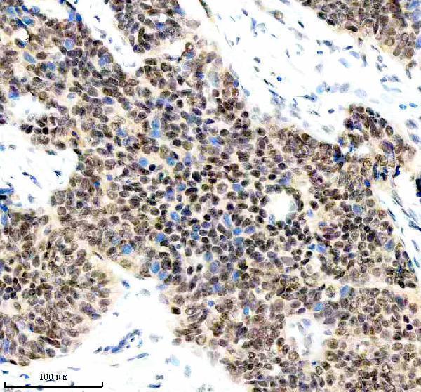
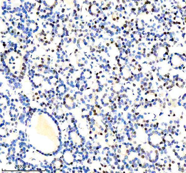
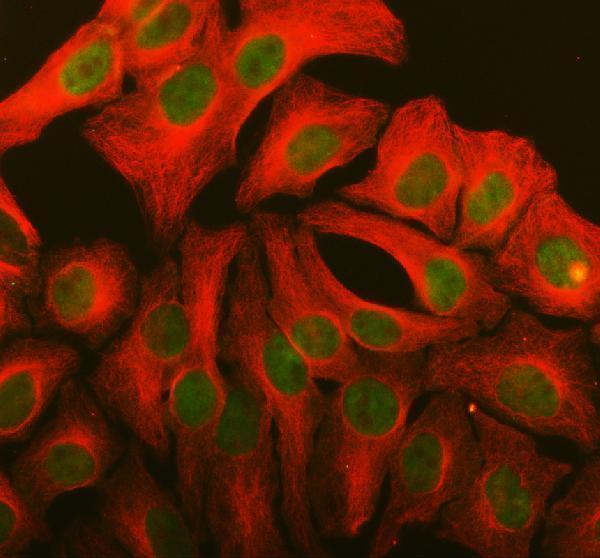
The IHC-validated anti-LARP7 antibody has paraffin-section images from human breast and thyroid cancers and rat testis, plus an IF/ICC image from HeLa cells (catalog images: A04811-1).

Real IHC data IHC analysis of LARP7 using anti-LARP7 antibody (A04811-1). LARP7 was detected in a paraffin-embedded section of human breast cancer tissue. Heat mediated antigen retrieval was performed in EDTA buffer (pH 8.0, epitope retrieval solution). The tissue section was blocked with 10% goat serum. The tissue section was then incubated with 1:100 rabbit anti-LARP7 Antibody (A04811-1) overnight at 4°C. Peroxidase Conjugated Goat Anti-rabbit IgG was used as secondary antibody and incubated for 30 minutes at 37°C. The tissue section was developed using HRP Conjugated Rabbit IgG Super Vision Assay Kit (Catalog # SV0002) with DAB as the chromogen.
Anti-LARP7 Antibody
Cat # A04811-1

A04811-1 is the sole SKU rendered; its IHC images show paraffin sections of human breast and thyroid cancers and rat testis (catalog IHC captions: A04811-1). Its IF/ICC image shows HeLa cells (catalog IF caption: A04811-1).

Which to pick: Choose A04811-1 for paraffin-section IHC: its images document EDTA retrieval at pH 8.0 and primary antibody at 1:100; the fixative is unreported (catalog IHC captions: A04811-1). For IF/ICC, the same polyclonal SKU has an image in HeLa cells (catalog IF caption: A04811-1; catalog: polyclonal). For cross-species work, A04811-1 lists human, mouse and rat reactivity, with IHC images supplied for human and rat samples (catalog reactivity and IHC captions: A04811-1).

Each figure is that product's own IHC / IF validation image from its datasheet.

References

  1. UniProt Consortium. UniProt entry Q4G0J3 (LARP7_HUMAN, La-related protein 7).
  2. Human Protein Atlas. LARP7 tissue IHC expression (reliability: Approved).
  3. Human Protein Atlas. LARP7 subcellular location (ICC-IF): Localized to the nucleoplasm and cytosol..
  4. Human Protein Atlas. LARP7 antibody validation summary (2 antibodies).
  5. LARP7 is a potential tumor suppressor gene in gastric cancer. Laboratory investigation; a journal of technical methods and pathology 2012 — PMC3446825.
  6. LARP7 in papillary thyroid carcinoma induces NIS expression through suppression of the SHH signaling pathway. Molecular medicine reports 2018 — PMC5983951.
  7. LARP7 suppresses P-TEFb activity to inhibit breast cancer progression and metastasis. eLife 2014 — PMC4126343.
  8. LARP7 enhances the potential of dental pulp stem cells to promote peripheral nerve repair. Stem cells (Dayton, Ohio) 2026 — PMC13223745.
  9. PubMed PMID:18483487 — UniProt-cited evidence.
  10. PubMed PMID:15815621 — UniProt-cited evidence.
  11. PubMed PMID:15489334 — UniProt-cited evidence.