This website uses cookies to ensure you get the best experience on our website.


- Table of Contents
Plan chromogenic IHC on paraffin sections using the catalog antibody at 1:100–1:300 (datasheet: IHC dilution). Interpret nuclear tissue staining (HPA tissue IHC) alongside the peroxisome matrix annotation (UniProt), with caution because HPA rates its tissue IHC reliability uncertain (HPA tissue IHC).

Expected localisation, validated protocols, controls and antibodies — the at-a-glance facts below, then the full design guide.
| Expected localisation | Nuclear staining observed; peroxisome matrix expected (HPA tissue IHC) (UniProt) | |
| Staining pattern | Nuclear staining in several tissues (HPA tissue IHC) | |
| Antigen retrieval | Citrate pH 6.0 HIER, 95–98 °C, 20 min (rule: cytoplasmic / membrane antigen) | |
| Positive control | Caudate+4 more · see all | |
| Negative control | Appendix+4 more · see all |
| Fixation | Matched tissue-IHC evidence does not establish the fixation claim. Validate the specimen-specific method before use. (selected-SKU IHC image A11643) | |
| Caveat | Nuclear IHC conflicts with peroxisomal location (HPA tissue IHC) (UniProt) | |
| Regulation | Staining regulation not specified (UniProt) | |
| Isoform / epitope | 2 isoforms; epitope impact undetermined (UniProt) |
The catalog antibody’s IHC-P protocol is accompanied by published LONP2 protocols for xenograft tumors (PMC13588115) and cervical cancer tissue microarrays (PMC5846714).
| Sample | Paraffin-embedded human lung carcinoma tissue; fixative not specified (datasheet A11643) |
| Fixation | Image fixative and duration unreported (datasheet A11643); verify before use. |
| Sectioning | 4–5 µm sections on charged slides (standard) |
| Deparaffinisation | Xylene, graded ethanol series to water (standard) |
| Antigen retrieval | Heat-induced epitope retrieval in citrate buffer, pH 6.0, 20 min at 95–98 °C (standard rule: cytoplasmic / membrane antigen) |
| Peroxidase block | 3% H2O2, 10 min, room temperature (standard) |
| Blocking | 10% normal serum of the secondary host, 30 min, room temperature (standard) |
| Primary antibody | Rabbit anti-LONP2, 1:100 - 1:300 (datasheet A11643) |
| Primary incubation | Overnight at 4 °C (standard) |
| Detection | HRP-polymer secondary, DAB chromogen 5–10 min (standard) |
| Counterstain | Hematoxylin, blue, dehydrate and mount (standard) |
| Expected result | LONP2-positive staining in glial cells of caudate (HPA tissue IHC: High). HPA tissue profile: Nuclear expression in several tissues. No signal in the no-primary control. |
LONP2 is a peroxisomal matrix protein with no transmembrane segment, so a convincing cellular pattern is cytoplasmic and granular where resolution permits (UniProt Q86WA8 localization and topology). HPA reports high IHC staining in glial, glandular, epithelial, glomerular and seminiferous-duct cells, but also reports nuclear staining in several tissues. Its tissue IHC reliability is Uncertain because the subcellular location conflicts with external data (HPA tissue IHC).
| Granular cytoplasmic signal appears in kidney glomerular cells or cervical glandular cells. | This compartment fits a peroxisomal matrix protein (UniProt Q86WA8 localization). HPA rates staining High in both named cell populations (HPA tissue IHC); the match supports further validation but does not establish antibody specificity. |
| Predominantly nuclear signal appears, with little convincing cytoplasmic staining. | HPA describes nuclear expression in several tissues, yet rates the tissue IHC evidence Uncertain because external subcellular data contradict it (HPA tissue IHC). Treat a nuclear-only result as unresolved, not confirmed LONP2 localization; check controls and seek independent evidence. |
| Strong staining appears in a cell population expected to be unstained, such as ovarian stromal cells. | HPA reports LONP2 as Not detected in ovarian stromal cells (HPA tissue IHC). Unexpected color could reflect cross-reactivity or endogenous detection activity; review the cell identity, compare adjacent tissue and inspect the no-primary control. |
| Color covers tissue broadly without a recognizable cellular pattern. | Diffuse background cannot establish the peroxisomal matrix localization reported for LONP2 (UniProt Q86WA8 localization). Uneven blocking, insufficient washing or detection background are general IHC possibilities; a no-primary control helps distinguish detection background from antibody-dependent staining. |
| No signal appears in kidney glomerular cells under the same run conditions. | HPA rates staining High in cells in glomeruli (HPA tissue IHC), so absence there calls for a run-level check. Confirm the relevant cells are present, then review retrieval, primary antibody dilution and chromogen development as general IHC variables. |
| Subcellular assignment | UniProt places LONP2 in the peroxisome matrix and lists no transmembrane segment (UniProt Q86WA8 localization and topology). HPA instead describes nuclear tissue staining and flags a contradiction with external localization data (HPA tissue IHC). Record the observed compartment before interpreting positivity. |
| Cell population and tissue | HPA rates glomerular cells High but liver cholangiocytes and pancreatic endocrine cells Low (HPA tissue IHC). UniProt reports high expression in liver, kidney and pancreas at the tissue level (UniProt Q86WA8 tissue specificity); that broader statement does not predict equal IHC intensity in every cell type. |
| Antibody validation | The listed rabbit polyclonal antibody HPA008862 has an Uncertain IHC validation status (HPA antibody record). A pattern that resembles HPA staining is therefore a comparison point, not independent proof of LONP2 specificity; interpret it alongside compartment and control results. |
| Isoforms and epitope coverage | UniProt lists two LONP2 isoforms and a chain spanning residues 2–852 (UniProt Q86WA8 isoforms and processing). The supplied records do not locate the antibody epitope or establish isoform-specific IHC detection; avoid assigning a staining difference to one isoform. |
| Situation | Likely cause | Next action |
|---|---|---|
| Only nuclei stain in an otherwise clean section. | HPA reports nuclear staining, but its tissue IHC reliability is Uncertain because that location conflicts with external data (HPA tissue IHC). | Document the nuclear pattern without calling it confirmed LONP2. Compare a no-primary control and an HPA High cell population, then seek independent localization evidence (HPA tissue IHC; UniProt Q86WA8 localization). |
| Kidney glomerular cells show no detectable staining. | Those cells are High in HPA tissue IHC, so the result differs from the reported pattern (HPA tissue IHC); the cause is not established by this record. | Check that glomeruli are in the section and that run controls developed. Review retrieval, antibody dilution and chromogen steps as general IHC troubleshooting before interpreting the sample as negative. |
| Ovarian stromal cells stain strongly. | HPA reports Not detected in ovarian stromal cells (HPA tissue IHC). Cross-reactivity or endogenous detection activity is possible, but neither is proven by the stain alone. | Verify cell identity and inspect a no-primary control. If color remains without primary antibody, troubleshoot the chromogenic detection system; if it disappears, reassess primary antibody specificity. |
| Background obscures individual cells across the section. | The distribution prevents comparison with HPA's cell-specific observations (HPA tissue IHC). General IHC sources of background include residual detection activity and inadequate blocking or washing. | Use a no-primary control to locate detection background. Review blocking, washing and detection conditions, then score only identifiable cells once the background allows a compartment call. |
| Liver or pancreas looks weaker than kidney. | UniProt describes high expression across these organs, while HPA scores liver cholangiocytes and pancreatic endocrine cells Low and kidney glomerular cells High (UniProt Q86WA8 tissue specificity; HPA tissue IHC). | Compare the specified cell populations rather than whole-section color. Report cell type and intensity together; do not infer a failed run solely from weaker staining in those HPA Low populations. |
| Can IF/ICC settle a nuclear-versus-peroxisomal IHC result? | The supplied HPA subcellular record has no main location or ICC-IF images for LONP2 (HPA subcellular record), so it cannot resolve the tissue IHC discrepancy. | Treat IF/ICC as a separate validation question and consult its dedicated guide. Here, document the IHC compartment and the conflict between UniProt peroxisomal localization and HPA's Uncertain nuclear tissue pattern (UniProt Q86WA8; HPA tissue IHC). |


Comprehensive Human Protein Atlas IHC scoring per tissue (reliability: Uncertain — Medium consistency between antibody staining and RNA expression data. Subcellular location is contradicted by external data.). Rows are taken directly from the HPA tissue chart — click any row's HPA link to view the source.
| Tissue | Cell type | Level | Evidence | Source |
|---|---|---|---|---|
| Caudate | Glial cells | High | Protein (IHC) | HPA → |
| Cervix | Glandular cells | High | Protein (IHC) | HPA → |
| Endometrium | Cells in endometrial stroma | Medium | Protein (IHC) | HPA → |
| Esophagus | Squamous epithelial cells | High | Protein (IHC) | HPA → |
| Fallopian tube | Glandular cells | High | Protein (IHC) | HPA → |
| Tissue | Cell type | Level | Evidence | Source |
|---|---|---|---|---|
| Appendix | Glandular cells | Not detected | Protein (IHC) | HPA → |
| Ovary | Ovarian stroma cells | Not detected | Protein (IHC) | HPA → |
| Small intestine | Glandular cells | Not detected | Protein (IHC) | HPA → |
| Spleen | Cells in red pulp | Not detected | Protein (IHC) | HPA → |
| Stomach | Glandular cells | Not detected | Protein (IHC) | HPA → |
Troubleshoot LONP2 chromogenic IHC by checking retrieval, compartment, controls and scoring; use the IF guidance only when planning a separate fluorescence experiment.
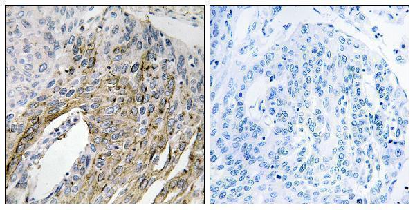
The catalog lists A11643 for IHC and IF/ICC in human, mouse, and rat (catalog: applications/reactivity). Its real IHC image shows paraffin-embedded human lung carcinoma tissue (catalog: IHC image caption).
A11643 will render with an IHC image of paraffin-embedded human lung carcinoma tissue and a peptide-blocked comparison (catalog: IHC image caption). IF and ICC are listed applications, with human, mouse, and rat reactivity; no IF image is supplied (catalog: applications/reactivity/IF image alts).
Which to pick: Choose A11643 for paraffin-section IHC because its own image caption documents that preparation; the fixative is unreported (catalog: IHC image caption). The same rabbit polyclonal antibody is listed for IF/ICC, although no IF image is supplied (catalog: host/dilution_raw/applications/IF image alts). Its listed reactivity covers human, mouse, and rat, while the supplied tissue image documents human tissue only (catalog: reactivity/IHC image caption).